principles of gene manipulation
Bạn đang xem bản rút gọn của tài liệu. Xem và tải ngay bản đầy đủ của tài liệu tại đây (13.8 MB, 320 trang )
Contents and supplementary information for:
Principles of
Gene Manipulation
Chapter 1 Gene manipulation: an all-embracing technique
Chapter 2
Basic techniques
- (POGC02.pdf, 1,560KB)
Chapter 3 Cutting and joining DNA molecules
Chapter 4 Basic biology of plasmid and phage vectors
Chapter 5 Cosmids, phasmids and other advanced vectors
Chapter 6 Cloning strategies
Additional updated information on
Cloning strategies
Chapter 7 Sequencing and
mutagenesis
Chapter 8 Cloning in bacteria other than
E. coli
Chapter 9 Cloning in
Saccharomyces cerevisiae
and other fungi
Chapter 10 Gene transfer to animal cells
Additional updated information on
Gene transfer to animal
cells
Chapter 11 Genetic manipulation of animals
Additional updated information on
Genetic manipulation of
animals
Chapter 12 Gene transfer to plants
Additional updated information on
Gene transfer to plants
Chapter 13 Advances in transgenic technology
Additional updated information on
Advances in transgenic
technology
or (POGC13.pdf - size: 353KB)
Chapter 14 Applications of recombinant DNA tecnology
Overview / Supplememtary Material / Related Titles / Related Websites / Ordering Information / Examination Copies / MCQ's /
New Edition
CHAPTER 1
Gene manipulation:
an all-embracing technique
Introduction
Occasionally technical developments in science
occur that enable leaps forward in our knowledge
and increase the potential for innovation. Molecular
biology and biomedical research experienced such
a revolutionary change in the mid-70s with the
development of gene manipulation. Although the
initial experiments generated much excitement, it is
unlikely that any of the early workers in the field
could have predicted the breadth of applications to
which the technique has been put. Nor could they
have envisaged that the methods they developed
would spawn an entire industry comprising several
hundred companies, of varying sizes, in the USA
alone.
The term gene manipulation can be applied to
a variety of sophisticated in vivo genetics as well as
to in vitro techniques. In fact, in most Western
countries there is a precise legal definition of gene
manipulation as a result of government legisla-
tion to control it. In the UK, gene manipulation is
defined as
the formation of new combinations of heritable
material by the insertion of nucleic acid molecules,
produced by whatever means outside the cell,
into any virus, bacterial plasmid or other vector
system so as to allow their incorporation into a
host organism in which they do not naturally
occur but in which they are capable of continued
propagation.
The definitions adopted by other countries are sim-
ilar and all adequately describe the subject-matter
of this book. Simply put, gene manipulation per-
mits stretches of DNA to be isolated from their host
organism and propagated in the same or a different
host, a technique known as cloning. The ability to
clone DNA has far-reaching consequences, as will
be shown below.
Sequence analysis
Cloning permits the isolation of discrete pieces of a
genome and their amplification. This in turn enables
the DNA to be sequenced. Analysis of the sequences
of some genetically well-characterized genes led to
the identification of the sequences and structures
which characterize the principal control elements
of gene expression, e.g. promoters, ribosome bind-
ing sites, etc. As this information built up it became
possible to scan new DNA sequences and identify
potential new genes, or open reading frames, because
they were bounded by characteristic motifs. Initially
this sequence analysis was done manually but to
the eye long runs of nucleotides have little meaning
and patterns evade recognition. Fortunately such
analyses have been facilitated by rapid increases
in the power of computers and improvements in soft-
ware which have taken place contemporaneously
with advances in gene cloning. Now sequences can
be scanned quickly for a whole series of structural
features, e.g. restriction enzyme recognition sites,
start and stop signals for transcription, inverted
palindromes, sequence repeats, Z-DNA, etc., using
programs available on the Internet.
From the nucleotide sequence of a gene it is easy
to deduce the protein sequence which it encodes.
Unfortunately, we are unable to formulate a set of
general rules that allows us to predict a protein’s
three-dimensional structure from the amino acid
sequence of its polypeptide chain. However, based
on crystallographic data from over 300 proteins,
certain structural motifs can be predicted. Nor does
an amino acid sequence on its own give any clue to
function. The solution is to compare the amino acid
sequence with that of other better-characterized pro-
teins: a high degree of homology suggests similarity
in function. Again, computers are of great value
since algorithms exist for comparing two sequences
or for comparing one sequence with a group of other
POGC01 9/11/2001 11:02 AM Page 1
2 CHAPTER 1
sequences simultaneously. The Internet has made
such comparisons easy because researchers can
access all the protein sequence data that are stored
in central databases, which are updated daily.
In vivo biochemistry
Any living cell, regardless of its origin, carries out a
plethora of biochemical reactions. To analyse these
different reactions, biochemists break open cells,
isolate the key components of interest and measure
their levels. They purify these components and try
to determine their performance characteristics. For
example, in the case of an enzyme, they might deter-
mine its substrate specificity and kinetic parameters,
such as K
m
and V
max
, and identify inhibitors and their
mode of action. From these data they try to build up
a picture of what happens inside the cell. However,
the properties of a purified enzyme in a test-tube
may bear little resemblance to its behaviour when
it shares the cell cytoplasm or a cell compartment
with thousands of other enzymes and chemical com-
pounds. Understanding what happens inside cells
has been facilitated by the use of mutants. These
permit the determination of the consequences of
altered regulation or loss of a particular compon-
ent or activity. Mutants have also been useful in
elucidating macromolecule structure and function.
However, the use of mutants is limited by the fact
that with classical technologies one usually has
little control over the type of mutant isolated and/or
location of the mutation.
Gene cloning provides elegant solutions to the above
problems. Once isolated, entire genes or groups of
genes can be introduced back into the cell type whence
they came or into different cell types or completely new
organisms, e.g. bacterial genes in plants or animals.
The levels of gene expression can be measured directly
or through the use of reporter molecules and can
be modulated up or down at the whim of the experi-
menter. Also, specific mutations, ranging from a
single base-pair to large deletions or additions, can
be built into the gene at any position to permit all
kinds of structural and functional analyses. Function
in different cell types can also be analysed, e.g. do
those structural features of a protein which result in
its secretion from a yeast cell enable it to be exported
from bacteria or higher eukaryotes? Experiments like
these permit comparative studies of macromolecu-
lar processes and, in some cases, gene cloning and
sequencing provides the only way to begin to under-
stand such events as mitosis, cell division, telomere
structure, intron splicing, etc. Again, the Internet has
made such comparisons easy because researchers can
access all the protein sequence data that are stored
in central databases, which are updated daily.
The original goal of sequencing was to determine
the precise order of nucleotides in a gene. Then the
goal became the sequence of a small genome. First it
was that of a small virus (φX174, 5386 nucleotides).
Then the goal was larger plasmid and viral genomes,
then chromosomes and microbial genomes until
ultimately the complete genomes of higher eukaryotes
(humans, Arabidopsis) were sequenced (Table 1.1).
Table 1.1 Increases in sizes of genomes sequenced.
Genome sequenced Year Genome size Comment
Bacteriophage fX174 1977 5.38 kb First genome sequenced
Plasmid pBR322 1979 4.3 kb First plasmid sequenced
Bacteriophage l 1982 48.5 kb
Epstein–Barr virus 1984 172 kb
Yeast chromosome III 1992 315 kb First chromosome sequenced
Haemophilus influenzae 1995 1.8 Mb First genome of cellular organism to be sequenced
Saccharomyces cerevisiae 1996 12 Mb First eukaryotic genome to be sequenced
Ceanorhabditis elegans 1998 97 Mb First genome of multicellular organism to be sequenced
Drosophila melanogaster 2000 165 Mb
Homo sapiens 2000 3000 Mb First mammalian genome to be sequenced
Arabidopsis thaliana 2000 125 Mb First plant genome to be sequenced
POGC01 9/11/2001 11:02 AM Page 2
Gene manipulation 3
Now the sequencing of large genomes has become
routine, albeit in specialist laboratories. Having the
complete genome sequence of an organism provides
us with fascinating insights into certain aspects
of its biology. For example, we can determine the
metabolic capabilities of a new microbe without
knowing anything about its physiology. However,
there are many aspects of cellular biology that can-
not be ascertained from sequence data alone. For
example, what RNA species are made when in the
cell or organism life cycle and how fast do they
turn over? What proteins are made when and how
do the different proteins in a cell interact? How does
environment affect gene expression? The answers to
these questions are being provided by the new dis-
ciplines of genomics, proteomics and environomics
which rely heavily on the techniques of gene mani-
pulation, which are discussed in later chapters. A
detailed presentation of whole-genome sequencing,
genomics and proteomics can be found in Primrose
and Twyman (2002).
The new medicine
The developments in gene manipulation that have
taken place in the last 25 years have revolutionized
the study of biology. There is no subject area within
biology where recombinant DNA is not being used
and as a result the old divisions between subject areas
such as botany, genetics, zoology, biochemistry, etc.
are fast breaking down. Nowhere has the impact of
recombinant DNA technology been greater than on
the practice of medicine.
The first medical benefit to arise from recombinant
DNA technology was the availability of significant
quantities of therapeutic proteins, such as human
growth hormone (HGH). This protein is used to treat
adolescents suffering from pituitary dwarfism to enable
them to achieve a normal height. Originally HGH was
purified from pituitary glands removed from cadavers.
However, a very large number of pituitary glands
are required to produce sufficient HGH to treat just
one child. Furthermore, some children treated with
pituitary-derived HGH have developed Creutzfeld–
Jakob syndrome. Following the cloning and expres-
sion of the HGH gene in Escherichia coli, it is possible
to produce enough HGH in a 10 litre fermenter to
treat hundreds of children. Since then, many differ-
ent therapeutic proteins have become available for
the first time. Many of these proteins are also manu-
factured in E. coli but others are made in yeast or
animal cells and some in plants or the milk of animals.
The only common factor is that the relevant gene
has been cloned and overexpressed using the tech-
niques of gene manipulation.
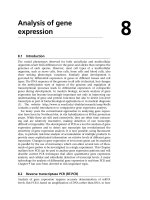
Medicine has benefited from recombinant DNA
technology in other ways (Fig. 1.1). New routes to
vaccines have been developed. The current hepatitis
B vaccine is based on the expression of a viral anti-
gen on the surface of yeast cells and a recombinant
vaccine has been used to eliminate rabies from foxes
in a large part of Europe. Gene manipulation can
Plants
Microbes
Therapeutic
small molecules
Diagnostic
proteins
Therapeutic
proteins
Microbes
Animals
Plants
Microbes
DNA
Vaccines
MEDICINE
Animal models
or human disease Pharamacogenomics
Profiling Cloned P450s
Genetic
disease
Infectious
disease
Diagnostic
nucleic
acids
Therapeutic
nucleic
acids
Vaccines
Gene therapy
Gene repair
Anti-sense drugs
Fig. 1.1 The impact of gene manipulation on the practice of medicine.
POGC01 9/11/2001 11:02 AM Page 3
4 CHAPTER 1
also be used to increase the levels of small molecules
within microbial cells. This can be done by cloning
all the genes for a particular biosynthetic pathway
and overexpressing them. Alternatively, it is pos-
sible to shut down particular metabolic pathways
and thus redirect particular intermediates towards
the desired end-product. This approach has been
used to facilitate production of chiral intermediates
and antibiotics. Novel antibiotics can also be created
by mixing and matching genes from organisms pro-
ducing different but related molecules in a technique
known as combinatorial biosynthesis.
Gene cloning enables nucleic acid probes to be
produced readily and such probes have many uses
in medicine. For example, they can be used to
determine or confirm the identity of a microbial
pathogen or to diagnose pre- or perinatally an
inherited genetic disease. Increasingly, probes are
being used to determine the likelihood of adverse
reactions to drugs or to select the best class of drug
to treat a particular illness (pharmacogenomics). A
variant of this technique is to use cloned cytochrome
P450s to determine how a new drug will be meta-
bolized and if any potentially toxic by-products will
result.
Nucleic acids are also being used as therapeutic
entities in their own right. For example, antisense
nucleic acids are being used to down-regulate gene
expression in certain diseases. In other cases, nucleic
acids are being administered to correct or repair
inherited gene defects (gene therapy/gene repair)
or as vaccines. In the reverse of gene repair, animals
are being generated that have mutations iden-
tical to those found in human disease. Note that
the use of antisense nucleic acids and gene therapy/
repair depends on the availability of information
on the exact cause of a disease. For most medical
conditions such information is lacking and cur-
rently available drugs are used to treat symptoms.
This situation will change significantly in the next
decade.
Biotechnology: the new industry
The early successes in overproducing mammalian
proteins in E. coli suggested to a few entrepreneurial
individuals that a new company should be formed to
exploit the potential of recombinant DNA technology.
Thus was Genentech born (Box 1.1). Since then
thousands of biotechnology companies have been
formed worldwide. As soon as major new develop-
ments in the science of gene manipulation are
reported, a rash of new companies are formed to
commercialize the new technology. For example,
many recently formed companies are hoping the
data from the Human Genome Sequencing Project
will result in the identification of a large number
of new proteins with potential for human therapy.
Others are using gene manipulation to understand
the regulation of transcription of particular genes,
arguing that it would make better therapeutic sense
to modulate the process with low-molecular-weight,
orally active drugs.
Although there are thousands of biotechno-
logy companies, fewer than 100 have sales of their
products and even fewer are profitable. Already
many biotechnology companies have failed, but
the technology advances at such a rate that there
is no shortage of new company start-ups to take
their place. One group of biotechnology companies
that has prospered is those supplying specialist
reagents to laboratory workers engaged in gene
manipulation. In the very beginning, researchers
had to make their own restriction enzymes and
this restricted the technology to those with pro-
tein chemistry skills. Soon a number of com-
panies were formed which catered to the needs of
researchers by supplying high-quality enzymes for
DNA manipulation. Despite the availability of these
enzymes, many people had great difficulty in clon-
ing DNA. The reason for this was the need for care-
ful quality control of all the components used in
the preparation of reagents, something researchers
are not good at! The supply companies responded
by making easy-to-use cloning kits in addition to
enzymes. Today, these supply companies can pro-
vide almost everything that is needed to clone,
express and analyse DNA and have thereby accel-
erated the use of recombinant DNA technology
in all biological disciplines. In the early days of
recombinant DNA technology, the development of
methodology was an end in itself for many academic
researchers. This is no longer true. The researchers
have gone back to using the tools to further our
POGC01 9/11/2001 11:02 AM Page 4
Gene manipulation 5
knowledge of biology, and the development of
new methodologies has largely fallen to the supply
companies.
The central role of E. coli
E. coli has always been a popular model system
for molecular geneticists. Prior to the development
of recombinant DNA technology, there existed a
large number of well-characterized mutants, gene
regulation was understood and there was a ready
availability of a wide selection of plasmids. Com-
pared with other microbial systems it was match-
less. It is not surprising, therefore, that the first
cloning experiments were undertaken in E. coli.
Subsequently, cloning techniques were extended to
a range of other microorganisms, such as Bacillus
subtilis, Pseudomonas sp., yeasts and filamentous
fungi, and then to higher eukaryotes. Curiously,
cloning in E. coli is technically easier than in any
other organism. As a result, it is rare for researchers
to clone DNA directly in other organisms. Rather,
DNA from the organism of choice is first mani-
pulated in E. coli and subsequently transferred back
to the original host. Without the ability to clone
and manipulate DNA in E. coli, the application of
recombinant DNA technology to other organisms
would be greatly hindered.
Table B1.1 Key events at Genentech.
1976 Genentech founded
1977 Genentech produced first human protein (somatostatin) in a microorganism
1978 Human insulin cloned by Genentech scientists
1979 Human growth hormone cloned by Genentech scientists
1980 Genentech went public, raising $35 million
1982 First recombinant DNA drug (human insulin) marketed (Genentech product licensed to Eli Lilly & Co.)
1984 First laboratory production of factor VIII for therapy of haemophilia. Licence granted to Cutter Biological
1985 Genentech launched its first product, Protropin (human growth hormone), for growth hormone deficiency in children
1987 Genentech launched Activase (tissue plasminogen activator) for dissolving blood clots in heart-attack patients
1990 Genentech launched Actimmune (interferon-g
1b
) for treatment of chronic granulomatous disease
1990 Genentech and the Swiss pharmaceutical company Roche complete a $2.1 billion merger
Biotechnology is not new. Cheese, bread and yoghurt
are products of biotechnology and have been known
for centuries. However, the stock-market excitement
about biotechnology stems from the potential of
gene manipulation, which is the subject of this book.
The birth of this modern version of biotechnology
can be traced to the founding of the company
Genentech.
In 1976, a 27-year-old venture capitalist called
Robert Swanson had a discussion over a few beers
with a University of California professor, Herb Boyer.
The discussion centred on the commercial potential
of gene manipulation. Swanson’s enthusiasm for the
technology and his faith in it was contagious. By
the close of the meeting the decision was taken to
found Genentech (Genetic Engineering Technology).
Though Swanson and Boyer faced scepticism from
both the academic and business communities they
forged ahead with their idea. Successes came thick
and fast (see Table B1.1) and within a few years
they had proved their detractors wrong. Over
1000 biotechnology companies have been set up in
the USA alone since the founding of Genentech
but very, very few have been as successful.
Box 1.1 The birth of an industry
POGC01 9/11/2001 11:02 AM Page 5
6 CHAPTER 1
Outline of the rest of the book
As noted above, E. coli has an essential role in recom-
binant DNA technology. Therefore, the first half of
the book is devoted to the methodology for manipu-
lating genes in this organism (Fig. 1.2). Chapter 2
covers many of the techniques that are common
to all cloning experiments and are fundamental to
the success of the technology. Chapter 3 is devoted
to methods for selectively cutting DNA molecules
into fragments that can be readily joined together
again. Without the ability to do this, there would
be no recombinant DNA technology. If fragments
of DNA are inserted into cells, they fail to replicate
except in those rare cases where they integrate into
the chromosome. To enable such fragments to be
propagated, they are inserted into DNA molecules
(vectors) that are capable of extrachromosomal re-
plication. These vectors are derived from plasmids
and bacteriophages and their basic properties are
described in Chapter 4. Originally, the purpose of
vectors was the propagation of cloned DNA but
today vectors fulfil many other roles, such as facil-
itating DNA sequencing, promoting expression of
cloned genes, facilitating purification of cloned gene
products, etc. The specialist vectors for these tasks
are described in Chapter 5. With this background in
place it is possible to describe in detail how to clone
the particular DNA sequences that one wants. There
are two basic strategies. Either one clones all the
DNA from an organism and then selects the very
small number of clones of interest or one amplifies
the DNA sequences of interest and then clones these.
Both these strategies are described in Chapter 6.
Once the DNA of interest has been cloned, it can
be sequenced and this will yield information on
the proteins that are encoded and any regulatory
signals that are present. There might also be a wish
to modify the DNA and/or protein sequence and
determine the biological effects of such changes.
The techniques for sequencing and changing cloned
genes are described in Chapter 7.
The role of vectors
Agarose gel electrophoresis
Blotting (DNA, RNA, protein)
Nucleic acid hybridization
DNA transformation & electroporation
Polymerase chain reaction (PCR)
Chapter 2
Restriction enzymes
Methods of joining DNA
Chapter 3
Basic properties of plasmids
Desirable properties of vectors
Plasmids as vectors
Bacteriophage λ vectors
Single-stranded DNA vectors
Vectors for cloning large DNA molecules
Specialist vectors
Over-producing proteins
Chapters 4 & 5
Cloning strategies
Cloning genomic DNA
cDNA cloning
Screening strategies
Expression cloning
Difference cloning
Chapter 6
Basic DNA sequencing
Analysing sequence data
Site-directed mutagenesis
Phage display
Chapter 7
Putting it
all together:
Cloning in
Practice
Basic
Techniques
Cutting &
Joining DNA
Vectors
Analysing &
Changing
Cloned
Genes
Fig. 1.2 ‘Roadmap’ outlining the basic
techniques in gene manipulation and
their relationships.
POGC01 9/11/2001 11:02 AM Page 6
Gene manipulation 7
In the second half of the book the specialist tech-
niques for cloning in organisms other than E. coli
are described (Fig. 1.3). Each of these chapters can
be read in isolation from the other chapters in this
section, provided that there is a thorough under-
standing of the material from the first half of the
book. Chapter 8 details the methods for cloning in
other bacteria. Originally it was thought that some
of these bacteria, e.g. B. subtilis, would usurp the
position of E. coli. This has not happened and gene
manipulation techniques are used simply to better
understand the biology of these bacteria. Chapter 9
focuses on cloning in fungi, although the emphasis
is on the yeast Saccharomyces cerevisiae. Fungi are
eukaryotes and are useful model systems for invest-
igating topics such as meiosis and mitosis, control of
cell division, etc. Animal cells can be cultured like
microorganisms and the techniques for cloning in
them are described in Chapter 10. Chapters 11 and
12 are devoted to the intricacies of cloning in animal
and plant representatives of higher eukaryotes and
Chapter 13 covers some cutting-edge techniques for
these same systems.
The concluding chapter is a survey of the dif-
ferent applications of recombinant DNA techno-
logy that are being exploited by the biotechnology
industry. Rather than going through application
after application, we have opted to show the inter-
play of different technologies by focusing on six
themes:
• Nucleic acid sequences as diagnostic tools.
• New drugs and new therapies for genetic diseases.
• Combating infectious disease.
• Protein engineering.
• Metabolic engineering.
• Plant breeding in the twenty-first century.
By treating the topic in this way we have been able to
show the interplay between some of the basic tech-
niques and the sophisticated analysis now possible
with genome sequence information.
Getting DNA into bacteria
Cloning in Gram-negative bacteria
Cloning in Gram-positive bacteria
Chapter 8
Why clone in fungi
Vectors for use in fungi
Expression of cloned DNA
Two hybrid system
Analysis of the whole genome
Chapter 9
Transformation of animal cells
Use of non-replicating DNA
Replication vectors
Viral transduction
Chapter 10
Transgenic mice
Other transgenic mammals
Transgenic birds, fish, Xenopus
Transgenic invertebrates
Chapter 11
Genetic
Manipulation
of Animals
Cloning in
Bacteria
Other Than
E.coli
Cloning in
Yeast &
Other
Fungi
Gene
Transfer
To Animal
Cells
Handling plant cells
Agrobacterium-mediated transformation
Direct DNA transfer
Plant viruses as vectors
Chapter 12
Genetic
Manipulation
of Plants
Inducible expression systems
Site-specific recombination
Gene inhibition
Insertional mutagenesis
Gene tagging
Entrapment constructs
Chapter 13
Advanced
Techniques
for Gene
Manipulation
in Plant and
Animals
Fig. 1.3 ‘Roadmap’ of the advanced
techniques in gene manipulation and
their application to organisms other
than E. coli.
POGC01 9/11/2001 11:02 AM Page 7
CHAPTER 2
Basic techniques
Introduction
The initial impetus for gene manipulation in vitro
came about in the early 1970s with the simultan-
eous development of techniques for:
• genetic transformation of Escherichia coli;
• cutting and joining DNA molecules;
• monitoring the cutting and joining reactions.
In order to explain the significance of these devel-
opments we must first consider the essential require-
ments of a successful gene-manipulation procedure.
The basic problems
Before the advent of modern gene-manipulation
methods there had been many early attempts at
transforming pro- and eukaryotic cells with foreign
DNA. But, in general, little progress could be made.
The reasons for this are as follows. Let us assume
that the exogenous DNA is taken up by the recipient
cells. There are then two basic difficulties. First,
where detection of uptake is dependent on gene
expression, failure could be due to lack of accurate
transcription or translation. Secondly, and more
importantly, the exogenous DNA may not be main-
tained in the transformed cells. If the exogenous
DNA is integrated into the host genome, there is no
problem. The exact mechanism whereby this integ-
ration occurs is not clear and it is usually a rare
event. However this occurs, the result is that the
foreign DNA sequence becomes incorporated into
the host cell’s genetic material and will subsequently
be propagated as part of that genome. If, however,
the exogenous DNA fails to be integrated, it will
probably be lost during subsequent multiplication of
the host cells. The reason for this is simple. In order
to be replicated, DNA molecules must contain an
origin of replication, and in bacteria and viruses there
is usually only one per genome. Such molecules are
called replicons. Fragments of DNA are not replicons
and in the absence of replication will be diluted out of
their host cells. It should be noted that, even if a DNA
molecule contains an origin of replication, this may
not function in a foreign host cell.
There is an additional, subsequent problem. If the
early experiments were to proceed, a method was
required for assessing the fate of the donor DNA. In
particular, in circumstances where the foreign DNA
was maintained because it had become integrated in
the host DNA, a method was required for mapping the
foreign DNA and the surrounding host sequences.
The solutions: basic techniques
If fragments of DNA are not replicated, the obvious
solution is to attach them to a suitable replicon.
Such replicons are known as vectors or cloning
vehicles. Small plasmids and bacteriophages are the
most suitable vectors for they are replicons in their
own right, their maintenance does not necessarily
require integration into the host genome and their
DNA can be readily isolated in an intact form. The
different plasmids and phages which are used as
vectors are described in detail in Chapters 4 and 5.
Suffice it to say at this point that initially plasmids
and phages suitable as vectors were only found in E.
coli. An important consequence follows from the use
of a vector to carry the foreign DNA: simple methods
become available for purifying the vector molecule,
complete with its foreign DNA insert, from trans-
formed host cells. Thus not only does the vector
provide the replicon function, but it also permits the
easy bulk preparation of the foreign DNA sequence,
free from host-cell DNA.
Composite molecules in which foreign DNA has
been inserted into a vector molecule are sometimes
called DNA chimeras because of their analogy with
the Chimaera of mythology – a creature with the
head of a lion, body of a goat and tail of a serpent.
The construction of such composite or artificial
POGC02 9/11/2001 11:01 AM Page 8
Basic techniques 9
recombinant molecules has also been termed genetic
engineering or gene manipulation because of the po-
tential for creating novel genetic combinations by
biochemical means. The process has also been termed
molecular cloning or gene cloning because a line of
genetically identical organisms, all of which contain
the composite molecule, can be propagated and grown
in bulk, hence amplifying the composite molecule
and any gene product whose synthesis it directs.
Although conceptually very simple, cloning of
a fragment of foreign, or passenger, or target DNA
in a vector demands that the following can be
accomplished.
• The vector DNA must be purified and cut open.
• The passenger DNA must be inserted into the
vector molecule to create the artificial recombinant.
DNA joining reactions must therefore be performed.
Methods for cutting and joining DNA molecules are
now so sophisticated that they warrant a chapter of
their own (Chapter 3).
• The cutting and joining reactions must be read-
ily monitored. This is achieved by the use of gel
electrophoresis.
• Finally, the artificial recombinant must be trans-
formed into E. coli or another host cell. Further details
on the use of gel electrophoresis and transformation
of E. coli are given in the next section. As we have
noted, the necessary techniques became available at
about the same time and quickly led to many cloning
experiments, the first of which were reported in
1972 ( Jackson et al. 1972, Lobban & Kaiser 1973).
Agarose gel electrophoresis
The progress of the first experiments on cutting and
joining of DNA molecules was monitored by velocity
sedimentation in sucrose gradients. However, this
has been entirely superseded by gel electrophoresis.
Gel electrophoresis is not only used as an analytical
method, it is routinely used preparatively for the
purification of specific DNA fragments. The gel is
composed of polyacrylamide or agarose. Agarose is
convenient for separating DNA fragments ranging
in size from a few hundred base pairs to about 20 kb
(Fig. 2.1). Polyacrylamide is preferred for smaller
DNA fragments.
The mechanism responsible for the separation
of DNA molecules by molecular weight during gel
electrophoresis is not well understood (Holmes
& Stellwagen 1990). The migration of the DNA
molecules through the pores of the matrix must play
an important role in molecular-weight separations
since the electrophoretic mobility of DNA in free
solution is independent of molecular weight. An
agarose gel is a complex network of polymeric
molecules whose average pore size depends on the
buffer composition and the type and concentration
of agarose used. DNA movement through the gel
was originally thought to resemble the motion of a
snake (reptation). However, real-time fluorescence
microscopy of stained molecules undergoing elec-
trophoresis has revealed more subtle dynamics
(Schwartz & Koval 1989, Smith et al. 1989). DNA
molecules display elastic behaviour by stretching in
the direction of the applied field and then contract-
ing into dense balls. The larger the pore size of the
–
+
21.226
kb pairs
7.421
5.804
5.643
4.878
3.530
Fig. 2.1 Electrophoresis of DNA in agarose gels. The direction
of migration is indicated by the arrow. DNA bands have been
visualized by soaking the gel in a solution of ethidium bromide
(see Fig. 2.3), which complexes with DNA by intercalating
between stacked base-pairs, and photographing the orange
fluorescence which results upon ultraviolet irradiation.
POGC02 9/11/2001 11:01 AM Page 9
10 CHAPTER 2
gel, the greater the ball of DNA which can pass
through and hence the larger the molecules which
can be separated. Once the globular volume of the
DNA molecule exceeds the pore size, the DNA
molecule can only pass through by reptation. This
occurs with molecules about 20 kb in size and it is
difficult to separate molecules larger than this with-
out recourse to pulsed electrical fields.
In pulsed-field gel electrophoresis (PFGE) (Schwartz
& Cantor 1984) molecules as large as 10 Mb can be
separated in agarose gels. This is achieved by caus-
ing the DNA to periodically alter its direction of
migration by regular changes in the orientation of
the electric field with respect to the gel. With each
change in the electric-field orientation, the DNA
must realign its axis prior to migrating in the new
direction. Electric-field parameters, such as the
direction, intensity and duration of the electric field,
are set independently for each of the different fields
and are chosen so that the net migration of the DNA
is down the gel. The difference between the direction
of migration induced by each of the electric fields is
the reorientation angle and corresponds to the angle
that the DNA must turn as it changes its direction of
migration each time the fields are switched.
A major disadvantage of PFGE, as originally
described, is that the samples do not run in straight
lines. This makes subsequent analysis difficult. This
problem has been overcome by the development of
improved methods for alternating the electrical field.
The most popular of these is contour-clamped homo-
geneous electrical-field electrophoresis (CHEF) (Chu
et al. 1986). In early CHEF-type systems (Fig. 2.2)
the reorientation angle was fixed at 120°. However,
in newer systems, the reorientation angle can be
varied and it has been found that for whole-yeast
chromosomes the migration rate is much faster with
an angle of 106° (Birren et al. 1988). Fragments of
DNA as large as 200–300 kb are routinely handled
in genomics work and these can be separated in a
matter of hours using CHEF systems with a reori-
entation angle of 90° or less (Birren & Lai 1994).
Aaij and Borst (1972) showed that the migration
rates of the DNA molecules were inversely propor-
tional to the logarithms of the molecular weights.
Subsequently, Southern (1979a,b) showed that
plotting fragment length or molecular weight
against the reciprocal of mobility gives a straight
line over a wider range than the semilogarithmic
plot. In any event, gel electrophoresis is frequently
performed with marker DNA fragments of known
size, which allow accurate size determination of an
unknown DNA molecule by interpolation. A par-
ticular advantage of gel electrophoresis is that the
DNA bands can be readily detected at high sensitiv-
ity. The bands of DNA in the gel are stained with
the intercalating dye ethidium bromide (Fig. 2.3),
and as little as 0.05 µg of DNA in one band can be
detected as visible fluorescence when the gel is
illuminated with ultraviolet light.
In addition to resolving DNA fragments of differ-
ent lengths, gel electrophoresis can be used to separ-
ate different molecular configurations of a DNA
molecule. Examples of this are given in Chapter 4
(see p. 44). Gel electrophoresis can also be used for
investigating protein–nucleic acid interactions in
the so-called gel retardation or band shift assay. It is
based on the observation that binding of a protein
to DNA fragments usually leads to a reduction in
120°
Migration of DNA
A–
B+
B–
A+
Fig. 2.2 Schematic representation of CHEF (contour-clamped
homogeneous electrical field) pulsed-field gel electrophoresis.
Fig. 2.3 Ethidium bromide.
HN
NH
N
⊕
Br
CH
5
2
2
2
–
POGC02 9/11/2001 11:01 AM Page 10
Basic techniques 11
electrophoretic mobility. The assay typically involves
the addition of protein to linear double-stranded DNA
fragments, separation of complex and naked DNA
by gel electrophoresis and visualization. A review of
the physical basis of electrophoretic mobility shifts and
their application is provided by Lane et al. (1992).
Nucleic acid blotting
Nucleic acid labelling and hybridization on mem-
branes have formed the basis for a range of experi-
mental techniques central to recent advances in our
understanding of the organization and expression
of the genetic material. These techniques may be
applied in the isolation and quantification of specific
nucleic acid sequences and in the study of their
organization, intracellular localization, expression
and regulation. A variety of specific applications
includes the diagnosis of infectious and inherited
disease. Each of these topics is covered in depth in
subsequent chapters.
An overview of the steps involved in nucleic acid
blotting and membrane hybridization procedures is
shown in Fig. 2.4. Blotting describes the immobiliza-
tion of sample nucleic acids on to a solid support,
generally nylon or nitrocellulose membranes. The
blotted nucleic acids are then used as ‘targets’ in
subsequent hybridization experiments. The main
blotting procedures are:
• blotting of nucleic acids from gels;
• dot and slot blotting;
• colony and plaque blotting.
Colony and plaque blotting are described in detail on
pp. 104–105 and dot and slot blotting in Chapter 14.
Southern blotting
The original method of blotting was developed by
Southern (1975, 1979b) for detecting fragments in
an agarose gel that are complementary to a given
RNA or DNA sequence. In this procedure, referred to
as Southern blotting, the agarose gel is mounted on
a filter-paper wick which dips into a reservoir con-
taining transfer buffer (Fig. 2.5). The hybridization
membrane is sandwiched between the gel and a
stack of paper towels (or other absorbent material),
which serves to draw the transfer buffer through the
gel by capillary action. The DNA molecules are car-
ried out of the gel by the buffer flow and immobilized
on the membrane. Initially, the membrane material
used was nitrocellulose. The main drawback with
this membrane is its fragile nature. Supported nylon
membranes have since been developed which have
greater binding capacity for nucleic acids in addition
to high tensile strength.
For efficient Southern blotting, gel pretreatment is
important. Large DNA fragments (> 10 kb) require a
longer transfer time than short fragments. To allow
Immobilization of nucleic acids
• Southern blot
• Northern blot
• Dot blot
• Colony/plaque lift
Pre-hybridization
Labelled DNA
or RNA probe
Removal of probe
prior to reprobing
Hybridization
Stringency washes
Detection
Fig. 2.4 Overview of nucleic acid
blotting and hybridization (reproduced
courtesy of Amersham Pharmacia
Biotech).
POGC02 9/11/2001 11:01 AM Page 11
12 CHAPTER 2
uniform transfer of a wide range of DNA fragment
sizes, the electrophoresed DNA is exposed to a short
depurination treatment (0.25 mol/l HCl) followed by
alkali. This shortens the DNA fragments by alkaline
hydrolysis at depurinated sites. It also denatures the
fragments prior to transfer, ensuring that they are in
the single-stranded state and accessible for probing.
Finally, the gel is equilibrated in neutralizing solution
prior to blotting. An alternative method uses posit-
ively charged nylon membranes, which remove the
need for extended gel pretreatment. With them the
DNA is transferred in native (non-denatured) form
and then alkali-denatured in situ on the membrane.
After transfer, the nucleic acid needs to be fixed to
the membrane and a number of methods are avail-
able. Oven baking at 80°C is the recommended
method for nitrocellulose membranes and this can
also be used with nylon membranes. Due to the
flammable nature of nitrocellulose, it is important
that it is baked in a vacuum oven. An alternative
fixation method utilizes ultraviolet cross-linking. It
is based on the formation of cross-links between a
small fraction of the thymine residues in the DNA
and positively charged amino groups on the surface
of nylon membranes. A calibration experiment must
be performed to determine the optimal fixation period.
Following the fixation step, the membrane is placed
in a solution of labelled (radioactive or non-radioactive)
RNA, single-stranded DNA or oligodeoxynucleotide
which is complementary in sequence to the blot-
transferred DNA band or bands to be detected.
Conditions are chosen so that the labelled nucleic
acid hybridizes with the DNA on the membrane.
Since this labelled nucleic acid is used to detect and
locate the complementary sequence, it is called the
probe. Conditions are chosen which maximize the
rate of hybridization, compatible with a low back-
ground of non-specific binding on the membrane
(see Box 2.1). After the hybridization reaction has
been carried out, the membrane is washed to remove
unbound radioactivity and regions of hybridization
Weight < 0.75 kg
Glass plate
Paper tissues
3 sheets filter paper
Membrane
Gel
Plastic tray
Fig. 2.5 A typical capillary blotting apparatus.
Rate enhancers Dextran sulphate and other polymers act as volume excluders to increase both the rate and the
extent of hybridization
Detergents and blocking agents Dried milk, heparin and detergents such as sodium dodecyl sulphate (SDS) have been used
to depress non-specific binding of the probe to the membrane. Denhardt’s solution
(Denhardt 1966) uses Ficoll, polyvinylpyrrolidone and bovine serum albumin
Denaturants Urea or formamide can be used to depress the melting temperature of the hybrid so that reduced
temperatures of hybridization can be used
Heterologous DNA This can reduce non-specific binding of probes to non-homologous DNA on the blot
continued
The hybridization of nucleic acids on membranes is a
widely used technique in gene manipulation and
analysis. Unlike solution hybridizations, membrane
hybridizations tend not to proceed to completion.
One reason for this is that some of the bound nucleic
acid is embedded in the membrane and is inaccessible
to the probe. Prolonged incubations may not generate
any significant increase in detection sensitivity.
The composition of the hybridization buffer can
greatly affect the speed of the reaction and the
sensitivity of detection. The key components of these
buffers are shown below:
Box 2.1 Hybridization of nucleic acids on membranes
POGC02 9/11/2001 11:01 AM Page 12
Basic techniques 13
are detected autoradiographically by placing the
membrane in contact with X-ray film (see Box 2.2).
A common approach is to carry out the hybridiza-
tion under conditions of relatively low stringency
which permit a high rate of hybridization, followed
by a series of post-hybridization washes of increasing
stringency (i.e. higher temperature or, more com-
monly, lower ionic strength). Autoradiography
following each washing stage will reveal any DNA
bands that are related to, but not perfectly comple-
mentary with, the probe and will also permit an
estimate of the degree of mismatching to be made.
Stringency control
Stringency can be regarded as the specificity with
which a particular target sequence is detected by
hybridization to a probe. Thus, at high stringency,
only completely complementary sequences will be
bound, whereas low-stringency conditions will allow
hybridization to partially matched sequences.
Stringency is most commonly controlled by the
temperature and salt concentration in the post-
hybridization washes, although these parameters
can also be utilized in the hybridization step.
In practice, the stringency washes are performed
under successively more stringent conditions
(lower salt or higher temperature) until the desired
result is obtained.
The melting temperature (T
m
) of a probe–target
hybrid can be calculated to provide a starting-point
for the determination of correct stringency. The
T
m
is the temperature at which the probe and
target are 50% dissociated. For probes longer than
100 base pairs:
T
m
= 81.5°C + 16.6 log M + 0.41 (% G + C)
where M = ionic strength of buffer in moles/litre.
With long probes, the hybridization is usually carried
out at T
m
− 25°C. When the probe is used to
detect partially matched sequences, the
hybridization temperature is reduced by 1°C
for every 1% sequence divergence between
probe and target.
Oligonucleotides can give a more rapid
hybridization rate than long probes as they can
be used at a higher molarity. Also, in situations
where target is in excess to the probe, for example
dot blots, the hybridization rate is diffusion-limited
and longer probes diffuse more slowly than
oligonucleotides. It is standard practice to use
oligonucleotides to analyse putative mutants
following a site-directed mutagenesis experiment
where the difference between parental and mutant
progeny is often only a single base-pair change
(see p. 132 et seq.).
The availability of the exact sequence of
oligonucleotides allows conditions for hybridization
and stringency washing to be tightly controlled so
that the probe will only remain hybridized when
it is 100% homologous to the target. Stringency is
commonly controlled by adjusting the temperature
of the wash buffer. The ‘Wallace rule’ (Lay Thein &
Wallace 1986) is used to determine the appropriate
stringency wash temperature:
T
m
= 4 × (number of GC base pairs) + 2 × (number
of AT base pairs)
In filter hybridizations with oligonucleotide probes,
the hybridization step is usually performed at 5°C
below T
m
for perfectly matched sequences. For every
mismatched base pair, a further 5°C reduction is
necessary to maintain hybrid stability.
The design of oligonucleotides for hybridization
experiments is critical to maximize hybridization
specificity. Consideration should be given to:
• probe length – the longer the oligonucleotide, the
less chance there is of it binding to sequences other
than the desired target sequence under conditions
of high stringency;
• oligonucleotide composition – the GC content
will influence the stability of the resultant hybrid
and hence the determination of the appropriate
stringency washing conditions. Also the presence
of any non-complementary bases will have an effect
on the hybridization conditions.
Box 2.1 continued
POGC02 9/11/2001 11:01 AM Page 13
14 CHAPTER 2
Fig. B2.1 Autoradiographs showing the detection of
35
S- and
3
H-labelled proteins in acrylamide gels with (+) and without
(−) fluorography. (Photo courtesy of Amersham Pharmacia Biotech.)
continued
The localization and recording of a radiolabel within
a solid specimen is known as autoradiography
and involves the production of an image in a
photographic emulsion. Such emulsions consist of
silver halide crystals suspended in a clear phase
composed mainly of gelatin. When a b-particle or
g-ray from a radionuclide passes through the
emulsion, the silver ions are converted to silver atoms.
This results in a latent image being produced, which
is converted to a visible image when the image is
developed. Development is a system of amplification
in which the silver atoms cause the entire silver halide
crystal to be reduced to metallic silver. Unexposed
crystals are removed by dissolution in fixer, giving
an autoradiographic image which represents the
distribution of radiolabel in the original sample.
In direct autoradiography, the sample is placed
in intimate contact with the film and the radioactive
emissions produce black areas on the developed
autoradiograph. It is best suited to detection of
weak- to medium-strength b-emitting radionuclides
(
3
H,
14
C,
35
S). Direct autoradiography is not suited
to the detection of highly energetic b-particles,
such as those from
32
P, or for g-rays emitted from
isotopes like
125
I. These emissions pass through and
beyond the film, with the majority of the energy
being wasted. Both
32
P and
125
I are best detected
by indirect autoradiography.
Indirect autoradiography describes the technique
by which emitted energy is converted to light by
means of a scintillator, using fluorography or
intensifying screens. In fluorography the sample
is impregnated with a liquid scintillator. The
radioactive emissions transfer their energy to the
scintillator molecules, which then emit photons which
expose the photographic emulsion. Fluorography
is mostly used to improve the detection of weak
b-emitters (Fig. B2.1). Intensifying screens are
Box 2.2 The principles of autoradiography
35
S
3
H
+ − + −
POGC02 9/11/2001 11:01 AM Page 14
Basic techniques 15
sheets of a solid inorganic scintillator which are
placed behind the film. Any emissions passing
through the photographic emulsion are absorbed
by the screen and converted to light, effectively
superimposing a photographic image upon the
direct autoradiographic image.
The gain in sensitivity which is achieved by use of
indirect autoradiography is offset by non-linearity
of film response. A single hit by a b-particle or g-ray
can produce hundreds of silver atoms, but a single
hit by a photon of light produces only a single silver
atom. Although two or more silver atoms in a silver
halide crystal are stable, a single silver atom is
unstable and reverts to a silver ion very rapidly.
This means that the probability of a second photon
being captured before the first silver atom has
reverted is greater for large amounts of radioactivity
than for small amounts. Hence small amounts of
radioactivity are under-represented with the use of
fluorography and intensifying screens. This problem
can be overcome by a combination of pre-exposing a
film to an instantaneous flash of light (pre-flashing)
and exposing the autoradiograph at −70°C.
Pre-flashing provides many of the silver halide
crystals of the film with a stable pair of silver atoms.
Lowering the temperature to −70°C increases the
stability of a single silver atom, increasing the time
available to capture a second photon (Fig. B2.2).
Fig. B2.2 The improvement in sensitivity of detection of
125
I-labelled IgG by autoradiography obtained by using an
intensifying screen and pre-flashed film. A, no screen and no pre-flashing; B, screen present but film not pre-flashed;
C, use of screen and pre-flashed film. (Photo courtesy of Amersham Pharmacia Biotech.)
A B C
Box 2.2 continued
POGC02 9/11/2001 11:01 AM Page 15
16 CHAPTER 2
The Southern blotting methodology can be extre-
mely sensitive. It can be applied to mapping restric-
tion sites around a single-copy gene sequence in a
complex genome such as that of humans (Fig. 2.6),
and when a ‘mini-satellite’ probe is used it can be
applied forensically to minute amounts of DNA (see
Chapter 14).
Northern blotting
Southern’s technique has been of enormous value,
but it was thought that it could not be applied
directly to the blot-transfer of RNAs separated by gel
electrophoresis, since RNA was found not to bind to
nitrocellulose. Alwine et al. (1979) therefore devised
a procedure in which RNA bands are blot-transferred
from the gel on to chemically reactive paper, where
they are bound covalently. The reactive paper is
prepared by diazotization of aminobenzyloxymethyl
paper (creating diazobenzyloxymethyl (DBM) paper),
which itself can be prepared from Whatman 540
paper by a series of uncomplicated reactions. Once
covalently bound, the RNA is available for hybrid-
ization with radiolabelled DNA probes. As before,
hybridizing bands are located by autoradiography.
Alwine et al.’s method thus extends that of Southern
and for this reason it has acquired the jargon term
northern blotting.
Subsequently it was found that RNA bands can
indeed be blotted on to nitrocellulose membranes
under appropriate conditions (Thomas 1980) and
suitable nylon membranes have been developed.
Because of the convenience of these more recent
methods, which do not require freshly activated paper,
the use of DBM paper has been superseded.
Western blotting
The term ‘western’ blotting (Burnette 1981) refers
to a procedure which does not directly involve nucleic
acids, but which is of importance in gene manipula-
tion. It involves the transfer of electrophoresed
protein bands from a polyacrylamide gel on to a
membrane of nitrocellulose or nylon, to which they
bind strongly (Gershoni & Palade 1982, Renart &
Sandoval 1984). The bound proteins are then avail-
Genomic DNA
Gene X
Restriction
endo-
nuclease
Gel
electro-
phoresis
Genomic DNA
Autoradio-
graphy
Photographic
film
Images correspond only to
fragments containing gene X
sequences – estimate
fragment sizes from mobility
Radioactive RNA or
denatured DNA containing
sequences complementary
to gene X (radioactive probe)
(1) Hybridize nitrocellulose
with radioactive probe
(2) Wash
Single stranded
DNA fragments
Agarose gel
Long DNA
fragments
DNA
fragments
Short DNA
fragments
(1) Denature in alkali
(2) Blot-transfer, bake
Nitrocellulose
–
+
Fig. 2.6 Mapping restriction sites
around a hypothetical gene sequence
in total genomic DNA by the Southern
blot method.
Genomic DNA is cleaved with a
restriction endonuclease into hundreds
of thousands of fragments of various
sizes. The fragments are separated
according to size by gel electrophoresis
and blot-transferred on to nitrocellulose
paper. Highly radioactive RNA or
denatured DNA complementary in
sequence to gene X is applied to the
nitrocellulose paper bearing the blotted
DNA. The radiolabelled RNA or DNA
will hybridize with gene X sequences
and can be detected subsequently by
autoradiography, so enabling the sizes
of restriction fragments containing
gene X sequences to be estimated from
their electrophoretic mobility. By
using several restriction endonucleases
singly and in combination, a map of
restriction sites in and around gene
X can be built up.
POGC02 9/11/2001 11:01 AM Page 16
Basic techniques 17
able for analysis by a variety of specific protein–ligand
interactions. Most commonly, antibodies are used to
detect specific antigens. Lectins have been used to
identify glycoproteins. In these cases the probe may
itself be labelled with radioactivity, or some other
‘tag’ may be employed. Often, however, the probe
is unlabelled and is itself detected in a ‘sandwich’
reaction, using a second molecule which is labelled,
for instance a species-specific second antibody, or
protein A of Staphylococcus aureus (which binds
to certain subclasses of IgG antibodies), or strept-
avidin (which binds to antibody probes that have
been biotinylated). These second molecules may
be labelled in a variety of ways with radioactive,
enzyme or fluorescent tags. An advantage of the
sandwich approach is that a single preparation of
labelled second molecule can be employed as a
general detector for different probes. For example,
an antiserum may be raised in rabbits which reacts
with a range of mouse immunoglobins. Such a
rabbit anti-mouse (RAM) antiserum may be radio-
labelled and used in a number of different applica-
tions to identify polypeptide bands probed with
different, specific, monoclonal antibodies, each mono-
clonal antibody being of mouse origin. The sand-
wich method may also give a substantial increase
in sensitivity, owing to the multivalent binding of
antibody molecules.
Alternative blotting techniques
The original blotting technique employed capillary
blotting but nowadays the blotting is usually accom-
plished by electrophoretic transfer of polypeptides
from an SDS-polyacrylamide gel on to the membrane
(Towbin et al. 1979). Electrophoretic transfer is also
the method of choice for transferring DNA or RNA
from low-pore-size polyacrylamide gels. It can also
be used with agarose gels. However, in this case,
the rapid electrophoretic transfer process requires
high currents, which can lead to extensive heating
effects, resulting in distortion of agarose gels. The
use of an external cooling system is necessary to
prevent this.
Another alternative to capillary blotting is vacuum-
driven blotting (Olszewska & Jones 1988), for which
several devices are commercially available. Vacuum
blotting has several advantages over capillary or
electrophoretic transfer methods: transfer is very
rapid and gel treatment can be performed in situ on
the vacuum apparatus. This ensures minimal gel
handling and, together with the rapid transfer, pre-
vents significant DNA diffusion.
Transformation of E. coli
Early attempts to achieve transformation of E. coli
were unsuccessful and it was generally believed that
E. coli was refractory to transformation. However,
Mandel and Higa (1970) found that treatment with
CaC1
2
allowed E. coli cells to take up DNA from bac-
teriophage λ. A few years later Cohen et al. (1972)
showed that CaC1
2
-treated E. coli cells are also effect-
ive recipients for plasmid DNA. Almost any strain of
E. coli can be transformed with plasmid DNA, albeit
with varying efficiency, whereas it was thought that
only recBC
−
mutants could be transformed with lin-
ear bacterial DNA (Cosloy & Oishi 1973). Later,
Hoekstra et al. (1980) showed that recBC
+
cells can
be transformed with linear DNA, but the efficiency is
only 10% of that in otherwise isogenic recBC
−
cells.
Transformation of recBC
−
cells with linear DNA is
only possible if the cells are rendered recombination-
proficient by the addition of a sbcA or sbcB muta-
tion. The fact that the recBC gene product is an
exonuclease explains the difference in transforma-
tion efficiency of circular and linear DNA in recBC
+
cells.
As will be seen from the next chapter, many bac-
teria contain restriction systems which can influence
the efficiency of transformation. Although the com-
plete function of these restriction systems is not yet
known, one role they do play is the recognition and
degradation of foreign DNA. For this reason it is
usual to use a restriction-deficient strain of E. coli as
a transformable host.
Since transformation of E. coli is an essential step
in many cloning experiments, it is desirable that it be
as efficient as possible. Several groups of workers
have examined the factors affecting the efficiency of
transformation. It has been found that E. coli cells
and plasmid DNA interact productively in an en-
vironment of calcium ions and low temperature
(0–5°C), and that a subsequent heat shock (37–45°C)
is important, but not strictly required. Several other
factors, especially the inclusion of metal ions in
POGC02 9/11/2001 11:01 AM Page 17
18 CHAPTER 2
addition to calcium, have been shown to stimulate
the process.
A very simple, moderately efficient transforma-
tion procedure for use with E. coli involves resus-
pending log-phase cells in ice-cold 50 mmol/l
calcium chloride at about 10
10
cells/ml and keeping
them on ice for about 30 min. Plasmid DNA (0. 1 µg)
is then added to a small aliquot (0.2 ml) of these now
competent (i.e. competent for transformation) cells,
and the incubation on ice continued for a further
30 min, followed by a heat shock of 2 min at 42°C.
The cells are then usually transferred to nutrient
medium and incubated for some time (30 min to
1 h) to allow phenotypic properties conferred by the
plasmid to be expressed, e.g. antibiotic resistance
commonly used as a selectable marker for plasmid-
containing cells. (This so-called phenotypic lag
may not need to be taken into consideration with
high-level ampicillin resistance. With this marker,
significant resistance builds up very rapidly, and
ampicillin exerts its effect on cell-wall biosynthesis
only in cells which have progressed into active
growth.) Finally the cells are plated out on selective
medium. Just why such a transformation procedure
is effective is not fully understood (Huang & Reusch
1995). The calcium chloride affects the cell wall and
may also be responsible for binding DNA to the cell
surface. The actual uptake of DNA is stimulated by
the brief heat shock.
Hanahan (1983) has re-examined factors that
affect the efficiency of transformation, and has devised
a set of conditions for optimal efficiency (expressed
as transformants per µg plasmid DNA) applicable to
most E. coli K12 strains. Typically, efficiencies of 10
7
to 10
9
transformants/µg can be achieved depending
on the strain of E. coli and the method used (Liu &
Rashidbaigi 1990). Ideally, one wishes to make a
large batch of competent cells and store them frozen
for future use. Unfortunately, competent cells made
by the Hanahan procedure rapidly lose their com-
petence on storage. Inoue et al. (1990) have optimized
the conditions for the preparation of competent cells.
Not only could they store cells for up to 40 days at
−70°C while retaining efficiencies of 1–5 ×10
9
cfu/µg,
but competence was affected only minimally by salts
in the DNA preparation.
There are many enzymic activities in E. coli which
can destroy incoming DNA from non-homologous
sources (see Chapter 3) and reduce the transforma-
tion efficiency. Large DNAs transform less effici-
ently, on a molar basis, than small DNAs. Even with
such improved transformation procedures, certain
potential gene-cloning experiments requiring large
numbers of clones are not reliable. One approach
which can be used to circumvent the problem of low
transformation efficiencies is to package recombin-
ant DNA into virus particles in vitro. A particular
form of this approach, the use of cosmids, is described
in detail in Chapter 5. Another approach is electro-
poration, which is described below.
Electroporation
A rapid and simple technique for introducing cloned
genes into a wide variety of microbial, plant and ani-
mal cells, including E. coli, is electroporation. This
technique depends on the original observation by
Zimmerman & Vienken (1983) that high-voltage
electric pulses can induce cell plasma membranes to
fuse. Subsequently it was found that, when sub-
jected to electric shock, the cells take up exogenous
DNA from the suspending solution. A proportion of
these cells become stably transformed and can be
selected if a suitable marker gene is carried on the
transforming DNA. Many different factors affect the
efficiency of electroporation, including temperature,
various electric-field parameters (voltage, resistance
and capacitance), topological form of the DNA,
and various host-cell factors (genetic background,
growth conditions and post-pulse treatment). Some
of these factors have been reviewed by Hanahan
et al. (1991).
With E. coli, electroporation has been found to give
plasmid transformation efficiencies (10
9
cfu/µg DNA)
comparable with the best CaC1
2
methods (Dower et al.
1988). More recently, Zhu and Dean (1999) have
reported 10-fold higher transformation efficiencies
with plasmids (9 × 10
9
transformants/µg) by co-
precipitating the DNA with transfer RNA (tRNA)
prior to electroporation. With conventional CaCl
2
-
mediated transformation, the efficiency falls off
rapidly as the size of the DNA molecule increases
and is almost negligible when the size exceeds 50 kb.
While size also affects the efficiency of electroporation
(Sheng et al. 1995), it is possible to get transforma-
tion efficiencies of 10
6
cfu/µg DNA with molecules
POGC02 9/11/2001 11:01 AM Page 18
Basic techniques 19
as big as 240 kb. Molecules three to four times this
size also can be electroporated successfully. This is
important because much of the work on mapping
and sequencing of genomes demands the ability
to handle large fragments of DNA (see p. 64 and
p. 126).
Transformation of other organisms
Although E. coli often remains the host organism of
choice for cloning experiments, many other hosts
are now used, and with them transformation may
still be a critical step. In the case of Gram-positive
bacteria, the two most important groups of organ-
isms are Bacillus spp. and actinomycetes. That B.
subtilis is naturally competent for transformation
has been known for a long time and hence the gen-
etics of this organism are fairly advanced. For this
reason B. subtilis is a particularly attractive alternat-
ive prokaryotic cloning host. The significant features
of transformation with this organism are detailed
in Chapter 8. Of particular relevance here is that
it is possible to transform protoplasts of B. subtilis, a
technique which leads to improved transformation
frequencies. A similar technique is used to transform
actinomycetes, and recently it has been shown that
the frequency can be increased considerably by first
entrapping the DNA in liposomes, which then fuse
with the host-cell membrane.
In later chapters we discuss ways, including elec-
troporation, in which cloned DNA can be introduced
into eukaryotic cells. With animal cells there is no
great problem as only the membrane has to be
crossed. In the case of yeast, protoplasts are required
(Hinnen et al. 1978). With higher plants one strat-
egy that has been adopted is either to package the
DNA in a plant virus or to use a bacterial plant
pathogen as the donor. It has also been shown that
protoplasts prepared from plant cells are competent
for transformation. A further remarkable approach
that has been demonstrated with plants and animals
(Klein & Fitzpatrick-McElligott 1993) is the use of
microprojectiles shot from a gun (p. 238).
Animal cells, and protoplasts of yeast, plant and
bacterial cells are susceptible to transformation by
liposomes (Deshayes et al. 1985). A simple transforma-
tion system has been developed which makes use of
liposomes prepared from a cationic lipid (Felgner
et al. 1987). Small unilamellar (single-bilayer) ves-
icles are produced. DNA in solution spontaneously
and efficiently complexes with these liposomes (in
contrast to previously employed liposome encapsida-
tion procedures involving non-ionic lipids). The
positively charged liposomes not only complex with
DNA, but also bind to cultured animal cells and are
efficient in transforming them, probably by fusion
with the plasma membrane. The use of liposomes as
a transformation or transfection system is called
lipofection.
The polymerase chain reaction (PCR)
The impact of the PCR upon molecular biology has
been profound. The reaction is easily performed, and
leads to the amplification of specific DNA sequences
by an enormous factor. From a simple basic prin-
ciple, many variations have been developed with
applications throughout gene technology (Erlich
1989, Innis et al. 1990). Very importantly, the PCR
has revolutionized prenatal diagnosis by allowing
tests to be performed using small samples of fetal tis-
sue. In forensic science, the enormous sensitivity of
PCR-based procedures is exploited in DNA profiling;
following the publicity surrounding Jurassic Park,
virtually everyone is aware of potential applica-
tions in palaeontology and archaeology. Many other
processes have been described which should pro-
duce equivalent results to a PCR (for review, see
Landegran 1996) but as yet none has found wide-
spread use.
In many applications of the PCR to gene mani-
pulation, the enormous amplification is secondary
to the aim of altering the amplified sequence. This
often involves incorporating extra sequences at the
ends of the amplified DNA. In this section we shall
consider only the amplification process. The applica-
tions of the PCR will be described in appropriate places.
Basic reaction
First we need to consider the basic PCR. The
principle is illustrated in Fig. 2.7. The PCR involves
two oligonucleotide primers, 17–30 nucleotides in
length, which flank the DNA sequence that is to be
amplified. The primers hybridize to opposite strands
of the DNA after it has been denatured, and are
POGC02 9/11/2001 11:01 AM Page 19
20 CHAPTER 2
orientated so that DNA synthesis by the polymerase
proceeds through the region between the two
primers. The extension reactions create two double-
stranded target regions, each of which can again be
denatured ready for a second cycle of hybridization
and extension. The third cycle produces two double-
stranded molecules that comprise precisely the
target region in double-stranded form. By repeated
cycles of heat denaturation, primer hybridization
and extension, there follows a rapid exponential
accumulation of the specific target fragment of
DNA. After 22 cycles, an amplification of about 10
6
-
fold is expected (Fig. 2.8), and amplifications of this
order are actually attained in practice.
In the original description of the PCR method
(Mullis & Faloona 1987, Saiki et al. 1988, Mullis
1990), Klenow DNA polymerase was used and,
because of the heat-denaturation step, fresh enzyme
had to be added during each cycle. A breakthrough
came with the introduction of Taq DNA polymerase
(Lawyer et al. 1989) from the thermophilic bacterium
Thermus aquaticus. The Taq DNA polymerase is
resistant to high temperatures and so does not need
to be replenished during the PCR (Erlich et al. 1988,
Sakai et al. 1988). Furthermore, by enabling the
extension reaction to be performed at higher tem-
peratures, the specificity of the primer annealing is
not compromised. As a consequence of employing
the heat-resistant enzyme, the PCR could be auto-
mated very simply by placing the assembled reaction
in a heating block with a suitable thermal cycling
programme (see Box 2.3).
5’+3’ Double stranded
3’– 5’ DNA target
Denaturation by
heat followed by
primer annealing
5’+3’
3’ 5’
and
3’– 5’
5’ 3’
5’ 3’
DNA synthesis
(primer extension)
3’
5’
and
5’
3’
3’ 5’
Denaturation by heat followed by primer
annealing and DNA synthesis
Cycle 2
5’ 3’
3’
5’
+
5’
3’
3’
5’
5’
3’
+
+
3’
5’
5’
3’
3’ 5’
Denaturation by heat followed by primer
annealing and DNA synthesis
Cycle 3
Cycle 1
5’ 3’
3’ 5’
5’
3’
3’ 5’
5’
3’
3’
5’
5’
3’
3’ 5’
5’ 3’
3’
5’
5’
3’
3’
5’
5’ 3’
3’
5’
5’ 3’
3’ 5’
Repeated cycles lead to exponential
doubling of the target sequence
Fig. 2.7 (left) The polymerase chain reaction. In cycle 1 two
primers anneal to denatured DNA at opposite sides of the
target region, and are extended by DNA polymerase to give
new strands of variable length. In cycle 2, the original strands
and the new strands from cycle 1 are separated, yielding a
total of four primer sites with which primers anneal. The
primers that are hybridized to the new strands from cycle 1
are extended by polymerase as far as the end of the template,
leading to a precise copy of the target region. In cycle 3,
double-stranded DNA molecules are produced (highlighted in
colour) that are precisely identical to the target region.
Further cycles lead to exponential doubling of the target
region. The original DNA strands and the variably extended
strands become negligible after the exponential increase of
target fragments.
POGC02 9/11/2001 11:01 AM Page 20
Basic techniques 21
Recent developments have sought to minimize
amplification times. Such systems have used small
reaction volumes in glass capillaries to give large
surface area-to-volume ratios. This results in almost
instantaneous temperature equilibration and minimal
annealing and denaturation times. This, accompan-
ied by temperature ramp rates of 10–20°C/s, made
possible by the use of turbulent forced hot-air sys-
tems to heat the sample, results in an amplification
reaction completed in tens of minutes.
While the PCR is simple in concept, practically
there are a large number of variables which can
influence the outcome of the reaction. This is espe-
cially important when the method is being used with
rare samples of starting material or if the end result
has diagnostic or forensic implications. For a detailed
analysis of the factors affecting the PCR, the reader
should consult McDowell (1999). There are many
substances present in natural samples (e.g. blood,
faeces, environmental materials) which can inter-
fere with the PCR, and ways of eliminating them
have been reviewed by Bickley and Hopkins (1999).
RT-PCR
The thermostable polymerase used in the basic PCR
requires a DNA template and hence is limited to the
amplification of DNA samples. There are numerous
instances in which the amplification of RNA would
be preferred. For example, in analyses involving the
diffierential expression of genes in tissues during
development or the cloning of DNA derived from an
mRNA (complementary DNA or cDNA), particularly
a rare mRNA. In order to apply PCR methodology
to the study of RNA, the RNA sample must first be
reverse-transcribed to cDNA to provide the necessary
DNA template for the thermostable polymerase. This
process is called reverse transcription (RT), hence
the name RT-PCR.
Avian myeloblastosis virus (AMV) or Moloney
murine leukaemia virus (MuLV) reverse transcrip-
tases are generally used to produce a DNA copy of
the RNA template. Various strategies can be adopted
for first-strand cDNA synthesis (Fig. 2.9).
Long accurate PCR (LA-PCR)
Amplification of long DNA fragments is desirable for
numerous applications of gene manipulation. The
basic PCR works well when small fragments are
amplified. The efficiency of amplification and there-
fore the yield of amplified fragments decrease signi-
ficantly as the size of the amplicon increases over 5 kb.
This decrease in yield of longer amplified fragments
is attributable to partial synthesis across the desired
sequence, which is not a suitable substrate for the
subsequent cycles. This is demonstrated by the pres-
ence of smeared, as opposed to discrete, bands on a gel.
Barnes (1994) and Cheng et al. (1994) examined
the factors affecting the thermostable polymeriza-
tion across larger regions of DNA and identified key
variables affecting the yield of longer PCR frag-
ments. Most significant of these was the absence of
a 3′–5′ exonuclease (proofreading) activity in Taq
polymerase. Presumably, when the Taq polymerase
misincorporates a dNTP, subsequent extension of
the strand either proceeds very slowly or stops
completely. To overcome this problem, a second
Fig. 2.8 Theoretical PCR amplification of a target fragment
with increasing number of cycles.
0
0
2
4
8
16
32
64
128
256
512
1024
2048
4096
8192
16,384
32,768
65,536
131,072
262,144
524,288
1,048,576
2,097,152
4,194,304
8,388,608
16,777,216
33,554,432
67,108,864
134,217,728
268,435,456
1
2
3
4
5
6
7
8
9
10
11
12
13
14
15
16
17
18
19
20
21
22
23
24
25
26
27
28
29
30
Number of double-stranded
target molecules
Cycle number
POGC02 9/11/2001 11:01 AM Page 21
22 CHAPTER 2
thermostable polymerase with proofreading capab-
ility is added. Thermostable DNA polymerases with
proofreading capabilities are listed in Table 2.1.
Key factors affecting the PCR
The specificity of the PCR depends crucially upon the
primers. The following factors are important in
choosing effective primers.
• Primers should be 17 to 30 nucleotides in length.
• A GC content of about 50% is ideal. For pri-
mers with a low GC content, it is desirable to
choose a long primer so as to avoid a low melting
temperature.
• Sequences with long runs (i.e. more than three or
four) of a single nucleotide should be avoided.
• Primers with significant secondary structure are
undesirable.
The reaction is assembled in a single tube, and then
placed in a thermal cycler (a programmable
heating/cooling block), as described below.
A typical PCR for amplifying a human genomic
DNA sequence has the following composition. The
reaction volume is 100 ml.
Input genomic DNA, 0.1–1 mg
Primer 1, 20 pmol
Primer 2, 20 pmol
20 mmol/l Tris-HCl, pH 8.3 (at 20°C)
1.5 mmol/l magnesium chloride
25 mmol/l potassium chloride
50 mmol/l each deoxynucleoside triphosphate
(dATP, dCTP, dGTP, dTTP)
2 units Taq DNA polymerase
A layer of mineral oil is placed over the reaction
mix to prevent evaporation.
The reaction is cycled 25–35 times, with the
following temperature programme:
Denaturation 94°C, 0.5 min
Primer annealing 55°C,1.5 min
Extension 72°C, 1 min
Typically, the reaction takes some 2–3 h overall.
Notes:
• The optimal temperature for the annealing step
will depend upon the primers used.
• The pH of the Tris-HCl buffer decreases markedly
with increasing temperature. The actual pH varies
between about 6.8 and 7.8 during the thermal cycle.
• The time taken for each cycle is considerably
longer than 3 min (0.5 + 1.5 + 1 min), depending
upon the rates of heating and cooling between steps,
but can be reduced considerably by using turbo
systems (p. 21).
• The standard PCR does not efficiently amplify
sequences much longer than about 3 kb.
Box 2.3 The polymerase chain reaction achieves enormous amplifications,
of specific target sequence, very simply
5’ AAAAAAA 3’ mRNA
1st strand cDNA
3’
Random primer
5’ AAAAAAA mRNA
1st strand cDNA
3’
3’
TTTTTTT 5’
Oligo (dT) primer
5’
AAAAAAA mRNA
1st strand cDNA
3’
3’
Sequence-specific primer
random primer
primer
Fig. 2.9 Three strategies for synthesis of
first-strand cDNA. (a) Random primer;
(b) oligo (dT) primer; (c) sequence-specific
primer.
POGC02 9/11/2001 11:01 AM Page 22
Basic techniques 23
• There should be no complementarity between the
two primers. The great majority of primers which
conform with these guidelines can be made to work,
although not all comparable primer sets are equally
effective even under optimized conditions.
In carrying out a PCR it is usual to employ a
hot-start protocol. This entails adding the DNA
polymerase after the heat-denaturation step of the
first cycle, the addition taking place at a temperature
at or above the annealing temperature and just prior
to the annealing step of the first cycle. The hot start
overcomes the problem that would arise if the DNA
polymerase were added to complete the assembly
of the PCR reaction mixture at a relatively low
temperature. At low temperature, below the desired
hybridization temperature for the primer (typically
in the region 45–60°C), mismatched primers will
form and may be extended somewhat by the poly-
merase. Once extended, the mismatched primer is
stabilized at the unintended position. Having been
incorporated into the extended DNA during the
first cycle, the primer will hybridize efficiently in
subsequent cycles and hence may cause the ampli-
fication of a spurious product.
Alternatives to the hot-start protocol include the
use of Taq polymerase antibodies, which are inactiv-
ated as the temperature rises (Taylor & Logan 1995),
and AmpliTaq Gold
TM
, a modified Taq polymerase
that is inactive until heated to 95°C (Birch 1996).
Yet another means of inactivating Taq DNA
polymerase at ambient temperatures is the SELEX
method (systematic evolution of ligands by expo-
nential enrichment). Here the polymerase is
reversibly inactivated by the binding of nanomolar
amounts of a 70-mer, which is itself a poor poly-
merase substrate and should not interfere with the
amplification primers (Dang & Jayasena 1996).
In order to minimize further the amplification of
spurious products, the strategy of nested primers may
be deployed. Here the products of an initial PCR
amplification are used to seed a second PCR ampli-
fication, in which one or both primers are located
internally with respect to the primers of the first
PCR. Since there is little chance of the spurious prod-
ucts containing sequences capable of hybridizing
with the second primer set, the PCR with these
nested primers selectively amplifies the sought-after
DNA.
As noted above, the Taq DNA polymerase lacks a
3′–5′ proofreading exonuclease. This lack appears
to contribute to errors during PCR amplification due
to misincorporation of nucleotides (Eckert & Kunkel
1990). Partly to overcome this problem, other
thermostable DNA polymerases with improved
fidelity have been sought, although the Taq DNA
polymerase remains the most widely used enzyme
for PCR. In certain applications, especially where
amplified DNA is cloned, it is important to check the
nucleotide sequence of the cloned product to reveal
any mutations that may have occurred during the
PCR. The fidelity of the amplification reaction can be
assessed by cloning, sequencing and comparing
several independently amplified molecules.
Real-time quantitative PCR
There are many applications of the PCR where it
would be advantageous to be able to quantify the
amount of starting material. Theoretically, there is
a quantitative relationship between the amount of
starting material (target sequence) and the amount
of PCR product at any given cycle. In practice,
replicate reactions yield different amounts of prod-
uct, making quantitation unreliable. Higuchi et al.
(1992, 1993) pioneered the use of ethidium bromide
to quantify PCR products as they accumulate. Ampli-
fication produces increasing amounts of double-
stranded DNA, which binds ethidium bromide,
resulting in an increase in fluorescence. By plotting
the increase in fluorescence versus cycle number it is
possible to analyse the PCR kinetics in real time. This
is much more satisfactory than analysing product
accumulation after a fixed number of cycles.
Table 2.1 Sources of thermostable DNA polymerases
with proofreading (3′–5′ exonuclease) activity.
DNA polymerase Source
Tma Thermotoga maritima
Deep Vent
TM
Pyrococcus sp.
Tli Thermococcus litoralis
Pfu Pyrococcus furiosus
Pwo Pyrococcus woesi
POGC02 9/11/2001 11:01 AM Page 23
24 CHAPTER 2
The principal drawback to the use of ethidium
bromide is that both specific and non-specific prod-
ucts generate a signal. This can be overcome by the
use of probe-based methods for assaying product
accumulation (Livak et al. 1995). The probes used
are oligonucleotides with a reporter fluorescent dye
attached at the 5′ end and a quencher dye at the 3′
end. While the probe is intact, the proximity of
the quencher reduces the fluorescence emitted by
the reporter dye. If the target sequence is present, the
probe anneals downstream from one of the primer
sites. As the primer is extended, the probe is cleaved
by the 5′ nuclease activity of the Taq polymerase
(Fig. 2.10). This cleavage of the probe separates
the reporter dye from the quencher dye, thereby
increasing the reporter-dye signal. Cleavage removes
the probe from the target strand, allowing primer
extension to continue to the end of the template
strand. Additional reporter-dye molecules are cleaved
from their respective probes with each cycle, effect-
ing an increase in fluorescence intensity proportional
to the amount of amplicon produced.
Instrumentation has been developed which
combines thermal cycling with measurement of
fluorescence, thereby enabling the progress of the
PCR to be monitored in real time. This revolutionizes
5’
3’
5’
Forward
primer
5’
3’
RQ
Probe
Reporter Quencher
5’
Reverse
primer
Binding of
primers and probe
5’
3’
5’
5’
3’
RQ
5’
Polymerization
5’
3’
5’
5’
3’
R
Q
5’
Strand
displacement
5’
3’
5’
5’
3’
R
Q
5’
Release of
reporter
5’
3’
5’
5’
3’
Q
5’
Polymerization
complete
R
Fig. 2.10 Real-time quantitative PCR. See
text for details.
POGC02 9/11/2001 11:01 AM Page 24