gene therapy for liver disease
Bạn đang xem bản rút gọn của tài liệu. Xem và tải ngay bản đầy đủ của tài liệu tại đây (237.46 KB, 29 trang )
An Introduction to Molecular Medicine and Gene Therapy. Edited by Thomas F. Kresina, PhD
Copyright © 2001 by Wiley-Liss, Inc.
ISBNs: 0-471-39188-3 (Hardback); 0-471-22387-5 (Electronic)
CHAPTER 7
Gene Therapy for Liver Disease
CHRISTY L. SCHILLING, MARTIN J. SCHUSTER, and GEORGE WU, M.D., PH.D.
BACKGROUND
The liver is a complex organ both in anatomy and function. These present challenges
as well as provide opportunities for gene therapy of liver disease. Anatomically, the
liver is a wedged-shaped, mutilobular, large organ. In adults, on the average, the liver
comprises 1.8 to 3.1% of total body weight. In children, the ratio is even larger, up
to 5.6% of body weight at birth. The liver receives blood from both the portal vein
and the hepatic artery, thus providing systemic ports of entry for therapeutic
approaches. The portal vein is the nutrient vessel carrying blood from the entire
capillary system of the digestive tract, spleen, pancreas, and gallbladder. The hepatic
artery provides an adequate supply of well-oxygenated blood to the liver. Innervation of the portal vein and hepatic artery alter the metabolic and hemodynamic
functions of the liver. The functional unit of the liver is the acinus, which is a small
parenchymal mass consisting of an arteriole, portal venule, bile ductule, and lymph
vessels. A zonal relation exists between the cells of the acini and their blood supply.
Different metabolic functions occur in the cells of each zone. For example, gluconeogenesis occurs in cells of zone 1, the area first to be supplied with fresh oxygenated blood. Cells of zone 3 actively metabolize alcohol and biotransform or
detoxify drugs. Thus, different zones of liver tissue may need to be targeted for
therapy of metabolic dysfunction. The recent discovery of hepatic stem cells and
cellular lineages also has great implications to liver gene therapy. These discoveries
indicate that cellular characteristics, phenotype, function, and metabolism are
unique to a cellular level in the liver as well as based on zonal location. Thus, the
liver exhibits both microheterogeneity and complexity at various levels that challenge the application of gene therapy to the organ.
INTRODUCTION
In the early years of gene therapy, the liver was not taken into consideration as a
153
154
GENE THERAPY FOR LIVER DISEASE
major target organ. In contrast to bone marrow and peripheral blood cells, liver cells
are not easily accessible and, in addition, there is no clearly separated pool of liver
stem cells. Nevertheless, more recently, certain characteristics of the liver have
drawn the attention of many researchers interested in gene therapy. The liver has
the ability to synthesize large amounts of different proteins and performs many
posttranslational modifications required for proper function of those proteins. It is
also able to regenerate after partial injury. Many systemic inherited disorders such
as hemophilia, familial hypercholesteremia, phenylketonuria, and other metabolic
diseases could be treated by addressing the underlying genetic defect in liver cells.
In addition, gene therapeutic strategies could theoretically be used to treat acquired
diseases such as viral infections of the liver. Infections by hepatitis B and C viruses
are major pulic health problems worldwide. For these reasons, the liver has become
an important target organ for gene therapy.
At the same time, certain circumstances make the liver an especially challenging
target for gene therapy. The liver is usually quiescent with respect to proliferation,
that is, having few dividing cells, and, therefore, not an ideal target for gene vectors
that require cell division. In addition, besides parenchymal hepatocytes, the liver
contains a number of other different types of cells. These facts should be considered
when choosing between different vectors and techniques of delivery of genes to liver
cells. Accordingly, the first part of this chapter will discuss the basic tools, focusing
on their application for hepatic gene delivery, while the second part will address the
clinical applications attempted so far.
GENERAL PRINCIPLES FOR HEPATIC GENE THERAPY
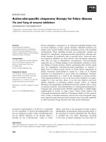
There are two basic approaches for gene transfer into hepatocytes: ex vivo and in
vivo strategies (Fig. 7.1). Ex vivo therapy requires the removal of a part of the liver.
To obtain hepatocytes, the removed tissue is treated with collagenase, and hepatocytes are separated from nonparenchymal cells by density gradient centrifugation.
Cells are then kept in culture and subjected to gene transfer by one of a variety of
methods. The population of cells is selected for those successfully genetically engineered and finally reinfused via the portal vein into the patient’s liver. However,
hepatocytes are not readily cultured. They undergo a few rounds of cell division but
not enough to substantially expand the population. Their viability is limited and culturing primary hepatocytes is hampered by some loss of differentiation. In addition,
an already ill patient may not be able to undergo the harvesting procedure.
While hepatocytes are kept in culture, several methods can be used to introduce
new genes. Deoxyribonucleic acid (DNA)-mediated techniques rely on commonly
used transfection methods such as calcium phosphate co-precipitation with DNA
and diethlyaminoethyl (DEAE) dextran complexed with DNA through electrostatic charges. These systems result in complexes that are taken up by the cell via endocytosis. Electroporation is another technique used to transfect cells.This involves the
exposure of cells to electrical pulses that render the plasma membrane momentarily
permeable. When performed in the presence of DNA, the membrane allows the
nucleic acid to enter the cells. All three of these methods result in low levels of transfection efficiency and transient expression of the therapeutic gene.Alternatively, different viral vectors as well as liposomes can be used for ex vivo gene transfer.
GENERAL PRINCIPLES FOR HEPATIC GENE THERAPY
155
Collagenase treatment
and hepatocyte separation
Culture
48hrs.
Reinfusion of genetically
altered hepatocytes
(a)
Construction of
gene vectors
Addition of
therapeutic gene
Liver specific infusion
into portal circulation
Recombinant
vector
Systemic infusion
(b)
FIGURE 7.1 Two basic methods for the delivery of genes to the liver. (a) Shows the ex
vivo approach. It requires the removal of part of the liver, usually the left lateral segment.
The liver tissue is treated with collagenase and hepatocytes are separated from nonparenchymal cells by density gradient centrifugation. Hepatocytes are then propagated in
culture and subjected to gene transfer. Finally successfully transformed cells are selected and
reinfused via a catheter into the portal circulation of the patient’s liver. (b) Shows the in vivo
approach. A gene vector, suitable for the delivery of genes to the liver is constructed. The
therapeutic gene is incorporated into this vector and the recombinant vector is infused into
the patient. Systemic infusion over a peripheral vein is appropriate for vectors that selectively target the liver; direct infusion into the portal circulation is preferrable for vectors
without liver targeting abilities.
For in vivo gene therapy, the therapeutic or normal gene is introduced directly
into the host. On one hand, in vivo gene therapy circumvents the need for the invasive procedures of harvesting and reimplantation and eliminates the need to culture
primary hepatocytes. On the other hand, it is necessary for any vehicle used for in
156
GENE THERAPY FOR LIVER DISEASE
vivo hepatic gene therapy to reach the liver efficiently. For systemic application,
the gene vectors are ideally targeted to the liver, avoiding broad biodistribution and
extrahepatic effects. Once inside the liver, a transgene has to pass through the fenestrations of endothelial cells to reach parenchymal liver cells, while simultaneously
avoiding clearance through phagocytosis by Kupffer cells. In vivo gene therapy can
also be mechanically directed to the liver by portal injection of the foreign gene
construct. Presently several viral systems as well as liposomal preparations and
protein–DNA conjugates have been used for in vivo gene therapy (Table 7.1).
Viral Vectors
Retrovirus Retrovirus can infect many different types of mammalian cells including liver cells. One limitation to the use of prototype retroviruses in hepatic gene
TABLE 7.1 Advantages and Disadvantages of Vehicles Concerning Liver-Directed
Gene Therapy
Vehicle
Retrovirus
Advantages
No immune/inflammatory
response
Absence of hepatic necrosis
Disadvantages
Requires dividing cells
Low expression in hepatic
cells in vivo
Integrates with stable
expression
Adenovirus
Targets hepatocytes
specifically
Expressed in nondividing
cells
Remains episomal
Transient expression
Inflammatory/immune
response
Injurious to hepatocytes
Adenoassociated virus
Expressed in nondividing
cells
Integrates with stable
expression
No inflammatory/immune
response
Small delivery capacity
Liposomes
DNA protected from
degradation
Large delivery capacity
Uptake by nonparenchymal
liver cells
Intracellular degradation in
lysosomes
No inflammatory/immune
response
Protein/DNA carriers
Liver specific
Large delivery capacity
No inflammatory/immune
response
Intracellular degradation in
lysosomes
Remains episomal
Transient expression
GENERAL PRINCIPLES FOR HEPATIC GENE THERAPY
157
therapy is that only dividing cells are efficiently transduced. To circumvent this
problem, researchers have performed partial hepatectomies before the administration of the retrovirus. Because the remaining liver tissue is induced to proliferate in
response to this injury, the percentage of transduced cells could be increased.
Adenovirus In early adenoviral constructs, in addition to expression of the
foreign gene, some viral genes were also expressed. The latter led to a virus-specific
immune response manifested by development of hepatitis and destruction of the
genetically altered hepatocytes. The expressed therapeutic protein usually became
undetectable after a maximum period of 4 weeks.The formation of neutralizing antibodies by B lymphocytes against viral proteins make a periodic readministration
less effective. This problem has been tackled by deleting additional viral genes to
minimize the expression of viral proteins. It has been shown that the therapeutic
gene expression level was increased in mouse liver while the immune response
previously seen was decreased. Adenoviral constructs have recently been prepared
in which all viral genes have been eliminated. Using a different approach, transient
administration of an immunosuppressive drug resulted in the long-term expression
of the adenoviral vector system. It has also been shown that it is possible to render
rats immunotolerant to adenoviral antigens by intrathymic injections and oral
administrations of adenoviral protein extracts or by neonatal administration of the
virus in utero, thereby increasing long-term expression and allowing readministration of adenoviral vectors.
Adenoassociated Virus Adenoassociated virus (AAV) can infect dividing as
well as nondividing cells making it a possible vector for use in organs such as the
liver. The rate of transduction in nondividing cells, however, is lower than that of
cells undergoing division. AAV transduces cells that are in S phase of the cell cycle.
Treatments that interfere with DNA metabolism, such as hydroxyurea or aphidicolin and topoisomerase inhibitors, markedly increased the number of recombinant AAV transduced cells. g-Irradiation has a similar effect on the efficiency of
this system. After localized irradiation to the liver, hepatocyte transduction was
increased up to 900-fold over hepatocytes of mice that were not irradiated. This is
probably due to the fact that the irradiation is cytotoxic, thereby stimulating division of the surviving cells.
Nonviral Vectors
Liposomes Liposomes are microscopic vesicles consisting of one or multiple
aqueous compartments. Liposome clearance from the circulation by the liver
is dependent on the size and surface composition of liposomes. Because the
fenestrations of the endothelial cells in the liver have a diameter of about 100 nm,
particles larger than 250 kD cannot pass into the space of Disse and, therefore, do
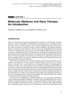
not interact significantly with hepatocytes (Fig. 7.2). For this reason, liposomes larger
than 100 nm are cleared by phagocytosis through Kupffer and endothelial cells.
Changing the size and lipid composition of the sphere can alter the biodistribution
to the different cell populations within the liver. This allows for the targeting
to either hepatocytes or Kupffer cells. One advantage of liposomes is the fact
that DNA can simply be incorporated in the aqueous phase or associated with the
158
GENE THERAPY FOR LIVER DISEASE
FIGURE 7.2 Liposomes are used as a device to deliver genes to hepatocytes. Liposomes
are microscopic vesicles consisting of lipid bilayers enclosing one or multiple aqueous compartments. DNA is incorporated in the aqueous phase or associated with the lipid material
after simply mixing with the lipid components. Liposomes enter the liver by the portal circulation. Their clearance from the circulation is largely dependent on their size and surface
composition. Because the fenestrations of the endothelial cells in the liver have a diameter
of about 100 nm, particles larger than 250 kD cannot pass into the space of Disse. Only small
liposomes can escape uptake by Kupffer and endothelial cells and interact with parenchymal
liver cells.
lipid material. In addition, the encapsulated gene is protected from enzymatic
degradation.
Cationic liposomes have been used to form DNA complexes in which the
DNA remains primarily on the outside of the microsphere. While this is an
advantage because the DNA that can be trapped within the vesicle is limited, it may
cause an aggregation of one or more liposomes and prevent uptake or promote
GENERAL PRINCIPLES FOR HEPATIC GENE THERAPY
159
phagocytosis by Kupffer cells. Liposomes are taken up by the cells via endocytosis
and eventually enter lysosomes. In lysosomes, enzymatic degradation of the
contents occurs and could decrease the efficiency of deliver of the therapeutic
gene to the nucleus. To circumvent this problem, liposomes have been developed that are pH sensitive, avoiding fusion with the lysosomes. Following internalization, these liposomes change their properties when they are exposed to the low
pH of endosomes. During endocytosis, they are able to destabilize the endosomal membrane or become fusogenic. In this way, the liposome may be able to
deliver its contents into the cytoplasm before the liposome is delivered to
lysosome.
Another means of improving the efficacy of liposomes to target parenchymal
liver cells is the incorporation of various ligands recognized by receptors on the
surface of hepatocytes. Examples of such targeting moieties are epidermal growth
factor, lactosylceramide, asialofetuin, lactose mono-fatty acid esters, and bgalactoside. For many preparations, uptake by endothelial or Kupffer cells compared to parenchymal cells is still predominant, and there is no unanimity on the
quantitative aspect of the differential uptake into different cell types in the liver.
Liposomes with galactose residues are also recognized by Kupffer cells via the galactose-particle receptor, and the distribution between parenchymal and nonparenchymal liver cells is strongly size dependent, with only very small liposomes
with limited loading capacity or vesicles containing lactosylceramide or lactose
mono-fatty acid esters preferentially directed to parenchymal cells.
Protein–DNA Complexes Soluble conjugates between naturally occurring
and recombinant proteins and DNA are attractive tools for gene therapy directed
to the liver. An example of the use of targeted delivery of protein–DNA complexes is the use of asialoglycoprotein receptors. The asialoglycoprotein receptor
is present in large numbers only on the plasma membrane of hepatocytes and binds
galactose-terminated glycoproteins and neoglycoproteins with high affinity. Bound
ligands are internalized by the cell via receptor-mediated endocytosis. Due to its
specificity, the asialoglycoprotein receptor (AsGPr) has been exploited as a means
to deliver drugs and DNA for therapeutic purposes, as well as diagnostic agents
to hepatocytes.
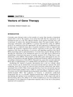
A system, based on asialoglycoprotein-poly-l-lysine conjugates has been developed to target DNA to the liver via the AsGPr (Fig. 7.3). The a1 acid glycoprotein, orosomucoid, was desialylated by treatment with neuraminidase to produce
asialoorosomucoid (ASOR), a high-affinity ligand for the AsGPr. Poly-l-lysine (PL)
was then covalently attached to the protein by carbodiimide-mediated amide bond
formation. The resulting ASOR-PL conjugate bound the negatively charged DNA
in a nondamaging electrostatic interaction and protected it from nuclease degradation. The complex was selectively and rapidly internalized into hepatocytes by
receptor-mediated endocytosis, and foreign genes were expressed in vitro and in
vivo. To further increase the persistence of foreign gene expression in vivo, a partial
hepatectomy, leading to stimulated hepatocyte replication was performed. The
underlying mechanism was shown to be the disruption of the microtubular network
necessary for the translocation of endosomes to lysosomes, which could also be
accomplished by colchicine administration.
160
GENE THERAPY FOR LIVER DISEASE
GP
AS
Covalently attached
Poly-L-lysine
positively charged
ASGP
Hepatocyte carrying the
liver specific ASGP-receptor
DNA-negatively
charged
ASGP
P
ASG
Endocytosis
Endosome
Receptor
recycling
P
ASGP
ASG
ASGP
receptor
Microtubular
network
Cytosol
Fusion of
endodome and lysosome
Lysosome
Lysosome
Release of DNA
at low pH
Nucleus
Degradation
Escape
of DNA
Transcription
of gene to mRNA
FIGURE 7.3 Use of asialoglycoprotein (ASGP) to target genes to the liver. The asialoglycoprotein receptor is present in large numbers only on the plasma membrane of hepatocytes
and binds galactose-terminated glycoproteins with high affinity. Positively charged polyl-lysine is covalently attached to ASGP by carbodiimide-mediated amide bond formation.
The resulting ASOR-PL conjugate binds the negatively charged DNA in a nondamaging electrostatic interaction. The complex is internalized into hepatocytes by receptor-mediated
endocytosis. After endocytosis the ligand dissociates from the receptor and the receptor recycles to the cell surface. The translocation of the endosome to the lysosome requires an intact
microtubular network. After fusion of endosome and lysosome, the DNA is released from its
carrier at low pH. Part of the DNA escapes the lysosome and reaches the nucleus where it
can be transcribed into mRNA.
CLINICAL APPLICATIONS OF LIVER-DIRECTED GENE THERAPY
161
CLINICAL APPLICATIONS OF LIVER-DIRECTED GENE THERAPY
Familial Hypercholesterolemia
Familial hypercholesterolemia (FH) is an autosomal dominant disorder that affects
one in every 500 people. It is caused by defects in the hepatic low-density lipoprotein (LDL) receptor gene. The reduced activity of the LDL receptor leads to
an inefficient clearance of LDL particles by the liver and therefore, a limited
metabolisim of LDL. Accordingly, this causes elevated serum LDL cholesterol
levels, which leads to premature coronary artery disease. Heterozygotes for FH
maintain only a portion of the normal LDL receptor function, and their serum LDL
levels are almost double that of normal individuals. Homozygotes, having two
mutant receptor genes, have only 0 to 20% of normal LDL receptor activity and
show extremely elevated serum cholesterol levels. Without treatment, this usually
leads to death by myocardial infarction before the age of 20.
The LDL receptor is, in fact, found on all cells. However, it is the hepatic expression of the receptor that plays the main role in regulating serum cholesterol levels.
The liver is the only organ that is capable of converting cholesterol to bile acids
and excreting them from the body. Pharmacological therapy for heterozygote FH
patients, who express the LDL receptor at a low level involves upregulation of LDL
receptor gene expression. Drugs, including 3-hydroxy-3-methylglutaryl coenzyme A
reductase inhibitors and bile acid binders, act to reduce intracellular hepatic free
cholesterol. This causes the LDL receptor gene expression to be influenced, accelerating LDL catabolism and, accordingly, reducing serum cholesterol. However, this
treatment, combined with strict dietary reduction of cholesterol intake, is only
feasible in the case of heterozygosity and does not reduce the serum cholesterol
level into the normal range. For those patients that lack expression of a functional
receptor due to homozygosity, or heterozygotes with an inefficient response to
pharmacological therapy, weekly plasmapheresis or liver transplantation are the
only alternatives. Both procedures are very expensive, and the latter is associated
with morbidity and mortality and limited organ supply. For these reasons, hepatic
gene therapy has been employed in an attempt to treat FH.
Early experiments in the Watanabe heritable hyperlipidemic (WHHL) rabbit, an
animal model for FH, demonstrated the possibility of successful ex vivo gene
therapy for FH. In these studies, hepatocytes were harvested, genetically modified
ex vivo with retroviruses that contained an intact LDL receptor gene, and transplanted back into the animal. Control experiments with mock transfected hepatocytes demonstrated no cholesterol lowering effect, but showed a transient increase
of the serum cholesterol levels probably due to the surgical procedure. Retroviral
transduced hepatocytes were shown to become stably engrafted into the animal’s
liver with a subsequent lowered serum cholesterol level. The effect was observed
for 6.5 months, the duration of the experiment. Subsequent experiments with dogs
and baboons also rendered encouraging results. In the case of the baboon, 1.5 years
after gene therapy, the transgene was still being expressed. The results of these early
experiments provided support for the efficacy of this treatment and paved the way
for human clinical trials.
A 28-year-old French Canadian woman was the first recipient of liver-directed
gene therapy. She was homozygous for a mutation in the LDL receptor gene, result-
162
GENE THERAPY FOR LIVER DISEASE
ing in the expression of a nonfunctional receptor. After suffering a myocardial
infarction at the age of 16, she had a coronary artery bypass at the age of 26. Her
baseline serum LDL concentration was 482 mg/dl (normal range 194 ± 34), and her
dyslipidemia did not respond to conventional drug therapy. The left lateral segment
of the patient’s liver, comprising about 15% of total mass, was removed and the
parenchymal liver cells were isolated. The cells were then transduced with a retroviral vector containing the full-length human LDL receptor gene under the control
of a chicken b-actin promoter and a cytomegalovirus (CMV) enhancer. To select for
successful transduction, cells were analyzed for the ability to uptake fluorescent
labeled LDL. Only genetically altered hepatocytes were reinfused into the portal
circulation (Fig. 7.4). The patient tolerated the procedures well without relevant side
effects.
Immediately following infusion of the genetically altered cells, the patient’s
serum LDL dropped by 180 mg/dl. A new baseline was established that was 17%
lower than before gene therapy. As her (LDL) decreased, her high-density lipoproteins (HDL) levels increased, improving her LDL/HDL ratio from 11 ± 0.4 to 7.9 ±
0.9. It is unclear as to why the HDL increased, although this same phenomenon has
been observed in patients that underwent orthotopic liver transplantation. The
patient also responded to the drug lovastatin, which prior to gene therapy had
no effect. Lovastatin is thought to deplete intracellular cholesterol, thereby upregulating expression of the LDL receptor. The recombinant receptor gene had no
transcriptional elements that could respond to cholesterol-mediated regulation.This
indicates that the response to lovastatin was related to posttranscriptional regulation, a mechanism demonstrated in previous studies. The response to lovastatin
diminished the patient’s serum LDL level further to 356 ± 22 mg/dl, and the effect
was meanwhile stable over a period of 2.5 years.
There was no immune response to the recombinant receptor. The patient’s sera
contained no antibodies to the recombinant receptor when a western blot analysis
was performed. Also, there was no evidence for autoimmune hepatitis following
gene therapy. In an extension of this study, four other FH individuals, including two
receptor-negative patients, were treated in a similar manner. Engraftment of successfully transduced hepatocytes as well as transgene expression was shown for all
patients, without significant side effects. Two out of four patients experienced a
significant improvement in their serum lipid profile, with a maximum reduction in
serum LDL of 150 mg/dl in one of the receptor-negative patients. None of the
patients developed an immune response to the transgene or to retroviral proteins.
Although gene transfer was demonstrated in all patients, the clinical impact on the
disease was low with serum cholesterol levels still exceedingly above the normal
range. In summary, this first human clinical trial showed the feasibility of ex vivo
gene therapy for FH but demonstrated the need for substantial modifications to
improve the percentage of transduced hepatocytes and the level and duration of
gene expression.
In an alternative approach, in vivo gene delivery was performed to treat WHHL
rabbits. The human LDL receptor gene was placed under the control of transcriptional elements from the mouse albumin gene, conferring efficient expression
in hepatocytes. The construct was conjugated via poly-l-lysine to ASOR, a highaffinity ligand for the ASOR receptor. Following systemic injection of this complex, analysis of WHHL rabbits revealed a rapid and liver-specific uptake of the
CLINICAL APPLICATIONS OF LIVER-DIRECTED GENE THERAPY
163
Left lateral segment
15%
Isolation of
hepatocyles
Surgical removal
of left lateral
liver segment
LTR
Retroviral vector
-actin
CMV
hLDL
enhancer promoter
Cell
culture
LTR
Infection
with recombinant
virus
Chromosomal DNA
LDL receptor
Reinfusion of
transduced
hepatocyles
Selection by
fluorescein labeled
LDL uptake
Serum
LDL Cholesterol
Harvest
of left
lateral
liver
segment
Transfusion
Application
of genetically of the HMG-CoAengineered reductase inhibitor,
hepatocyles
lovastatin
FIGURE 7.4 Gene therapy for LDL receptor deficiency. The left lateral liver segment of a
patient homozygous for a mutation in the LDL receptor gene is removed and hepatocytes
are isolated. The cells are transduced in culture with a retroviral vector containing the fulllength human LDL receptor gene under the control of a chicken b-actin promoter and an
cytomegalovirus (CMV) enhancer. The successfully transduced cells are selected by the use
of fluorescein-labeled LDL. Only genetically altered hepatocytes are reinfused into the portal
circulation of the patient. The patients baseline serum LDL concentration was 482 mg/dl.
Immediately following infusion of the transduced hepatocytes, the patients serum LDL
dropped by 180 mg/dl. In addition the patient now responded to lovastatin, a HMG-CoA
reductase inhibitor, which prior to gene therapy had no effect. The observed reduction in the
patients serum LDL level is meanwhile stable over a period of 2.5 years.
164
GENE THERAPY FOR LIVER DISEASE
DNA–protein conjugate, followed by expression of the transgene. The animals
experienced an immediate, but transient, decrease in total serum cholesterol by
153 ± 53 mg/dl. In control experiments, animal injected with a construct carrying the
CAT (chloramphenicol acetyltransferase) reporter gene instead of LDL receptor
gene showed CAT expression, but no diminuation of serum cholesterol levels. In
this study the expression was only 2 to 4% of the endogenous level of LDL receptor expression, and the effect on the serum lipid profile lasted less than one week.
These initial results were encouraging because of the specificity of the delivery.
However, the low levels and short duration of recombinant gene expression were
disappointing.
In recent animal studies, recombinant adenoviruses were used for in vivo liverdirected transfer of the LDL receptor gene. It was possible to restore LDL receptor expression in WHHL rabbits and LDL receptor knock-out mice, leading to
substantial reductions in serum cholesterol levels. However, the expression of the
recombinant receptor as well as the effect on the lipid profile has been only transient. This was due to the immune response that the host mounted against a lowlevel expression of viral proteins, with the subsequent destruction of the genetically
altered cells. Especially in receptor-negative subjects, the expression of an LDL
receptor could also trigger an immune response against the neoprotein, which would
further reduce the expression of the transgene. To circumvent this problem, another
group of researchers delivered the very low density lipoprotein (VLDL) receptor
gene to the liver of LDL receptor knock-out mice using recombinant adenoviruses.
Since the VLDL receptor is already expressed in extrahepatic tissue, there is no
immune response to the receptor after hepatic expression. Also the VLDL receptor binds LDL with a low affinity. It mediates the uptake of VLDL, the precursor
of LDL, and, therefore, results in a decrease of serum cholesterol.
Hemophilia B
Hemophilia B is an X-linked recessive coagulation disorder caused by a deficiency
or functional defect of blood clotting factor IX. The condition can be life threatening without regular infusions of factor IX concentrates in patients with evidence of
bleeding. Extensive testing of these products can eliminate impurities, but this form
of therapy still bears the risk of transfusion-transmitted viruses such as hepatitis C
and human immunodeficiency virus (HIV). In addition, the half life of factor IX is
only 24 h and, therefore, makes repeated transfusions often necessary. The liver is
the primary source for circulating factor IX and the prime target for a gene therapeutic approach to treat hemophilia B.
To date, attempts have been made in animal systems using the ex vivo approach.
The problems with these therapies are similar to those that have been encountered
with correcting other disorders: (1) the concentrations of circulating factor IX are
low and (2) there is a loss of gene expression over time. The latter is due to loss of
transduced cells or inactivation of the expression vectors.
There is a well-characterized canine model that has been used in preclinical trials
for hemophilia B. These dogs have no detectable factor IX activity due to a missense mutation in the catalytic domain.A retrovirus vector that contained the canine
factor IX gene under the control of retroviral promoter and enhancer elements was
used for direct delivery to the dogs liver via infusion into the portal circulation.
CLINICAL APPLICATIONS OF LIVER-DIRECTED GENE THERAPY
165
Analysis by ELISA and a biological assay demonstrated that plasma levels of 2 to
10 ng/ml of factor IX were achieved. In a normal canine, the level is about 11.5 mg/ml.
While the levels of circulating factor IX did not reach that of wild-type dogs, there
was a dramatic improvement in the biochemical parameters of hemostasis. This was
demonstrated by measuring the whole blood clotting time (WBCT), which in
normal dogs is 6 to 8 min. In dogs that have hemophilia B, the WBCT was about 45
to 50 min. After undergoing gene therapy this time was reduced more than 50%
with times in the range of 18 to 22 min. Although the concentration of factor IX was
as little as 0.1% of normal values, there was a dramatic improvement in clotting
times. Also, encouraging is the fact that this effect remained stable for over 9 months
(Fig. 7.5).
Adenoviral vectors that express canine factor IX have also been used to treat
hemophilia B dogs. Viral particles (2.4 ¥ 1012) were infused into the portal
vasculature of the dogs. The animals produced 2 to 3 times the wild-type level of
factor IX. However, the effect was only transient. The increase in factor IX concentration did normalize their clotting times, but the levels and clinical parameters
returned to pretreatment levels in 2 months. While repeated administrations could
be considered, it is possible that an immune response could develop with subsequent treatment.
Another group of researchers tried using adenoassociated viral (AAV) vectors
to express human factor IX in mouse livers. They simply injected the mice in a tail
vein with the recombinant vector after g-irradiation was applied to the liver. As previously discussed, this treatment probably stimulates cells to divide, thereby improving the efficacy of adenoassociated viral gene therapy. The concentration of human
factor IX in mice transduced with the AAV vector was between 0.1 and 1 ng/ml. This
result is similar to that observed in the dog model. The normal values for human
factor IX was 5 mg/ml, while levels of about 100 ng/ml would prevent chronic disease.
a1-Antitrypsin Deficiency
a1-Antitrypsin (AAT) is a serum glycoprotein, predominantly synthesized in the
liver and secreted into the blood. It is a protease inhibitor whose function is
essential in protecting the alveolar surface of the lung from destructive protease
activity. Its major substrate, neutrophil elastase (NE), is released by neutrophils
during phagocytosis, membrane perturbation, or cell lysis and cleaves connective
tissue matrix proteins located in alveolar walls. In normal individuals the levels
of AAT are sufficient to neutralize circulating NE. The different forms of AAT
deficiency result in reduced plasma levels of the protease inhibitor and in the failure
of NE to be neutralized. This is manifested in a high risk for the early development of pulmonary emphysema, due to proteolysis of the pulmonary extracellular
matrix.
The normal gene for AAT is designated M and accounts for 95% of alleles in the
caucasian American population. The most common mutants, called Z and S occur
with an allelic frequency of 1 to 2% and 2 to 4%, respectively, in this population. In
contrast Asians and African Americans are minimally affected. Homozygous individuals for the Z allele have only 10 to 15% circulating AAT levels bearing a certain
risk for pulmonary emphysema. Homozygous individuals for the S allele and MS or
MZ heterozygotes are phenotypically normal. However, some SZ heterozygotes
166
GENE THERAPY FOR LIVER DISEASE
LTR
Factor IX
LTR
Retrovirus carrying
the factor IX gene
Infusion into factor IX
deficient dog via
portal circulation
Factor IX Expression
Infection of Hepatocytes
and chromosomal integration
of the Factor IX gene
11mg/ml
10ng/ml
Normal
Dog
Treated
Dog
Factor IX
deficient
dog
Clotting Time (min)
Test for gene expression
and clinical effects
50
Treated
Dog
Normal Dog
10
2
4
6
8
10
Month
Retrovirus
injection
FIGURE 7.5 Gene therapy for factor IX deficiency. A recombinant retrovirus vector is constructed that contains the canine factor IX gene under the control of retroviral promoter and
enhancer elements (LTR). This vector is infused into the portal circulation of dogs that have
no detectable factor IX activity. The retrovirus is taken up by liver cells and the provirus
DNA integrates into the chromosomal DNA. Analysis of the dogs’ plasma by ELISA reveals
plasma factor IX levels of 2 to 10 ng/ml. A normal canine has a plasma factor IX level of
about 11.5 mg/ml. While the levels of circulating factor IX in the treated dog does not reach
that of wild-type dogs, there was a dramatic improvement in the whole blood clotting time.
could display an increased risk for the manifestation of pulmonary emphysema.
Homozygosity for the so-called null allele results in a complete lack of AAT in the
plasma, and these patients are extremely likely to develop emphysema. The same
is true for heterozygotes bearing an S or Z allele with the null allele. A number of
CLINICAL APPLICATIONS OF LIVER-DIRECTED GENE THERAPY
167
different mutations are responsible for the null allele, ranging from point mutations
to complete deletions. About 10% of individuals homozygous for the Z allele bear
the additional risk of significant clinical liver injury, probably due to the accumulation of misfolded AAT in the ER of hepatocytes.
The current treatment for AAT deficiency consists of weekly intravenous applications or intratracheal inhalation of human AAT, produced from serum. Recombinant human AAT, synthesized in bacteria or yeast has the disadvantage of a
shorter half-life and increased renal clearance due to improper posttranslational
glycosylation. While the administration of human AAT has been shown to raise the
serum AAT activities in patients, the response is only temporary, and a significant
impact on the prevention of pulmonary damage has yet to be proven for the intravenous as well as the intratracheal application. a1-Antitrypsin deficiency is another
candidate disease for gene replacement therapy, whereby ideally, the correct gene
could be delivered to hepatocytes and offer a long-term stable production of AAT.
Attempts to correct this disorder have been studied on dogs where the introduction of the correct gene was performed in an ex vivo manner. After transplantation
of retroviral transduced hepatocytes, the cells achieved peak production of human
AAT in vivo at day 10 posttransplantation. However, these levels dropped and
became undetectable around day 47.
Another group of investigators attempted an in vivo approach using small liposomes as the method of gene delivery. A plasmid containing the full-length human
a1-antitrypsin gene was encapsulated in small liposomes and was intravenously
injected into mice. A single dose of liposomal-delivered plasmid induced the production of human AAT in mouse hepatocytes and resulted in measurable levels of
human AAT in mouse plasma, still detectable after 11 days. In control experiments,
the injection of free plasmid did not result in measurable AAT expression (Fig. 7.6).
Interestingly, there was no additive effect when additional doses of the liposome
complex were delivered. However, partial hepatectomy performed 3 h after the
intravenous application of the liposomal formulation increased human AAT plasma
levels significantly. On day 11 after the intravenous (IV) injection, human AAT
levels had increased 6.4 times compared to animals injected without the performance of partial hepatectomy. It is unclear why the repetitive application did not
further increase the gene expression. Also, it is not completely understood why the
stimulation of cell proliferation by partial hepatectomy increased gene expression.
Presumably, this may be due to mechanisms that alter the compartmentalization
of liposomal-delivered DNA within the cells, allowing escape from the lysosomal
degradative pathway.
Crigler–Najjar Syndrome (Bilirubin UDP b-D Glucuronosyltransferase
Deficiency)
Bilirubin is the principal degradation product of heme. The enzyme that catalyzes
the coupling of bilirubin with glucuronic acid is bilirubin UDP-glucuronosyltransferase (B-UGT). The prototype of an inherited bilirubin conjugation disorder is
Crigler–Najjar (CN) syndrome type I. Patients with this recessively inherited disease
are characterized by high serum levels of unconjugated bilirubin, with little or no
conjugated pigment in the bile. They do not respond to enzyme induction therapy
with phenobarbitol and suffer a variety of neurological damages such as motor
168
GENE THERAPY FOR LIVER DISEASE
FIGURE 7.6 Gene therapy for a1-antitrypsin (AAT) deficiency. A plasmid that contains
the full-length human AAT gene is encapsulated in small liposomes. The liposomes are
injected into the tail vein of a mouse. A single dose of liposomal-delivered plasmid induces
the production of human AAT in mouse hepatocytes and results in measurable levels of
human AAT in mouse plasma, lasting 11 days. If a partial hepatectomy is performed 3 h after
the intravenous application of the liposomal formulation, AAT plasma levels are significantly
higher.
abnormalities, hearing loss, kernicterus, and finally death. At present, the only definitive treatment for this disorder is liver transplantation. A similar defect exists in
Gunn rats, which are homozygous for the mutation and, therefore, show no hepatic
B-UGT activity. These rats exhibit lifelong hyperbilirubinemia and develop bilirubin encephalopathy. They provide a model system for studies on the efficacy of gene
therapy for Crigler–Najjar syndrome type I.
An example of transient in vivo correction of this defect has been made by targeted delivery of the human B-UGT gene to the liver of Gunn rats using asialoglycoprotein poly-l-lysine DNA conjugates as previously described. As a strategy
to prolong the duration of targeted gene expression, advantage was taken of the
fact that the translocation of endosomes to lysosomes as part of the endocytotic degradative pathway requires an intact microtubular network. Colchicine, a
CLINICAL APPLICATIONS OF LIVER-DIRECTED GENE THERAPY
169
microtubule disruptive agent, was administered 30 min prior to the injection of the
ASOR–DNA complex to prevent the translocation of the endosomal vesicles containing the ligand to lysosomes. Targeted delivery of B-UGT under these conditions
resulted in the persistence of the delivered DNA in the liver for 10 weeks. Bilirubin glucuronides were excreted in the bile and serum bilirubin levels decreased by
25 to 35% in 2 to 4 weeks and remained reduced for a period of 8 weeks. Without
treatment with colchicine, the DNA remained in the liver for only 2 days and there
was no effect on serum bilirubin levels.
These studies used concentrations of colchicine that would be toxic to humans.
There are other drugs that could produce the same effect yet are safe for application in clinical human trials. Alternatively, to avoid side effects and broad biodistribution, colchicine could be delivered in a liver-specific manner. In this way,
microtubular disruption provided a noninvasive method for prolonging the effect
of this liver-specific method of gene therapy.
As discussed previously, recombinant adenoviruses are efficient in transferring
foreign genes to quiescent, nondividing cells and high levels of gene expression can
be achieved using this vector system. However, since they do not integrate their
DNA into the host genome, subsequent administrations will be necessary. Therefore, the immune response, usually evoked after the initial injection has yet to be
circumvented. Gunn rats were used to address this problem. Previously delivering
the human B-UGT gene via recombinant adenovirus has proven to be effective for
a short period. Treated animals showed excretion of bilirubin glucuronides and a
70% reduction of serum bilirubin levels. This effect was only transient due to the
immune response mounted against adenoviral antigens, expressed by transduced
hepatocytes. The same effect was not seen in subsequent applications to the same
animals due to neutralizing antibodies. A group of researchers investigated whether
the administration of recombinant adenovirus during the neonatal period could
induce a tolerance to the recombinant adenovirus. Gunn rats (1 to 3 days old) were
injected with 1 ¥ 108 plaque forming units (pfu) of recombinant adenovirus carrying the human B-UGT gene. Subsequent injections were administered 56 and 112
days later. Control experiments were performed using recombinant adenovirus that
contain the Lacz reporter gene. Animals that received the B-UGT, but not those
that received Lacz, had a reduction of serum bilirubin levels by 70 to 76% as compared to untreated animals. There was a gradual increase of serum bilirubin levels
by day 53, but the second and third injection of recombinant adenovirus had an
additive effect on serum bilirubin levels. Analysis also showed that antibodies and
cytotoxic lymphocyte activity to the recombinant adenovirus were not detectable.
This demonstrates that injecting the recombinant adenovirus during the neonatal
stage tolerized the animals and permitted long-term therapy with repeated
administrations.
One concern with this treatment is the question if the induction of tolerance
against the recombinant adenovirus could result in tolerance to wild-type virus as
well. Adenoviral infections are common throughout the life span of a human being,
usually manifested as self-limited, uncomplicated disease. The same group of
researchers injected two doses of wild-type virus into Gunn rats previously tolerized with three doses of recombinant adenoviruses starting in the neonatal period.
The animals elicited a cytotoxic T-lymphocyte immune response after the first injection of wild-type virus, which was further increased after the second injection. Inter-
170
GENE THERAPY FOR LIVER DISEASE
estingly, the animals continued to express the transferred B-UGT gene and did not
experience an increase in unconjugated serum bilirubin levels.
Gene Therapy for Viral Infections
so
l
cle
U
NA
mR
Ribosome binding site
Antisense
oligonucleotides
is e
G G CC CU G A
U G G
A
C T T GU A A C
CA T T G A C
airing
e-p
as
B
es
nt
Cap
Poly-A-Tail
n se o
ligonucleotid
G
C
A UU
A A
T U
A U C A AAA
A
A
C
AA
to
Nu
Cy
C
G
U
G
A C
A TT
UG C
GA
A
GC
C
Chromosomal DNA
us
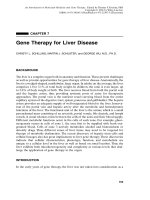
In contrast to many other gene therapeutic strategies, where replacement of a defective gene is the predominant goal, the therapy of viral infections by means of gene
therapeutic technology is to inhibit viral replication, transcription, or translation of
viral genes or assembly of viral particles. If the nucleic acid sequence of a viral gene
is known, antisense oligonucleotides consisting of short single strands of DNA can
be designed to bind the corresponding messenger ribonucleic acid (mRNA) (e.g.,
the sense strand) by complementary base pairing. This can result in direct inhibition of translation or cleavage of the RNA component of RNA–DNA hybrids by
intracellular RNase H (Fig. 7.7). Antisense oligonucleotides are usually 15 to 20
bases long and made by the use of an automated DNA synthesizer.
Ribosomes
DN
A
A
AA
a
AA
A
RNA
DNA Hyb
rid
RNase H
Block of
translation
Degradation of
RNA-DNA hybrid
by RNase H
FIGURE 7.7 Antisense oligonucleotides. Chromosomal DNA is transcribed into messenger RNA (mRNA), containing a cap at the 5¢ and a poly-A-tail at the 3¢ terminus. Messenger RNA leaves the nucleus for the cytosol where translation into proteins takes place. Translation is performed by ribosome and requires a ribosome binding site. Antisense oligonucleotides consist of a short single strand of DNA. If the nucleic acid sequence of a viral gene
is known, they can be designed to bind the viral mRNA by complementary base pairing.
This results in direct inhibition of translation or cleavage of the RNA component of the
RNA–DNA hybrid by RNase H. Replication of hepatitis B and C virus depends on an RNA
intermediate. Therefore antisense oligonucleotides can interfere with viral replication.
CLINICAL APPLICATIONS OF LIVER-DIRECTED GENE THERAPY
171
A related strategy uses ribozymes to suppress viral replication or transcription
of viral genes. Ribozymes are RNA molecules with a catalytic moiety capable of
cleaving target RNA molecules surrounded by RNA arms able to bind to the target
sequence by complementary base pairing similar to antisense oligonucleotides
(Fig. 7.8). Theoretically, one ribozyme can cleave many target RNA molecules.
Transfection of a vector containing the sequence of a ribozyme could result in
the generation of many copies of therapeutic ribozyme molecules within target
cells.
Another antiviral strategy consists of the use of dominant negative polypeptides,
designed to interact with their native counterparts, thereby interrupting viral assembly or enzyme function.
Chronic Viral Hepatitis
There are at least five different viruses causing hepatitis in human. Hepatitis A virus
and hepatitis E virus, contagious predominantly through a fecal-oral route, cause
s
t
AA
Cy
CA
A
Antisense region II
Catalytic domain
GC
A
AG
A
G
G
CG
A
AG
UU
C
AU
A U
G CG U
C CG C
U
A
mRN C U C A G
G
GU A
UCC CA
G
UAC
CU U G A G
Base
CG A A
Cleavage
Site
A
UU
A
RN
m
UG
Ribozyme
molecules
AA
Nu
ol
cl
C A
G
U AG
G
G
eu
A C
B AT T
CU C
A
G
C
U
NA
pairing
UG
AC
al D
os
C
som
hro mo
A
A
Antisense region I
Cleavage
CA
U
AAA
UACUCCGUC
CGCU
GAA
A A UG A
GG
CA
U C UCCGC A A U
AG
AG
GC
G
U
U
AA A
CUUA
Recycling of
ribozymes
Release
substrates
FIGURE 7.8 Ribozymes. Chromosomal DNA is transcribed into messenger RNA
(mRNA), containing a cap at the 5¢ and a poly-A-tail at the 3¢ terminus. Messenger RNA
leaves the nucleus for the cytosol where translation into proteins takes place. Ribozymes
are RNA molecules with a catalytic moiety capable of cleaving target RNA molecules. The
catalytic domain is surrounded by two RNA arms designated as antisense regions. The
antisense regions are designed to bind the target sequence by complementary base pairing.
After cleavage the substrate is released and the ribozyme recycles to cleave other target
molecules. Ribozymes can cleave mRNA molecules as well as viral RNA involved in viral
replication.
172
GENE THERAPY FOR LIVER DISEASE
acute self-limited disease. Three other well-characterized viruses, hepatitis B virus
(HBV), hepatitis C virus (HCV), and hepatitis D virus (HDV) are known to cause
persistent infection and chronic disease of the liver.
Hepatitis B Virus HBV is a small DNA virus with a partially double-stranded
circular DNA molecule of about 3200 base pairs. It belongs to a group of hepatotropic DNA viruses (hepadnaviruses) that includes the hepatitis virus of the
woodchuck, ground squirrel, Pekin duck, and heron. The virus consists of an outer
envelope and an internal core (nucleocapsid). The envelope is composed mainly of
hepatitis B surface antigen (HBsAg). The nucleocapsid contains hepatitis core
antigen (HBcAg), a DNA polymerase/reverse trancriptase, and the viral genome.
Different from all other known mammalian DNA viruses, hepadnaviruses replicate
via reverse transcription of an RNA intermediate, in a manner endogenous to the
life cycle of RNA retroviruses (e.g., HIV). Based on this fundamental step in the
replication of the virus, antiviral strategies aimed at the reverse transcription of
HIV RNA or at HIV reverse transcriptase are also potentially useful against HBV
infection.
A number of antisense sequences that are capable of inhibiting the replication
of hepatitis B and hepatitis C viruses in vitro have been identified. Efficacy has also
been observed with an antisense phosphorothioate DNA in vivo. However, because
oligonucleotide uptake by cells is generally low, and susceptibility to degradation in
plasma can be quite high, some form of targeting would be desirable for successful
use of antisense strategies for therapy of viral hepatitis in vivo. A system, based on
asialoglycoprotein-poly-l-lysine conjugates, was used to prepare ASOR-PL complexes with an 21-mer antisense oligonucleotide complementary to the sequence of
the polyadenylation signal of the HBV genome. By using a radioactive end-labeled
species, it was determined that the oligo alone was taken up with a rate of
0.05 pmol/h/million cells by two hepatoma cell lines, HepG2 (AsGPr positive) or SK
Hep1 (AsGPr negative). However, the uptake of oligo conjugated to ASOR-PL
was 10 times faster into HepG2 cells but was not changed in SK Hep1 cells. Coincubation with an excess asialoorosomucoid blocked the uptake. To show whether
the targeted antisense has antiviral activity, the HepG2 2.2.15 cell line was used. This
cell line possesses AsGPrs, is stably transfected with the complete HBV genome,
and secretes viral antigens as well as infectious virus particles. Administration of
complexed antisense DNA blocked the expression of HBsAg in these cells, and
reduced the replication of viral DNA by about 80% compared to untreated controls. A complexed oligonucleotide with random sequence had no effect, and the
antisense oligo DNA alone decreased the expression of surface antigen and viral
replication by only approximately 30%.
In a subsequent investigation, ASOR-PL complexed to a 21-mer phosphorothioate antisense oligonucleotide against the polyadenylation region and adjacent
upstream sequences of WHV was used to treat WHV-infected woodchucks. Animals
were injected intravenously with ASOR-PL complexes containing 0.4 mg antisense
for 5 consecutive days (total dose 2 mg/animal, 0.1 mg/kg/day). Although there was
no difference in the levels of surface antigen between treated and untreated animals,
a significant decrease in viral burden was observed. Treated animals showed a 1 to
2 log decrease in circulating viral DNA, 25 days posttreatment. The decline lasted
for approximately 2 weeks, after which there was a gradual rise in DNA levels.
CLINICAL APPLICATIONS OF LIVER-DIRECTED GENE THERAPY
173
Antisense alone or a complex containing a random oligo DNA of the same size
and linkage failed to have any significant effect on viral DNA levels.
Targeted pretreatment of hepatocytes with the above antisense oligonucleotide
complexed to ASOR-PL was used to prevent subsequent infection with HBV.
Usually, it cannot be anticipated when an acute exposure to HBV will occur.
However, after liver transplantation in patients infected with HBV, the grafts are
invariably reinfected. Furthermore, there is an accelerated course in most cases.
Protection of the graft by pretreatment could prevent reinfection. Pretreatment of
Huh7 cells (AsGPr positive) with ASOR-PL antisense complexes before lipofection with an HBV plasmid (6.5 million copies of plasmid per cell) inhibited the
amount of newly synthesized, core-associated viral DNA in Huh7 cells to undetectable levels, or less than 0.1 pg, as assessed by quantitative PCR. HBsAg, secreted
by the cells into the medium, was inhibited in a dose-dependent manner by a
maximum of 97%, and the inhibition lasted for 6 days. Pretreatment with unconjugated antisense or complexed random oligo showed no significant effects.
Very recently, a related targeting device, consisting of human adenovirus particles conjugated to N-acetyl-glucosamine-modified bovine serum albumin, streptavidin, and PL, was used to deliver phophorothioate-modified 16-mer antisense
oligonucleotides to hepatocytes via the AsGPr. The oligonucleotide was directed
against the encapsulation signal of the core gene. Chicken hepatoma cells (LHM)
were transfected by complexed HBV–DNA. When the cells were treated with complexed oligonucleotide before and after treatment with complexed HBV–DNA,
an approximately 80% inhibition of core-particle-associated HBV–DNA level was
observed.
Another antiviral strategy consists of the use of dominant negative polypeptides,
designed to interact with their native counterparts, thereby interrupting viral assembly or enzyme function. Mutants of HBV core protein were shown to inhibit wildtype viral replication by interference with nucleocapsid formation.
Hepatitis C Virus HCV contains a single-stranded RNA genome of positive
polarity and is about 9500 bp in length. Its replication requires a negative stranded
RNA intermediate synthesized by the viral RNA dependent–RNA polymerase. The
viral genome encodes a single polyprotein of 3010 to 3033 amino acids in length.
Posttranslational processing results in the RNA binding nucleocapsid protein C, the
envelope proteins E1 and E2, and the nonstructural proteins NS1 to NS5, including
RNA-dependent RNA polymerase. At both termini of the RNA genome exist conserved sequences called noncoding regions (NCR), involved in RNA replication,
translation initiation, and presumably RNA packaging.
Presently, animal models are limited to chimpanzees. For this reason, in vitro
studies using artificial reporter constructs frequently are employed to investigate
new treatment involving gene therapy for hepatitis C. In an early investigation,
hepatitis C virus cDNA was cloned and used for screening highly conserved regions
of the hepatitis C genome for potential target sequences in an antisense approach.
After transcription with T7 RNA polymerase, HCV RNA was purified and mixed
with a 10-fold molar excess with sense or antisense oligonucleotides. These mixtures
were used for in vitro translation in a rabbit reticulocyte lysate in the presence of
35
S-methionine to synthesize HCV proteins. Sense oligonucleotides showed no significant inhibitory effect on HCV protein synthesis as measured by the incorpora-
174
GENE THERAPY FOR LIVER DISEASE
tion of 35S-methionine. In contrast, an antisense oligonucleotide directed against the
5¢ NCR inhibited in vitro translation more than 50%. Another antisense oligonucleotide directed against the start codon of the HCV core gene inhibited in vitro
translation up to 97%. Interestingly, antisense oligonucleotides directed against
further downstream sequences had no inhibitory effect on translation, presumably
due to the inefficiency blocking ribosomal translocation during translation. It is
noteworthy that a molar ratio of oligonucleotide to HCV RNA of 10 to 1 was necessary to achieve the reported effects.
In subsequent studies, the ability of antisense oligonucleotides to inhibit translation in cell culture was investigated. Human hepatoma cell lines were transfected
with plasmids carrying conserved HCV target regions either downstream of a
CMV promoter or upstream of a luciferase reporter gene. Four different antisense
oligonucleotides that were directed against the 5¢ NCR were co-transfected with the
reporter construct. At a concentration of 0.3 mM (~3 mg per 35 mm cell culture dish)
two showed an inhibitory effect of 95% on luciferase activity. It is important to note
that sense oligonucleotides also inhibited luciferase expression up to 30%.
Ribozymes have been shown to be effective against hepatitis B and hepatitis C
viral RNA. Until now experiments using ribozyme technology have been demonstrated to cleave HBV RNA in vitro, but no suppression of HBV replication or
HBV protein translation have been reported in cell systems or in vivo.
For HCV, suppression of viral gene expression in cells by ribozymes was successfully demonstrated. Again a plasmid carrying an HCV-luciferase reporter gene
was constructed with the 5¢ NCR and part of the core gene placed between a CMV
promoter and the luciferase gene. Additionally, four vectors carrying the sequence
for hammerhead ribozymes directed against the 5¢ NCR or core region were used
to synthesize ribozyme molecules for in vitro studies. After in vitro transcription of
HCV-luciferase RNA, the different ribozyme molecules were investigated for their
cleavage activity. The human hepatoma cell line Huh7 was then used to investigate
the in vivo activity. Cells were co-transfected with ribozyme RNA and HCVluciferase RNA at molar ratios of 0 : 1, 3 : 1, 10 : 1, and 30 : 1, the first ratio serving as
the control. Two of the ribozymes, directed against the 5¢ NCR and core region,
respectively, suppressed luciferase activity by 73% (ribozyme : reporter gene ratio
10 : 1) and 55% (30 : 1), respectively. Control experiments with ribozymes harboring
a mutation in their catalytic region did not show any inhibitory effect at the same
molar ratio. Co-transfection of the HCV reporter plasmid and eukaryotic expression vectors encoding the two most promising ribozymes with a 20-fold molar excess
of the ribozyme vector showed suppression of luciferase activity by approximately
50 and 40%. Control experiments with ribozymes not directed against HCV or
co-transfection of a vector carrying the luciferase gene without upstream HCV
sequences proved the specificity of the observed effect.
Finally, cell lines constitutively producing the two most promising ribozymes
after stable transfection with the ribozyme carrying vectors were investigated.
Ribozyme expressing cells were transiently transfected with the HCV-luciferase
reporter plasmid and showed an inhibition of luciferase activity of 30 and 50%
compared to parental cells transiently transfected with the reporter construct.
When a conventional luciferase reporter plasmid was transiently transfected,
ribozyme-expressing cell lines and parental cells showed no difference in luciferase
activity.
CLINICAL APPLICATIONS OF LIVER-DIRECTED GENE THERAPY
175
Hepatocellular Carcinoma
Hepatocellular carcinoma (HCC) is one of the most common malignancies affecting man and causes an estimated one million deaths per year worldwide. Identified
major risk factors are chronic infection with hepatitis B or C virus, liver cirrhosis,
especially due to alcohol abuse or genetic hemochromatosis, and repeated exposure
to aflatoxin. Surgery is the only curative therapy for HCC. However, due to the
extent of the tumor and associated cirrhosis at the time of diagnosis, it is inappropriate in the majority of patients. The search for new therapies has not yet resulted
in a significant improvement of the extremely poor prognosis of patients with
unresectable HCC.
Compared to the above-mentioned disorders, gene therapy for HCC faces additional challenges. For example, it should be noted that tumors are diverse, and a
single malignancy does not contain a homogenous population of cells. Tumor cells
can be diverse in reference to cell surface receptors as well as cell turnover. Solid
tumors contain rapidly dividing cells as well as quiescent cells. Perhaps the most difficult task is the fact that many HCC are multilocular or metastatic at the time of
diagnosis, requiring systemic treatment. Until now gene therapeutic trials for HCC
have been investigated in animal models and have not reached the state of clinical
trials.
At the present time, most of the studies on gene therapy for HCC attempt to
increase the immunogenicity of the tumor. This can be accomplished by transferring a gene that codes for a neoantigen into tumor cells or by amplifying or evoking
an immune response against the malignant cells through the introduction of genes
coding for a cytokine. Alternatively, the “suicide-gene” approach, in which a gene,
coding for an enzyme, is introduced into tumor cells to convert a harmless prodrug
into a cytotoxic agent inside of tumor cells making the tumor sensitive to exposure
to prodrug.
In one of the first studies, recombinant retroviruses were constructed, carrying
the varicella-zoster virus thymidine kinase (VZV-tk) gene transcriptionally
regulated by either the hepatoma-associated a-fetoprotein or the liver-associated
albumin promoter sequences. Cells expressing VZV-tk became selectively sensitive
to the harmless prodrug araM which is converted to the cytotoxic araATP by VZVtk, producing a cell-specific cytotoxic effect. With the inclusion of the a-fetoprotein
promoter, the expression of the VZV-tk should only occur in HCC cells producing
a-fetoprotein and not in normal a-fetoprotein negative hepatocytes (Fig. 7.9).
In subsequent studies HCC cells were transduced by the use of an adenoviral
vector containing the herpes simplex virus thymidine kinase (HSV-tk) gene,
rendering cells sensitive to the prodrug gancyclovir, which is also converted by the
thymidine kinase into a toxic triphosphate form. After implantation of gene-transduced tumor cells into nude mice, complete regression of these tumors could be
achieved by gancyclovir exposure. It was also possible to demonstrate an antitumor
effect by the direct injection of the adenoviral vector into preestablished tumors. In
addition, since the HSV-tk gene was under the control of an a-fetoprotein promoter,
only tumors expressing a-fetoprotein could be successfully treated and, therefore,
all other cells are spared. It was shown that the transduction of only a small number
of tumor cells can result in almost a complete regression of the mass. The explanation for this observation is called the “bystander” effect and most likely due to
176
GENE THERAPY FOR LIVER DISEASE
Retroviral vector containing VZV-TK
under control of albumin promoter
LTR
Alb.-promoter
VZV-TK
Retroviral vector containing VZV-TK
under control of AFP promoter
LTR
LTR
AFP-promoter
VZV-Tk
LTR
Infection of cells
Hepatocytes
HCC cells
Cells express
VZV-TK from
AFP promoter
Non-liver cells
Hepatocytes
No expression
No expression
Addition of araAMP
araAMP
Tk
Cells express
VZV-TK from
AFP promoter
Addition of araAMP
araATP araAMP
Cells die
HCC cells
araATP
Cells grow
araAMP
araATP araAMP
Cells grow
Tk
araATP
Cells die
FIGURE 7.9 Suicide gene approach. Recombinant retroviruses are constructed, carrying
the varicella-zoster virus thymidine kinase (VZV-tk) gene under control of either the albumin
(alb, left part) or the a-fetoprotein promoter (right part). Hepatocytes or HCC cells express
albumin and therefore express VZV-tk from the albumin promoter. Nonliver cells do not
express VZV-tk from the albumin promoter (left part). HCC cells express a-fetoprotein and
therefore express VZV-tk from the a-fetoprotein promoter. Hepatocytes do not express
VZV-tk from the a-fetoprotein promoter (right part). Cells expressing VZV-tk become selectively sensitive to the harmless prodrug araM, which is converted to the cytotoxic araATP by
VZV-tk.
immunological mechanisms evoked by the death of the transduced tumor cells or
by the release of the cytotoxic triphosphate into the extracellular space.
In an alternative approach, a retrovirus vector expressing the TNF-a gene
was used to transduce hepatocellular carcinoma cells. The use of albumin or afetoprotein regulatory elements results again in a liver cell or HCC cell specific gene
expression. Neither the infection nor the expression of TNF-a had any cytotoxic
effect on the proliferation or the viability of the cells in vitro, compared to the
unmodified parental HCC cells. This was true for both of the TNF-a encoding
retrovirus vectors, as well as for a control retrovirus vector, containing only the
neomycin resistance gene.After subcutaneous injection of the transduced HCC cells
into mice, only 1 of 20 animals developed a tumor, whereas 10 of 10 and 8 of 10
mice injected with the parental HCC cells or the control vector-infected HCC cells,
CLINICAL APPLICATIONS OF LIVER-DIRECTED GENE THERAPY
177
respectively, developed tumors. The former group of 19 animals, which had
not experienced any tumor growth after injection with TNF-a-transduced HCC
cells, showed a partial resistance to the parental tumor cells. This was demonstrated
by a rechallenge with the same number of parental HCC cells implanted in the
vicinity of the previous injection site, which resulted in the development
of subcutaneous tumors in only 4 of 19 animals. However, there is no unanimity
about the involved immunological mechanisms: neither the prevention of chemotactic recruitment and migration of macrophages nor the depletion of CD4 or
CD8 T lymphocytes nor a sublethal dose of whole-body radiation before the injection of the tumor cells prevented the effect of TNF-a. On the other hand, the
method was shown to be effective in nude mice, and therefore, appeared to be independent of an intact T-lymphocyte function. The involvement of macrophages
as well as T lymphocytes was demonstrated by immunohistochemical analysis.
However, it remains unclear what mechanisms of the host response are critical to
the rejection or growth of the transduced cells. It is reasonable to assume, that local
production of TNF-a induces indirect immunological mechanisms leading to the
rejection of parental tumor cells, and it would be of major interest if the same effect
could be observed after a rechallenge of the resistant animals with tumor cells at a
distant site.
In contrast to tumor models currently employed, the usual clinical situation
requires the treatment of an established tumor. To address this problem other experiments went further in demonstrating that TNF-a-transduced HCC cells can
prevent the tumor growth of previously implanted unmodified HCC cells. All
animals given unmodified cells, or cells infected with the control vector at the second
injection, developed tumors, but only 6 of 20 mice that received TNF-a-transduced
HCC cells developed tumors at the site of the prior injection.
Most HCC are multilocular or metastatic at the time of diagnosis, requiring
systemic treatment. The major limitation of many trials in gene therapy for the
treatment of cancer is the lack of systemic effect of the applied strategy. The only
study to date showing a regression of a disseminated intrahepatic tumor used
the vascular delivery of retrovirus-producing cells encoding interleukin-2 or -4 by
intrasplenic injection, and, thereby demonstrated the efficacy against multilocular
but not systemic disease.
Alcoholic Liver Disease
Innovative approaches in gene therapy allow biomedical research investigations in
behavioral-induced diseases. Alcoholic liver disease is such an example. The chronic
consumption of alcohol in certain individuals leads to liver diseases resulting in liver
failure. To date, therapy for alcoholic liver disease is the cessation of alcohol consumption and in the case of end-stage liver disease (liver failure) liver transplantation. Liver transplantation is a difficult option due to the shortage of donor organs.
Thus, new options for therapy are needed. Recent studies have provided new
insights in the pathogenic mechanisms of alcoholic liver disease. These studies have
shown that two mediators are independently important for the induction of liver
fibrosis due to ethanol (see Fig. 7.10). These mediators are TNF-a and TGF-b and
are targets for gene therapy approaches to prevent liver fibrosis due to ethanol
consumption.