Basic mass spectrometry pot
Bạn đang xem bản rút gọn của tài liệu. Xem và tải ngay bản đầy đủ của tài liệu tại đây (1.23 MB, 76 trang )
Basic mass
spectrometry
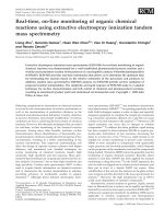
Approach of Proteomics
Sample pre-treatment
Separation of proteins
Digestion of separated proteins
Mass spectrometry analysis
Identification via
Database searchDefinition of Mass Spectrometer
An electronic instrument that analyze
An electronic instrument that analyze
substances according to the mass
substances according to the mass
-
-
to
to
-
-
charge (m/z)
charge (m/z)
ratio of constituent atoms, groups of atoms or
ratio of constituent atoms, groups of atoms or
molecules present.
molecules present.
Mass Spectrometer
Definition of Mass Spectrometer
Analytes are converted to gas phase ions.
The ions are separated by their mass-to-charge
ratios (m/z) and are detected
Relative ion current (signal) is plotted versus
m/z to produce a mass spectrum
What do you get from an MS detector?
Compound identity
molecular weight
molecular structure
Quantitation of detected compounds
molecular weight
molecular structure
Major components of MS
Sample
Sample
Introduction
Introduction
System
System
Ion
Ion
Source
Source
Analyzer
Analyzer
Detector
Detector
Interface
Interface
Vacuum
Vacuum
System
System
Mass Spectrometry
Two major components:
Ionization Source: Sample of interest is ionized and
then desorbed in to the gas phase
Mass Analyzer: acts to guide the gas-phase ions
through the instrument to the detector. At the
detector, the ions mass-to-charge (m/z) ratios are
measured.
Ionization Methods
The two most common methods to ionize biological
molecules:
- MALDI: Matrix-Assisted Laser Desorption
Ionization.
- ESI: ElectroSpray Ionization
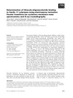
ElectroSpray Ionization
ESI greatly enhanced the ability to characterize protein and
peptides by MS.
ESI requires the sample of interest to be in solution- flow in to the
ionization source region.
To ionize the sample, high voltage is applied, result in the sample
becoming positively or negatively charged; however, positive
ionization is used primarily in the analysis proteins and peptides.
As it exits the spray tip, the solution produces submicrometer-sized
droplets containing both solute and analyte ions. The sample is
then desorbed of solute prior to entering the analyzer region.
Desorption is achieved by evaporation of the solvent by passing
the sample through a heated capillary or a curtain of drying gas,
typically nitrogen.
What distinguished ESI from other ionization methods is its ability
to produce multiply charged ions from large biological molecules.
ESI (A) and MALDI (B)
Matrix-Assisted Laser Desorption
Ionization
MALDI is another “soft” ionization process that generates
ions by irradiating a solid mixture with a pulsed laser beam.
The solid mixture is comprised of the analyte of interest
dissolved in an organic matrix compound.
The laser pulse both inderectly ionizes and desorbs the
analyte molecules from the solid mixture.
MALDI source region at a relatively high pressure, mass
analyzer region at a low pressure.
MALDI can produce both positive and negative ions. Positive
ions are formed by the acceptance of a proton as the analyte
leaves the matrix.
In MALDI, peptide and large biomolecular ions are singly
charged. MALDI is typically interfaced with mass analyzers
with large m/z ranges such as TOF spectrometers.
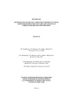
Desorption ElectroSpray Ionization
A new method of desorption ionization is not yet applied to
proteomic technology.
Desorption ElectroSpray Ionization (DESI) allows the direct
analysis of surfaces by MS.
Electrosprayed droplets are directed toward a surface to be
analyzed. The droplets produce gaseous ions of the material
on the surface and these ions are sampled with a mass
analyzer.
The mass analyzer is equipped with an atmospheric interface
connected via a flexible extanded ion transfer line.
Show the capability of analyzing a range of compounds from
nonpolar small molecules to polar peptides and proteins.
The potential exists for direct monitoring of proteins on the
surface of cells in culture from tissue sections.
Desorption Electrospray Ionization
Mass
Spectrometry
Terminology
Mass
Nominal Mass
The mass of an ion with a given empirical formula calculated using
the integer mass numbers of the most abundant isotope of
each element
Ex : M=249 C
20
H
9
+ or C
19
H
7
N+ or C
13
H
19
N
3
O
2
+
Exact Mass
The mass of an ion with a given empirical formula calculated using
the exact mass of the most abundant isotope of each element
Ex : M=249 C
20
H
9
+ 249.0070
C
19
H
7
N+ 249.0580
C
13
H
19
N
3
O
2
+ 249.1479
The fundamentals of accurate mass
carbon has a mass of 12.0000
hydrogen has a mass of 1.0078
oxygen has a mass of 15.9949
nitrogen has a mass of 14.0031
It is possible to have combinations of atoms which
have the same nominal (or integer) mass but different
accurate mass.
If such compounds can be mass measured with
sufficient accuracy it is possible to determine
elemental composition.
Importance of both MS and
chromatographic resolution
Courtesy of R. Willoughby
et. Al.
“A Global View of LC/MS” 1998
Resolution
Resolution, equally called Resolving Power, of a mass
spectrometer is a measure of its ability to separate adjacent ions.
At higher resolution, small mass differences may be detected.
249
249.0070 249.0580 249.1479
3 different compounds
Same nominal mass
Low resolution
3 different compounds
3 different exact masses
High resolution
C
20
H
9
+
C
19
H
7
N+
C
13
H
19
N
3
O
2
+
C
20
H
9
+ C
19
H
7
N+ C
13
H
19
N
3
O
2
+
Determining resolution
2 adjacent ion peaks
with a 10% valley max
Double Ion method
R =
m
ave
∆
m
r
Full Width at Half Maximum
(FWHM)
or at 5% of the peak height
Single Ion method
R =
m
∆
m
∆
m
r
m
ave
Where m is the m/z centroid of
the peak and ∆m is the width of
the peak at 5% or more commonly, 50% of
maximum
Resolution = Time of flight
2*(Time Spread)
Vpusher
Time = 0
Contributions to time spread include:
Spatial distribution of ions in pusher
Energy/velocity spread of ions in pusher
Resolution example
Mass range
Mass Range
is usually defined by the lower
and upper m/z value observable by a mass analyzer.
If your compound mass does not fall in the mass
range
of the mass analyzer, it will not be detected !
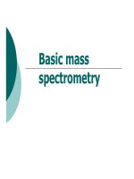
More than one charge (multiply charge ion) allows the
ion to be measured at a lower m/z.
Mass range: Multiply charged
molecules
650 700 750 800 850 900 950 1,000
0
2,000
4,000
6,000
8,000
m/z
Intensity
[M+18H]
18+
[M+17H]
17+
[M+16H]
16+
[M+15H]
15+
[M+14H]
14+
[M+13H]
13+
Positive ion mode,
ESI, 2 ul/min,
50% MeOH:49%
H2O:1% HOAc
5 µmol Cytochrome c (horse heart)
MW = 12,360.9
Mass accuracy
Ability of a mass analyzer to assign the mass
of an ion close to its true value (exact mass)
∆m
accuracy
= m
real
- m
measured
ppm = 10
6
* ∆m
accuracy
/ m
measured
High mass accuracy (exact mass measurement)
is usually associated to high resolution analyzers
Goals :
- Unknown compound determination
Exact mass helps to define its atomic/elemental composition
- Target analysis
Exact mass proves the presence of a particular
ion in a mixture
∆
∆∆
∆m accuracy