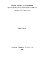
Gene delivery using cell penetrating peptides-zeolitic imidazolate frameworks
Bạn đang xem bản rút gọn của tài liệu. Xem và tải ngay bản đầy đủ của tài liệu tại đây (3.4 MB, 10 trang )
Microporous and Mesoporous Materials 300 (2020) 110173
Contents lists available at ScienceDirect
Microporous and Mesoporous Materials
journal homepage: />
Gene delivery using cell penetrating peptides-zeolitic
imidazolate frameworks
Hani Nasser Abdelhamid a, b, *, Moataz Dowaidar c, Mattias H€allbrink c, Ülo Langel c, **
a
Department of Materials and Environmental Chemistry, Stockholm University, Svante Arrhenius V€
ag 16C, Stockholm, SE-106 91, Sweden
Advanced Multifunctional Materials Laboratory, Department of Chemistry, Assiut University, Assiut, 71515, Egypt
c
Department of Biochemistry and Biophysics, Stockholm University, Svante Arrhenius V€
ag 16B, Stockholm, SE-10691, Sweden
b
A R T I C L E I N F O
A B S T R A C T
Keywords:
Cell-penetrating peptides
Metal-organic frameworks
Hierarchical porous materials
Zeolitic imidazolate frameworks
Gene delivery
Cell-penetrating peptides (CPPs), and metal-organic frameworks (MOFs) are promising as next-generation for the
delivery of gene-based therapeutic agents. Oligonucleotide (ON)-mediated assembly of nanostructures composed
of hierarchical porous zeolitic imidazolate framework (ZIF-8), and nanoparticles such as graphene oxide (GO),
and magnetic nanoparticles (MNPs) for gene therapy are reported. Five different types of non-viral vectors (ZIF8, RhB@ZIF-8, BSA@ZIF-8, MNPs@ZIF-8, and GO@ZIF-8), and three gene therapeutic agents (plasmid, splice
correction oligonucleotides (SCO), and small interfering RNA (siRNA)) were investigated. The polyplexes were
characterized and applied for gene transfection. The materials show very low toxicity with high efficiency for
luciferase transfection. ZIF-8 enhances the transfection of plasmid, SCO, siRNA of CPPs by 2–8 folds. The
mechanism of the cell uptakes was also highlighted. Data reveal cell internalization via scavenger class A
(SCARA).
1. Introduction
Gene therapy, which involves the delivery of exogenous nucleic acids
to target cells, has been considered a promising strategy to prevent and
treat a myriad of diseases, including cancer, cystic fibrosis, inflamma
tory and infectious diseases, cardiovascular diseases, Duchenne’s
muscular dystrophy, AIDS, beta-thalassemia, and diabetes [1,2]. How
ever, the direct delivery of nucleic acid is very limited due to its insta
bility in physiological conditions and its inability to penetrate the
plasma membrane. Thus, viral and non-viral vectors were applied as
carriers. Viral vectors lack security and thus are less favorable compared
to non-viral vectors. Features of non-viral vectors including their flexi
bility in packaging nucleic acids and ease of production offer additional
advantages. However, many of these vectors showed low transfection
efficiency. Thus, peptide-based gene delivery vectors, including
cell-penetrating peptide (CPPs), or protein transduction domains (PTDs)
[3], are promising due to their high safety, flexibility in conjugating
nucleic acids, and simple synthesis [4]. They ensure-invasive delivery of
therapeutic or diagnostic molecules into mammalian cells [5]. They are
good capping systems and may provide specific intracellular compart
ments without macrophage recognition and subsequent phagocytosis,
cross endothelial and epithelial barriers and enter the cytoplasm of
target cells [6]. Peptides are relatively small, low-cost, and are stable in
a wide range of biological conditions [7]. Some of these peptides have
entered into Phase I, Phase II, and Phase III clinical trials [8]. However,
loading CPPs with gene therapeutic agents is a major challenge.
Metal-organic frameworks MOFs are hybrid porous materials con
sisting of metal centers and organic linkers [9–11]. They have been
widely used for several applications such as biomedicine [12–21],
biotechnology [22,23], and analytical chemistry [24,25]. MOFs (e.g.
UiO-66, Universiteteti Oslo) was reported for co-delivery of cisplatin
and siRNAs [26]. UiO-66 enhances the therapeutic efficacy and over
coming drug resistance in ovarian cancer cells [26]. The drug release
from MOFs can be stimuli via metal-ion/ligand, strand/anti-strand, or
light [27]. MOFs can be loaded with nucleic acids through metal
–phosphate coordination interaction [26], intrinsic, multivalent coor
dination between DNA backbone phosphate and unsaturated zirconium
sites on MOFs [28], or encapsulation [29]. MOFs offered high density
(~2500 strands/particle) [28], and their surface can be easily func
tionalized with terminal phosphate-modified oligonucleotides [30].
They can be easily conjugated with nanoparticles that offer
multi-functionalities [31]. Although issues such as cost of synthesis,
* Corresponding author. Department of Materials and Environmental Chemistry, Stockholm University, Svante Arrhenius v€
ag 16C, Stockholm, SE-106 91, Sweden.
** Corresponding author.
E-mail addresses: , (H.N. Abdelhamid), (Ü. Langel).
/>Received 13 January 2020; Received in revised form 5 March 2020; Accepted 12 March 2020
Available online 14 March 2020
1387-1811/© 2020 The Authors. Published by Elsevier Inc. This is an open access article under the CC BY license ( />
H.N. Abdelhamid et al.
Microporous and Mesoporous Materials 300 (2020) 110173
Fig. 1. Schematic representation for the synthesis of ZIF-8 composite and their application for gene delivery.
Fig. 2. Characterization of ZIF-8 composite using (a) XRD, and (b-c) SEM images for GO@ZIF-8 (b) and MNPs@ZIF-8 (c). Scale bars represent 100 nm.
biodegradability, biocompatibility, and toxicity limit MOF applicability
[32].
Zeolitic imidazolate framework-8 (ZIF-8) is microporous MOFs built
from zinc nodes and 2-methylimidazole (Hmim) [33–39]. Zn-based
MOFs have also been reported as nanocarriers due to the low toxicity of
zinc ion [40]. ZIF-8 was applied to deliver anticancer drugs such as
curcumin (CCM) [41], doxorubicin (DOX) [42,43], camptothecin (CPT)
[44], and CpG (stand for 50 —C—phosphate—G—30 , C, and G represent
cytosine and guanine) oligodeoxynucleotides [45]. ZIF-8 can be conju
gated with other nanoparticles such as Fe3O4@PAA\AuNCs\ZIF-8 of
fering tri-modal cancer imaging (magnetic resonance, computed X-ray
tomography and fluorescence imaging) and chemotherapy into a single
system [43]. In vivo anticancer experiments indicate that CCM@ZIF-8
NPs exhibit higher antitumor efficacy compared to free CCM [41]. En
gineering MOFs with proteins do not only improve the specificity and
affinity but also increase the material biocompatibility [31].
Here, we presented simple oligonucleotides (ONs) loading, and
efficient release via a synergistic combination of the advantages of ZIF-8
nanoparticles as chemically tunable nanocarriers with those of CPPs,
PepFects (PF), as a capping system. PepFects (PF) are transportan 10
(TP10) analogs with an N-terminal fatty acid moiety [46]. ZIF-8 and
their composite with rhodamine B dye, bovine serum albumin (BSA),
2
H.N. Abdelhamid et al.
Microporous and Mesoporous Materials 300 (2020) 110173
Fig. 3. Zeta potential measurements for (a) ZIF-8, (b) RhB@ZIF-8, (c) MNPs@ZIF-8, and GO@ZIF-8 before and after modifications with CPPs, pGL3, SCO, and siRNA.
magnetic nanoparticles (MNPs), and graphene oxide (GO) were evalu
ated. Three ONs gene therapeutic agents; plasmid (pGL3), splice
correction oligonucleotides (SCO), and small interfering RNA (siRNA)
were investigated using HeLa cells, HeLa puLc 705 cells, and U-87
MG-luc2 cancer cells, respectively. ZIF-8 and its composite improve cell
transfection, and enhance cellular uptake of ONs with high
biocompatibility.
2.2. Synthesis of ZIF-8, RhB@ZIF-8, BSA@ZIF-8, MNPs@ZIF-8, and
GO@ZIF-8
A Zn(NO3)2⋅6H2O solution (0.84 M), and Hmim (3.0 M) were pre
pared by dissolving 25 g, and 62.5 g in 100 mL, and 250 mL, of deionized
water, respectively. MNPs, GO, and BSA solutions were prepared by
dispersion of 1 mg in 1 mL of deionized water.
In a glass scintillation vial, 0.10 mL of TEA was added to 0.8 mL of
the Zn(NO3)2⋅6H2O solution (0.67 mmol) [47]. Then, 4.0 mL of the RhB
solution (8 μmol), BSA, MNPs, or GO (1 mg mLÀ 1) was added, followed
by the addition of the Hmim solution (6.9 mmol, 6 mL). The reaction
solutions were stirred for 1 h before collection using centrifugation (13,
000 rpm, 30 min). The products were washed using water and ethanol
(2 � 40 mL) and dried overnight in an oven at 85 � C.
The solutions of ZIF-8, RhB@ZIF-8, BSA@ZIF-8, MNPs@ZIF-8, and
GO@ZIF-8 (10 mg) were prepared via dispersion in 10 mL of deionized
water using ultrasonication.
2. Materials and methods
Zinc nitrate hexahydrate (Zn(NO3)2⋅6H2O), 2-methylimidazole
(Hmim), and triethylamine (TEA) were purchased from Sigma Aldrich
(Germany). Rhodamine B (RhB), and bovine serum albumin (BSA)
encapsulated ZIF-8 (RhB@ZIF-8, or BSA@ZIF-8) were prepared
following literature [47]. Natural graphite ( 20 ỵ 84 mesh, 99.9%) was
obtained from Alfa Aesar (Great Britain). Bare magnetic nanoparticles
(MNPs) [48,49], and graphene oxide (GO) [50–55] were synthesized
following literature. Phosphorothioate 20 -O-methyl RNA oligonucleo
tides with Cy5 labeling at 50 -end were purchased from Microsynth AG,
Switzerland.
2.3. Cell culture
HeLa cells, HeLa puLc 705 cells, U-87 MG-luc2 (7000 cells of 100 μL
per well, 96 well plates) were cultured at 37 � C and 5% CO2 in 0.1 mM
non-essential amino acids Dulbecco’s modified Eagles medium (DMEM)
supplemented with 10% (v/v) FBS, 1 mM L-glutamine, 100 U mLÀ 1
penicillin, and 100 mg mLÀ 1 streptomycin (Invitrogen, Sweden).
2.1. Synthesis of peptides
CPPs; PF14 (Stearyl-AGYLLGKLLOOLAAAALOOLL-NH2), and PF221
(Stearyl-FLKLLKKFLFLKLLKKFL-amide), were synthesized on an auto
mated Syro II multiple peptide synthesizer (MultiSynTech, Witten,
Germany), using standard solid-phase Fmoc protocols applying Rinkamide Chem matrix resin (PCAS BioMatrix, Canada). The crude pep
tides were purified using high-pressure liquid chromatography (HPLC,
Germany).
2.4. Water-soluble tetrazolium salt-1 (WST-1) toxicity assay
The material biocompatibility was measured for HeLa cells (7000
cells/well) using cell proliferation reagent (WST-1, Roche Diagnostics
Scandinavia AB, Sweden). The cells were incubated with peptide–
plasmid complexes modified ZIF-8. The wells containing the treated
cells were incubated for another 24 h. The cytotoxicity was measured by
3
H.N. Abdelhamid et al.
Microporous and Mesoporous Materials 300 (2020) 110173
Fig. 4. TEM images of ZIF-8 composite for a, c, (e) PF14, and b, d, f PF221 using a-b pGL3, c-d SCO, and e-f siRNA. Scale bars represent 200 nm.
following the absorbance at 450 nm on Sunrise™-Tecan microplate
absorbance.
handled using the same procedure as that for the SCO experiments
described above.
2.5. Transfection using pGL3 luciferase plasmid, splice correction
oligonucleotides (SCO), and siRNA
2.6. Confocal laser scanning microscopy
One day before the experiment 7,000 HeLa cells were seeded in
serum-containing media containing PF14-Alexa 568-705ASO (MR10,
100 nM of SCO) complexes. The treated with Fast-dio membrane stain
according to the manufacturer’s instruction Confocal microscopy was
performed using a Leica DM/IRBE 2 epifluorescence microscope
controlled using Micro-ManagerRef (Leica, Mannheim, Germany). Argon
ion laser was used to excite fluorescein at 488 nm. Emission was
recorded between 500 and 550 nm. Images were analyzed using Fiji
(ImageJ) software. Image stacks were filtered with the 3d mean filter
using 2 � 2 � 2 settings.
Synthesis of CPPs–ZIF-8-plasmid complexes were performed in a
small vial, 80 μL of MQ water, 4 μL of ZIF-8 (1 mg mLÀ 1) Leica DM/IRBE
2 epifluorescence microscope controlled using Micro-ManagerRef, and 4
μL of pGL3 (205 ng μLÀ 1) were mixed. Finally, PF221 or PF14 (10.5 μL)
was added. The vials were incubated at room temperature for 2 h.siRNA
and PF14 were mixed in a molar ratio of 1:20 with and without ZIF-8
composite.
HeLa cells (7000 cells per 100 μL, 96 well plates) were used to
evaluate pGL3 luciferase-expressing plasmid (Promega, USA, Qiagen
Plasmid Midi kit, Qiagen, USA). pGL3-ZIF-8-CPPs (CPPs: PF221, and
PF14, 10 μL) was added to HeLa cells. After incubation for 24 h, the
medium was decanted. The cells were lysed in cell lysis buffer (Promega,
USA) and were measured using GLOMAX™ luminometer (Promega,
USA).
The biological activity of SCO-ZIF-8-CPPs (CPPs: PF221, and PF14)
was determined using HeLa puLc 705 cells (7000 cells/100 μL). The
media of cells were aspirated from each well and followed by the
addition of the lysis solution (10 μL, 0.2% Triton X-100 in HKR buffer).
Finally, luciferase activity was measured using Promega’s luciferase
assay system on GLOMAX™ 96 microplate luminometer (Promega,
Sweden).
U-87 MG-luc2 cells (7000 cells/100 μL) were cultured and investi
gated for the transfection of siRNA. After the cells have been treated
with PF14-siRNA or PF14-siRNA-ZIF-8 (10 μL) for 24 h, the plates were
2.7. Characterization techniques
X-ray diffraction (XRD) patterns were recorded using a PANalytical
X’Pert Pro diffractometer equipped with a Pixel detector using Cu Kα1
radiation (λ ¼ 1.5406 Å, current of 40 mA, an accelerating voltage of 40
kV and a source slit of 10 mm). Transmission electron microscopy (TEM)
was performed on a JEM-2100 instrument (JEOL, Japan) at an accel
erating voltage of 200 kV. Scanning electron microscopy (SEM) images
were recorded using a JSM-7000F instrument (JEOL, Japan) at an
accelerating voltage of 15.0 kV. Zeta potentials were recorded using the
Zetasizer Nano Z system (Malvern Panalytical Ltd, UK).
4
H.N. Abdelhamid et al.
Microporous and Mesoporous Materials 300 (2020) 110173
Fig. 5. TEM images of RhB@ZIF-8 composite for (a, c, e) PF14, and (b, d, f) PF221 using (a-b) pGL3, (c-d) SCO, and (e-f) siRNA. Scale bars represent 200 nm.
2.8. Statistical analysis
hydrogen bonding. Adsorption of pGL3, SCO, or siRNA was achieved via
soaking ZIF-8 composite in aqueous solutions of those gene therapeutic
agents.
The charge of the materials was determined using Zeta potential
(Fig. 3). All ZIF-8, except for GO@ZIF-8, have positive zeta potentials
(Fig. 3). The nanocomposites based on SCO for all ZIF-8 materials are
positive charge materials. In contrast, all materials based on siRNA have
high zeta potential with negative values (Fig. 3). The negative charges
are due to the high phosphate group content of siRNA.
The morphology of the formed complexes is characterized using TEM
images (Figs. 4–5, Figs. S2–S3), and SEM image (Fig. S4). Data show the
formation of protein corona of CPPs and gene therapeutic agents on the
external surface of ZIFs nanocomposites. The results reveal ZIF-8
nanoparticle with a size of 25–150 nm (Figs. 4–5, Figs. S2–S3). The
images reveal a layer of CPPs and gene therapeutic agents with a size of
6 � 3–10 � 3 nm indication the formation of a core-shell structure
(Fig. S5).
Data were generated from at least three independent experiments,
and statistically analyzed using a two-way ANOVA test (the program of
Graphpad Prism) for the statistical significance: *p < 0.05; **p < 0.01;
***p < 0.001; ****p < 0.0001.
3. Results and discussion
Schematic representation for the synthesis of hierarchical porous
ZIF-8 composite with RhB, BSA, MNPs, and GO is shown in Fig. 1.
Adding triethylamine (TEA) to an aqueous solution of Zn2ỵ leads to the
formation of ZnO (Fig. 1). The molecules (RhB, and BSA), or nano
particles (MNPs, and GO, Fig. S1) are adsorbed into the formed ZnO that
convert into hierarchical porous ZIF-8 after the addition of Hmim
(Fig. 1). XRD patterns confirm the formation of a pure phase of ZIF-8
composited, called RhB@ZIF-8, BSA@ZIF-8, MNPs@ZIF-8, and
GO@ZIF-8 (Fig. 2a). SEM images show the formation of ZIF-8 nano
particles with a particle size of 25–100 nm (Fig. 2b and c).
The formation of polyplex consist of CPPs (PF14, and PF221), ZIF-8
composite (RhB@ZIF-8, BSA@ZIF-8, MNPs@ZIF-8, and GO@ZIF-8),
and gene therapeutic agents (pGL3, SCO, and siRNA) take place via
non-covalent interactions (Fig. 1). PF14 and PF221 were chosen because
it is an efficient delivery reagent for pGL3, SCOs, and siRNA [56]. The
complexation takes place via the step-by-step addition of each reagent.
The interactions inside the polyplex are mainly electrostatic forces and
3.1. Cell viability
The WST-1 assay was used to evaluate the cytotoxicity of the formed
complexes in HeLa cells (Fig. 6). As shown in Fig. 6, the cytotoxicity of
the materials concentration-independent for the investigated molar
ratio and charge ratio. There is no observation for any toxicity for ZIF-8
composite with and without CPPs (Fig. 6). Biocompatibility of ZIF-8
toward six different cell lines representing various body parts (kidney,
5
H.N. Abdelhamid et al.
Microporous and Mesoporous Materials 300 (2020) 110173
Fig. 6. WST-1 cell viability using HeLa cells for (a-b) pGL3 using charge ratio of (a) 5, and (b) 10, and (c-d) SCO using molar ratio of (c) 10, and (d) 20.
skin, breast, blood, bones, and connective tissue) revealed that ZIF-8
showed insignificant cytotoxicity up to a threshold value of 30 μg
mLÀ 1 [57]. It was also reported that ZIF8-CpG ODNs complexes showed
no cytotoxicity compared to ZIF-8 alone [45]. These results indicate that
the platform of ZIF-8 and CPPs are biocompatible materials and can be
applied as carriers for gene-based therapeutic agents.
compared to the complexes without ZIF-8 nanocomposites. ZIF-8 im
proves the transfection of PF14 that showed higher transfection
compared to commercial transfection agent Lipofectamine™ 2000 [56].
Gene silencing or knockdown using small/short interfering RNA
(siRNA) for the prepared materials tested (Fig. 8). siRNA is a doublestranded RNA molecule which is similar to miRNA. It prevents trans
lation of mRNA after transcription via interfering with the comple
mentary nucleotide sequences [61]. Cell transfection using siRNA (10
and 25 nM) indicates significant gene knockdown using CPPs-ZIF8
vectors (Fig. 8).
3.2. Cell transfection using pGL3, SCO, and siRNA
To investigate the performance of the prepared materials ZIF-8
nanocomposite, three gene therapeutic agents called pGL3, splice
correction oligonucleotides, and small interfering RNA were
investigated.
The prepared materials; ZIF-8, RhB@ZIF-8, BSA@ZIF-8, MNPs@ZIF8, and GO@ZIF-8, were applied to introduce luciferase reporter genes as
a plasmid DNA (pGL3) into the HeLa cells for PF14, and PF221 (Fig. 7).
Two different ratios of the plasmid: ZIF-8 composite of 1:0.5 and 1:1
were investigated. To account for transfection variation among wells, a
control plasmid containing pGL3-PF14, or pGL3-PF221were transfected
for normalization purposes. Results show no transfection efficiency in
the absence of CPPs (Fig. 7). ZIF-8 nanocomposites increase the effi
ciency of pGL3-PF14 by 2–4 folds. While ZIF-8 nanocomposites improve
the transfection of pGL3-PF221 by 3-8-fold (Fig. 7). Results reveal that
CPPs-ZIF8 is robust enough to have luciferase signals significantly
higher than the background (Fig. 7).
Transfection of splice correction oligonucleotides (50 -CCU CUU ACC
UCA GUU ACA- Cy5-labeled) using the prepared materials is also
investigated (Figs. S6–S9). The used SCOs are antisense oligonucleotides
(ONs) with 18 bases in length [58–60]. The complexes of the ternary
component i.e. SCO, ZIF-8 nanocomposite, and CPPs were tested for
HeLa cell line with a recombinant plasmid pLuc 705 that carry the
luciferase gene interrupted by a mutated human beta-globin intron 2
IVS2-705. Results demonstrate that complexes containing ZIF-8 nano
composites have higher transfection efficacy for oligonucleotides as
3.3. Mechanism of uptakes
Finally, we elucidated the intracellular fate of the CPPs-ZIF8 after
incubation using uptake of Scavenger class A (SCARA, Fig. 9), and
confocal microscopy (Fig. 10). A study using curcumin loaded ZIF-8
showed intracellular distribution via the clathrin-mediated endocy
tosis to the lysosomal pathway [62]. In general, there are several pro
posed mechanisms for gene therapeutic agents. Among those proposed
mechanisms, endocytosis, energy-independent translocation [3], acti
vation of sphingomyelinase which converts sphingomyelin into cer
amide [63], and presence of scavenger class A (SCARA) may explain the
cell uptakes of the formed complexes (Fig. 9). SCARA is a receptor that
can bind and endocytose acetylated low-density lipoprotein [64]. The
experiment was performed via incubation HeLa cells with inhibitor
(Dextran sulfate (Dex), polyinosinic acid (Poly I) or fucoidan (Fuc)) and
the corresponding control reagents (Chondroitin sulfate (Chon), poly
cytidylic acid (Poly C) or galactose (Gal)) (Fig. 9). As shown in Fig. 9, no
transfection was observed in the presence of the inhibitors. On the other
side, the transfection was successfully observed in the presence of the
control reagents that confirm the high impact of SCARAin the uptake of
PF14–SCO-ZIF-8.
The cell after transfection was also observed under a confocal mi
croscope (Fig. 10). There are no notable changes in cell morphology. The
6
H.N. Abdelhamid et al.
Microporous and Mesoporous Materials 300 (2020) 110173
Fig. 7. a-b) pGL3 transfection of HeLa cells for PF14 using plasmid: ZIF-8 composite of (a) 1:0.5, and (b) 1:1.
Fig. 9. Cell uptakes based on the mechanism of scavenger receptor class.
Fig. 8. Transfection of U-87 MG-luc2 cells with siRNA.
7
H.N. Abdelhamid et al.
Microporous and Mesoporous Materials 300 (2020) 110173
Fig. 10. Confocal microscopy images of Hela-705 cells incubated for 24 h with PF14-Alexa 568–705ASO (MR10, 100 nM ASO) complexes (in red) for (a) PF-14, (b)
PF14-ZIF8, (c) PF14–RhB@ZIF8, (d) PF14-BSA@ZIF8, (e) PF14-MNPs@ZIF8, and (f) PF14-GO@ZIF8. Cell-membranes are stained with Fast-dio stain (in green). (For
interpretation of the references to color in this figure legend, the reader is referred to the Web version of this article).
red color corresponding oligonucleotides indicate the presence of the
gene therapeutic agent inside the cell revealing successful cell trans
fection (Fig. 10). Results indicate that most of the oligonucleotides
entered the cells by endocytosis that release the gene after degradation
in the cytosol (Fig. 1). It is important to keep in mind that ZIF-8 nano
particles can be dissolved in the acidic environment of the cancer cells
(pH 5.5) [28]. These results confirm that CPPs-ZIF8 is an effective gene
vector for the internalization of a gene therapeutic agent into the cells.
hyperthermia.
Declaration of competing interest
None.
CRediT authorship contribution statement
Hani Nasser Abdelhamid: Conceptualization, Methodology,
Writing - original draft. Moataz Dowaidar: Data curation. Mattias
€llbrink: Visualization. Ülo Langel: Supervision, Writing - review &
Ha
editing.
4. Conclusions
Based on our presented findings, a broad gene vector based on ZIF-8
nanoparticles with tunable properties can be used to prepare effective
materials for gene delivery. Our protocol offers facile integration of
multifunctional components via inclusion and post-synthetic modifica
tions of ZIF-8. These materials may be useful for multi-targeted medical
applications in both diagnosis and therapy with exceptional high cell
biocompatibility. The synthesized materials appear promising due to the
presence of other components such as dye, protein, magnetic nano
particles, and graphene oxide. These reagents offered potential appli
cations of the composites as a multifunctional platform for cancer
theranostics involving magnetic resonance imaging, drug delivery, and
Acknowledgments
This work was funded by the Swedish Research Council (VR-NT), and
from the Innovative Medicines Initiative Joint Undertaking under grant
agreement no. 115363 sources of which are composed of financial
participation from the European Union’s Seventh Framework Pro
gramme (FP7/2007–2013) and EFPIA companies, Swedish Cancer
Foundation, Sweden, and from the European Regional Development
Fund through the Center of Excellence in Molecular Cell Engineering
8
H.N. Abdelhamid et al.
Microporous and Mesoporous Materials 300 (2020) 110173
(2014-2020.4.01.15–0013), and by the Estonian Research Council
(IUT20-26). Many thanks to Prof. X. Zou for providing part of the
necessary tools to do these measurements in her lab at SU, Sweden.
[22] R.K. Keservani, A.K. Sharma, Nanoparticulate Drug Delivery Systems, Apple
Academic Press, 2019. />id¼xSiFDwAAQBAJ&printsec¼frontcover&hl¼ar#v¼onepage&q&f¼false.
accessed January 31, 2019.
[23] H.N. Abdelhamid, H.-F. Wu, Nanoparticles advanced drug delivery for cancer cells,
in: R.K. Keservani, A.K. Sharma (Eds.), Nanoparticulate Drug Deliv. Syst., 2019,
pp. 121–144.
[24] H.N. Abdelhamid, Nanoparticle-based surface assisted laser desorption ionization
mass spectrometry: a review, Microchim. Acta. 186 (2019) 682, />10.1007/s00604-019-3770-5.
[25] H.N. Abdelhamid, Nanoparticles assisted laser desorption/ionization mass
spectrometry, in: Handb. Smart Mater. Anal. Chem., John Wiley & Sons, Ltd,
Chichester, UK, 2019, pp. 729–755, />ch23.
[26] C. He, K. Lu, D. Liu, W. Lin, Nanoscale metal–organic frameworks for the Codelivery of cisplatin and pooled siRNAs to enhance therapeutic efficacy in drugresistant ovarian cancer cells, J. Am. Chem. Soc. 136 (2014) 5181–5184, https://
doi.org/10.1021/ja4098862.
[27] J.S. Kahn, L. Freage, N. Enkin, M.A.A. Garcia, I. Willner, Stimuli-Responsive DNAfunctionalized metal-organic frameworks (MOFs), Adv. Mater. 29 (2017),
1602782, />[28] Z. Wang, Y. Fu, Z. Kang, X. Liu, N. Chen, Q. Wang, Y. Tu, L. Wang, S. Song, D. Ling,
H. Song, X. Kong, C. Fan, Organelle-specific triggered release of
immunostimulatory oligonucleotides from intrinsically coordinated
DNA–metal–organic frameworks with soluble exoskeleton, J. Am. Chem. Soc. 139
(2017) 15784–15791, />[29] R. Ricc�
o, W. Liang, S. Li, J.J. Gassensmith, F. Caruso, C. Doonan, P. Falcaro,
Metal–organic frameworks for cell and virus biology: a perspective, ACS Nano 12
(2018) 13–23, />[30] S. Wang, C.M. McGuirk, M.B. Ross, S. Wang, P. Chen, H. Xing, Y. Liu, C.A. Mirkin,
General and direct method for preparing oligonucleotide-functionalized
metal–organic framework nanoparticles, J. Am. Chem. Soc. 139 (2017)
9827–9830, />[31] L. He, M. Brasino, C. Mao, S. Cho, W. Park, A.P. Goodwin, J.N. Cha, DNAassembled core-satellite upconverting-metal-organic framework nanoparticle
superstructures for efficient photodynamic therapy, Small 13 (2017), 1700504,
/>[32] S. Beg, M. Rahman, A. Jain, S. Saini, P. Midoux, C. Pichon, F.J. Ahmad, S. Akhter,
Nanoporous metal organic frameworks as hybrid polymer–metal composites for
drug delivery and biomedical applications, Drug Discov. Today 22 (2017)
625–637, />[33] K.S. Park, Z. Ni, A.P. Cote, J.Y. Choi, R. Huang, F.J. Uribe-Romo, H.K. Chae,
M. O’Keeffe, O.M. Yaghi, A.P. C^
ot�
e, J.Y. Choi, R. Huang, F.J. Uribe-Romo, H.
K. Chae, M. O’Keeffe, O.M. Yaghi, A.P. Cote, J.Y. Choi, R. Huang, F.J. Uribe-Romo,
H.K. Chae, M. O’Keeffe, O.M. Yaghi, Exceptional chemical and thermal stability of
zeolitic imidazolate frameworks, Proc. Natl. Acad. Sci. U.S.A. 103 (2006)
10186–10191, />[34] H.N. Abdelhamid, M. Dowaidar, M. H€
allbrink, Ü. Langel, Cell penetrating peptideshierarchical porous zeolitic imidazolate frameworks nanoparticles: an efficient
gene delivery platform, SSRN Electron. J. (2019), />ssrn.3435895.
[35] H.N. Abdelhamid, A.M. El-Zohry, J. Cong, T. Thersleff, M. Karlsson, L. Kloo, X. Zou,
Towards implementing hierarchical porous zeolitic imidazolate frameworks in dyesensitized solar cells, R. Soc. Open Sci. 6 (2019), 190723, />rsos.190723.
[36] L. Valencia, H.N. Abdelhamid, Nanocellulose leaf-like zeolitic imidazolate
framework (ZIF-L) foams for selective capture of carbon dioxide, Carbohydr.
Polym. 213 (2019) 338–345, />[37] A.F. Abdel-Magied, H.N. Abdelhamid, R.M. Ashour, X. Zou, K. Forsberg,
Hierarchical porous zeolitic imidazolate frameworks nanoparticles for efficient
adsorption of rare-earth elements, Microporous Mesoporous Mater. 278 (2019)
175–184, />[38] S. Sultan, H.N. Abdelhamid, X. Zou, A.P. Mathew, CelloMOF: nanocellulose
enabled 3D printing of metal-organic frameworks, Adv. Funct. Mater. (2018),
1805372, />[39] H.N. Abdelhamid, X. Zou, Template-free and room temperature synthesis of
hierarchical porous zeolitic imidazolate framework nanoparticles and their dye
and CO 2 sorption, Green Chem. 20 (2018) 1074–1084, />C7GC03805D.
[40] M.-X. Wu, Y.-W. Yang, Metal-organic framework (MOF)-Based drug/cargo delivery
and cancer therapy, Adv. Mater. 29 (2017), 1606134, />adma.201606134.
[41] M. Zheng, S. Liu, X. Guan, Z. Xie, One-step synthesis of nanoscale zeolitic
imidazolate frameworks with high curcumin loading for treatment of cervical
cancer, ACS Appl. Mater. Interfaces 7 (2015) 22181–22187, />10.1021/acsami.5b04315.
[42] H. Ren, L. Zhang, J. An, T. Wang, L. Li, X. Si, L. He, X. Wu, C. Wang, Z. Su,
Polyacrylic acid@zeolitic imidazolate framework-8 nanoparticles with ultrahigh
drug loading capability for pH-sensitive drug release, Chem. Commun. 50 (2014)
1000–1002, />[43] R. Bian, T. Wang, L. Zhang, L. Li, C. Wang, A combination of tri-modal cancer
imaging and in vivo drug delivery by metal–organic framework based composite
nanoparticles, Biomater. Sci. 3 (2015) 1270–1278, />C5BM00186B.
[44] J. Zhuang, C.H. Kuo, L.Y. Chou, D.Y. Liu, E. Weerapana, C.K. Tsung, Optimized
metal-organic-framework nanospheres for drug delivery: evaluation of small-
Appendix A. Supplementary data
Supplementary data to this article can be found online at https://doi.
org/10.1016/j.micromeso.2020.110173.
References
[1] B.G. Nordestgaard, S.J. Nicholls, A. Langsted, K.K. Ray, A. Tybjærg-Hansen,
Advances in lipid-lowering therapy through gene-silencing technologies, Nat. Rev.
Cardiol. 15 (2018) 261–272, />[2] A. Khvorova, J.K. Watts, The chemical evolution of oligonucleotide therapies of
clinical utility, Nat. Biotechnol. 35 (2017) 238–248, />nbt.3765.
[3] M. Lindgren, M. H€
allbrink, A. Prochiantz, Ü. Langel, Cell-penetrating peptides,
Trends Pharmacol. Sci. 21 (2000) 99–103, />(00)01447-4.
[4] Z. Kang, Q. Meng, K. Liu, Peptide-based gene delivery vectors, J. Mater. Chem. B
(2019), />[5] S. Dissanayake, W.A. Denny, S. Gamage, V. Sarojini, Recent developments in
anticancer drug delivery using cell penetrating and tumor targeting peptides,
J. Contr. Release 250 (2017) 62–76, />jconrel.2017.02.006.
[6] C.P. Cerrato, K. Künnapuu, Ü. Langel, Cell-penetrating peptides with intracellular
organelle targeting, Expet Opin. Drug Deliv. 14 (2017) 245–255, />10.1080/17425247.2016.1213237.
[7] A. Komin, L.M. Russell, K.A. Hristova, P.C. Searson, Peptide-based strategies for
enhanced cell uptake, transcellular transport, and circulation: mechanisms and
challenges, Adv. Drug Deliv. Rev. 110–111 (2017) 52–64, />10.1016/j.addr.2016.06.002.
[8] G. Guidotti, L. Brambilla, D. Rossi, Cell-penetrating peptides: from basic Research
to clinics, Trends Pharmacol. Sci. 38 (2017) 406–424, />tips.2017.01.003.
[9] H.-C. Zhou, J.R. Long, O.M. Yaghi, Introduction to metal-organic frameworks,
Chem. Rev. 112 (2012) 673–674, />€
[10] O.K. Farha, A. Ozgür
Yazaydın, I. Eryazici, C.D. Malliakas, B.G. Hauser, M.
G. Kanatzidis, S.T. Nguyen, R.Q. Snurr, J.T. Hupp, De novo synthesis of a
metal–organic framework material featuring ultrahigh surface area and gas storage
capacities, Nat. Chem. 2 (2010) 944–948, />[11] H.-C. Zhou, S. Kitagawa, Metal–organic frameworks (MOFs), Chem. Soc. Rev. 43
(2014) 5415–5418, />[12] H.N. Abdelhamid, Zinc hydroxide nitrate nanosheets conversion into hierarchical
zeolitic imidazolate frameworks nanocomposite and their application for CO2
sorption, Mater. Today Chem. 15 (2020), 100222, />mtchem.2019.100222.
[13] M.N. Goda, H.N. Abdelhamid, A.E.-A.A. Said, Zirconium oxide sulfate-carbon
(ZrOSO4@C) derived from carbonized UiO-66 for selective production of dimethyl
ether, ACS Appl. Mater. Interfaces 12 (2020) 646–653, />acsami.9b17520.
[14] A.A. Kassem, H.N. Abdelhamid, D.M. Fouad, S.A. Ibrahim, Metal-organic
frameworks (MOFs) and MOFs-derived CuO@C for hydrogen generation from
sodium borohydride, Int. J. Hydrogen Energy 44 (2019) 31230–31238, https://
doi.org/10.1016/j.ijhydene.2019.10.047.
[15] H.N. Abdelhamid, Surfactant assisted synthesis of hierarchical porous metalorganic frameworks nanosheets, Nanotechnology 30 (2019), 435601, https://doi.
org/10.1088/1361-6528/ab30f6.
[16] H.N. Abdelhamid, M. Wilk-Kozubek, A.M. El-Zohry, A. Bermejo G�
omez,
A. Valiente, B. Martín-Matute, A.-V. Mudring, X. Zou, Luminescence properties of a
family of lanthanide metal-organic frameworks, Microporous Mesoporous Mater.
279 (2019) 400–406, />[17] H.E. Emam, H.N. Abdelhamid, R.M. Abdelhameed, Self-cleaned photoluminescent
viscose fabric incorporated lanthanide-organic framework (Ln-MOF), Dyes
Pigments 159 (2018) 491–498, />[18] H.N. Abdelhamid, Nanoparticle assisted laser desorption/ionization mass
spectrometry for small molecule analytes, Microchim. Acta. 185 (2018) 200,
/>[19] H.N. Abdelhamid, A. Bermejo-G�
omez, B. Martín-Matute, X. Zou, A water-stable
lanthanide metal-organic framework for fluorimetric detection of ferric ions and
tryptophan, Microchim. Acta. 184 (2017) 3363–3371, />s00604-017-2306-0.
[20] Y. Yang, K. Shen, J. Lin, Y. Zhou, Q. Liu, C. Hang, H.N. Abdelhamid, Z. Zhang,
H. Chen, A Zn-MOF constructed from electron-rich π-conjugated ligands with an
interpenetrated graphene-like net as an efficient nitroaromatic sensor, RSC Adv. 6
(2016) 45475–45481, />[21] Q. Yao, A. Bermejo G�
omez, J. Su, V. Pascanu, Y. Yun, H. Zheng, H. Chen, L. Liu, H.
N. Abdelhamid, B. Martín-Matute, X. Zou, Series of highly stable isoreticular
lanthanide metal–organic frameworks with expanding pore size and tunable
luminescent properties, Chem. Mater. 27 (2015) 5332–5339, />10.1021/acs.chemmater.5b01711.
9
H.N. Abdelhamid et al.
[45]
[46]
[47]
[48]
[49]
[50]
[51]
[52]
[53]
[54]
[55]
[56]
Microporous and Mesoporous Materials 300 (2020) 110173
molecule encapsulation, ACS Nano 8 (2014) 2812–2819, />nn406590q.
H. Zhang, W. Chen, K. Gong, J. Chen, Nanoscale zeolitic imidazolate framework-8
as efficient vehicles for enhanced delivery of CpG oligodeoxynucleotides, ACS
Appl. Mater. Interfaces 9 (2017) 31519–31525, />acsami.7b09583.
M. M€
ae, S. EL Andaloussi, P. Lundin, N. Oskolkov, H.H.J. Johansson, P. Guterstam,
U. Langel, Ü. Langel, A stearylated CPP for delivery of splice correcting
oligonucleotides using a non-covalent co-incubation strategy, J. Contr. Release 134
(2009) 221–227, />H.N. Abdelhamid, Z. Huang, A.M. El-Zohry, H. Zheng, X. Zou, A Fast and scalable
approach for synthesis of hierarchical porous zeolitic imidazolate frameworks and
one-pot encapsulation of target molecules, Inorg. Chem. 56 (2017) 9139–9146,
/>H.N. Abdelhamid, H.F. Wu, Thymine chitosan nanomagnets for specific
preconcentration of mercury (II) prior to analysis using SELDI-MS, Microchim.
Acta. (2017), />J. Gopal, H.N. Abdelhamid, P.-Y. Hua, H.-F. Wu, Chitosan nanomagnets for
effective extraction and sensitive mass spectrometric detection of pathogenic
bacterial endotoxin from human urine, J. Mater. Chem. B. 1 (2013) 2463, https://
doi.org/10.1039/c3tb20079e.
K.H. Hussein, H.N. Abdelhamid, X. Zou, H.-M. Woo, Ultrasonicated graphene oxide
enhances bone and skin wound regeneration, Mater. Sci. Eng. C 94 (2019)
484–492, />R.M. Ashour, H.N. Abdelhamid, A.F. Abdel-Magied, A.A. Abdel-khalek, M. Ali,
A. Uheida, M. Muhammed, X. Zou, J. Dutta, Rare earth ions adsorption onto
graphene oxide nanosheets, Solvent Extr. Ion Exch. 35 (2017) 91–103, https://doi.
org/10.1080/07366299.2017.1287509.
H.N. Abdelhamid, H.-F. Wu, Reduced graphene oxide conjugate thymine as a new
probe for ultrasensitive and selective fluorometric determination of mercury(II)
ions, Microchim. Acta 182 (2015) 1609–1617, />H.N. Abdelhamid, M.S. Khan, H.F. Wu, Graphene oxide as a nanocarrier for
gramicidin (GOGD) for high antibacterial performance, RSC Adv. 4 (2014)
50035–50046, />H. Nasser Abdelhamid, B.-S. Wu, H.-F. Wu, Graphene coated silica applied for high
ionization matrix assisted laser desorption/ionization mass spectrometry: a novel
approach for environmental and biomolecule analysis, Talanta 126 (2014) 27–37,
/>P.-Y. Hua, M. Manikandan, H.N. Abdelhamid, H.-F. Wu, Graphene nanoflakes as an
efficient ionizing matrix for MALDI-MS based lipidomics of cancer cells and cancer
stem cells, J. Mater. Chem. B. 2 (2014) 7334–7343.
K. Ezzat, S. EL Andaloussi, E.M. Zaghloul, T. Lehto, S. Lindberg, P.M.D. Moreno, J.
R. Viola, T. Magdy, R. Abdo, P. Guterstam, R. Sillard, S.M. Hammond, M.J.
[57]
[58]
[59]
[60]
[61]
[62]
[63]
[64]
10
A. Wood, A.A. Arzumanov, M.J. Gait, C.I.E. Smith, M. H€
allbrink, Ü. Langel, S.E.
L. Andaloussi, E.M. Zaghloul, T. Lehto, S. Lindberg, P.M.D. Moreno, J.R. Viola,
T. Magdy, R. Abdo, P. Guterstam, R. Sillard, S.M. Hammond, M.J.A. Wood, A.
A. Arzumanov, M.J. Gait, C.I.E. Smith, M. H€
allbrink, Ü. Langel, PepFect 14, a novel
cell-penetrating peptide for oligonucleotide delivery in solution and as solid
formulation, Nucleic Acids Res. 39 (2011) 5284–5298, />nar/gkr072.
M. Hoop, C.F. Walde, R. Ricc�
o, F. Mushtaq, A. Terzopoulou, X.-Z. Chen, A.
J. DeMello, C.J. Doonan, P. Falcaro, B.J. Nelson, J. Puigmartí-Luis, S. Pan�e,
Biocompatibility characteristics of the metal organic framework ZIF-8 for
therapeutical applications, Appl. Mater. Today. 11 (2018) 13–21, />10.1016/j.apmt.2017.12.014.
M. Dowaidar, H.N. Abdelhamid, M. H€
allbrink, X. Zou, Ü. Langel, Graphene oxide
nanosheets in complex with cell penetrating peptides for oligonucleotides delivery,
Biochim. Biophys. Acta Gen. Subj. 1861 (2017) 2334–2341, />10.1016/j.bbagen.2017.07.002.
M. Dowaidar, H.N. Abdelhamid, M. H€
allbrink, K. Kurrikoff, K. Freimann, X. Zou,
Ü. Langel, K. Kurrikoff, X. Zou, Ü. Langel, Magnetic nanoparticle assisted selfassembly of cell penetrating peptides-oligonucleotides complexes for gene
delivery, Sci. Rep. 7 (2017) 9159, />M. Dowaidar, H. Nasser Abdelhamid, M. H€
allbrink, Ü. Langel, X. Zou, Chitosan
enhances gene delivery of oligonucleotide complexes with magnetic
nanoparticles–cell-penetrating peptide, J. Biomater. Appl. 33 (2018) 392–401,
/>A.R. Thierry, S. Abes, S. Resina, A. Travo, J.P. Richard, P. Prevot, B. Lebleu,
Comparison of basic peptides- and lipid-based strategies for the delivery of splice
correcting oligonucleotides, Biochim. Biophys. Acta Biomembr. 1758 (2006)
364–374, />A. Tiwari, A. Singh, N. Garg, J.K. Randhawa, Curcumin encapsulated zeolitic
imidazolate frameworks as stimuli responsive drug delivery system and their
interaction with biomimetic environment, Sci. Rep. 7 (2017), 12598, https://doi.
org/10.1038/s41598-017-12786-6.
R. Wallbrecher, T. Ackels, R.A. Olea, M.J. Klein, L. Caillon, J. Schiller, P.H. Bov�
eeGeurts, T.H. van Kuppevelt, A.S. Ulrich, M. Spehr, M.J.W. Adjobo-Hermans,
R. Brock, Membrane permeation of arginine-rich cell-penetrating peptides
independent of transmembrane potential as a function of lipid composition and
membrane fluidity, J. Contr. Release 256 (2017) 68–78, />j.jconrel.2017.04.013.
K. Ezzat, H. Helmfors, O. Tudoran, C. Juks, S. Lindberg, K. Padari, S. El-Andaloussi,
M. Pooga, U. Langel, Scavenger receptor-mediated uptake of cell-penetrating
peptide nanocomplexes with oligonucleotides, Faseb. J. 26 (2012) 1172–1180,
/>