Aesthetic Microtia Reconstruction with Medpor doc
Bạn đang xem bản rút gọn của tài liệu. Xem và tải ngay bản đầy đủ của tài liệu tại đây (616.58 KB, 9 trang )
Aesthetic Microtia Reconstruction with
Medpor
Thomas Romo III, M.D.
1
and Shari D. Reitzen, M.D.
2
ABSTRACT
The complex architecture of the auricle makes it one of the most challenging
structures for the reconstructive surgeon to re-create. Overlying the ear’s unique cartilage
framework are layers of varied soft tissues forming a three-dimensional organ, which is
distinctively positioned on the head. Arguably, the most challenging auricle to reconstruct
is third-degree microtia due to a near-total absence of native tissue and a need for lifelong
durability of the reconstruction. Many methods of reconstruction have been studied;
autogenous costal cartilage reconstruction has been one of the more traditional methods,
with favorable long-term results reported by several surgeons. However, this technique
requires tremendous artistic and technical skill on the part of the surgeon-sculptor to
construct a realistic-appearing ear. High-density porous polyethylene (Medpor) is a stable,
alloplastic implant that can integrate with host tissues, is resistant to infection, and has
been successfully applied to reconstruction of the head and neck. For auricular recon-
struction, Medpor—enveloped in a temporoparietal fascial flap with full-thickness skin
graft coverage—is a durable and aesthetically gratifying alternative in microtic patients.
This alternative surgical technique reduces surgical time and morbidity, standardizes results
among surgeons, and facilitates an aesthetic, natural-appearing reconstruction of the
auricle.
KEYWORDS: Medpor auricular framework, microtia, temporoparietal flap
The external ear is a critical component of the
overall aesthetic balance and contour of the face. Its
characteristic three-dimensional topography consists of
interrelated length, width, and lateral pro jection, such
that even slight alterations in the size, shape, location, or
position of the ear are easily recognized, especially when
compared with an opposite ‘‘normal’’ ear. The auricle
itself is intricately formed from consistently located,
precisely oriented topographic ‘‘peaks and valleys’’ and
may in fact represent the most detailed structure of the
body. For these reasons, surgical reconstruction of the
auricle is a challenging, and time-consuming endeavor.
There are two categories of auricular deformity:
acquired and congenital. Acquired defects may in-
clude burns , trauma, loss due to tumor resectio n, or
animal and human bites. Con genital malformations of
the ear include microtia, lop ear, cup ear, and prom-
inent ear. Mic rotia represen ts a sign ificant challenge
due to the lack of native tissue and absence of
preexisting structure to simulate pos ition, and will
be the mai n focus of this article. Approaches toward
reconstructi on, focusing on traditional costal cartilage
techniques, as well as alloplastic alternatives, are
discussed.
1
Division of Facial Plastic & Reconstructive Surgery, Lenox Hill
Hospital, New York, New York;
2
Department of Otolaryngology-
Head & Neck Surgery, New York University Medical Center, New
York, New York.
Address for correspondence and reprint requests: Thomas Romo,
III, M.D., Division of Facial Plastic & Reconstructive Surgery, Lenox
Hill Hospital, 135A East 74th Street, New York, NY 10021.
Aesthetic Reconstruction of Head and Neck Defects; Guest Editors,
Manoj T. Abraham, M.D., F.A.C.S., Keith E. Blackwell, M.D.
Facial Plast Surg 2008;24:120–128. Copyright # 2008 by Thieme
Medical Publishers, Inc., 333 Seventh Avenue, New York, NY 10001,
USA. Tel: +1(212) 584-4662.
DOI 10.1055/s-2008-1037453. ISSN 0736-6825.
120
AESTHETIC CONCERNS/GOALS
Due to its intricate surface topography and conspicuous
location, the ear is an unforgiving structure when
undergoing aesthetic reconstructi on. A full under-
standing of th e external anatomy and cephalometric
parameters of the ear is essential for a successful out-
come. The structural framework, composed of auricular
carti lage, consists of discrete co mpone nts wit h a char-
acter istic shape and position: the h elix and antihelix,
central conchal complex, and an inferiorly placed lo-
bule.
1,2
The most basic princip les of ear reconstruction
were defined by Tolleth.
3
He defined the four prima ry
lines o f the ear, which give it a natural appearan ce. T he
first line consists of the most lat eral aspect of the helix
and defi nes the overall shape of t he ear. The second line
is the medial aspect of t he helix and defines the helical
rim. This line provides important stru cture as it divides
the cymbum and the cavum conchae. T he third line
repre sents the con chal bowl, tragus, and antitragus, and
the fourth line outlines the fossa triangulari s. With this
basic outline i n mind, the car ving of cartilage or
synthetic implants will reflect the natural contours of
the ear.
In addition to the configuration of the auricle
itself, its location relative to other facial structures is of
equal importance. The ear’s location, pos terior setback,
and protrusion are all critical aspects of a nor mal-
appearing ear.
1–4
These factors, and left-right symme-
try, are most evident on the frontal view of a patient,
where a comp arison between a nor mal ear and a
reconstructed ear is most app arent. The superiormost
aspect of the helix generally sits at the level of the
supra orbital rim (in most patients this corresponds to
the tail of the eyebrow), whereas the inferior most
aspect of the lobule corresponds to the base of th e
columella. The average width of the ear is 55% of its
length (55 to 65 mm). Thi s length is also the distance
from the lateral orbital rim to the ear. With the head
positioned in the Frankfort horizontal plane, the ear is
positioned 15 to 20 degrees posterior to the vertical
axis. The auriculoc eph alic angle is between 25 and 35
degrees.
There are three types of soft tissue overly ing the
structural fra mework.
1
The anterior and lateral surfa-
ces of the ear consist of thin skin , without sub cuta-
neous adipose tis sue, and closely adhere to the
cartilage. Poster omedially, the soft tissue envelope is
looser and mobile over the cartilage . In addition, the
helical rim in this region contains fibroareolar subcu-
taneous tissue that contribu tes to the bulk of the
structure. Finally, the lo bular tissue consist s of firm,
globular fatty tissues and ov erlying soft skin. It is
important to replicate these properties of the soft
tissue envelope when attempting an adequate repre-
sentation of the ear.
MICROTIA
Microtia occurs at an incidence of 1 in 7000 to 8000
live births, with a higher incidence at altitudes higher
than 2000 m, and within Hispanic and Asian popula-
tions.
1,4–6
Microtia is twice as common in males. It is
mainly unilateral (4:1) and somewhat more common in
the right ear (3:2). Most of the time, it is associated with
conductive hearing loss. It is important to fit patients
with bone-conducting hearing aids for normal cognitive
development, as surgical hearing restoration cannot be
accomplished for a minimum of 6 to 8 years, depending
on the physical and psychological growth of the child.
Aural atresia, which is often associated with microtia, is
usually repaired after the auricular rec onstruction to
ensure an undisturbed blood supply.
7
On evaluation of
a newborn with a microtic ear, the care provider should
assess for other congenital malformations, including
otocraniofacial syndromes (hemifacial microsomia, mac-
rostomia, cleft lip/palate), urogenital syndromes, cardiac
defects, and otocervical syndromes.
The most common classification scheme for mi-
crotia is that of Weerda.
8
First-degree dysplasias consist
of a well-formed auricle with recognizable but minor
deformities; skin or cartilage is usually not required for
reconstruction. Second-degr ee dysplasias contain some
recognizable structures with rudimentary, misshapen
pinnae; a partial reconstruction with skin/cartilage is
needed. Total auricular reconstruction is required for
third-degree dysplasias, where there is severe attenuation
of the pinnae and the ear has no recognizable features.
TOTAL AURICULAR RECONSTRUCTION
Microtia surgery, for third-degree dysplasias, requires
total auricular reconstruction. The reconstructed auricle
must endure for the lifetime of the individual. There
have been many attempts over the years to accomplish
this goal. Materials have included bone (tibial, iliac,
mastoid), xenogenous sources (ox, calf cartilage), autog-
enous cartilage, allograft cartilage, and more recently,
alloplastic implants.
1,6
Methods and surgical techniques
over the years have been based largely on the work of
Tanzer and Brent using autogenous costal cartilage.
5,9–13
In Tanzer’s long-term follow-up study of 44
reconstructed auricles with autogenous costal cartilage
over 6 to 19 years, he reported no diminution in height;
no softening, shrinkage, or exposure of the framework;
excellent resistance to trauma; and no function al de-
formity of the chest.
11
However, Tanz er did encounter
noticeable blurring of the reconstructed auricle’s contour
in five patients, exposure of the framework in five
patients, extrusion of metal sutures in the framework
in 11 patients, hypersensitive chest scars in three pa-
tients, a hypertrophic chest scar in one patient, and seven
patients with a depression in the region of the chest
incision.
AESTHETIC MICROTIA RECONSTRUCTION WITH MEDPOR/ROMO, REITZEN 121
Similar reviews by Brent of over 1000 recon-
structed patients have reported a low complication rate
of 0.25%.
9,10
These reconstructed ears were able to
withstand direct trauma and retained their form over a
follow-up of 1 to 19 years.
Generally, the Tanzer-Brent technique requires
four surgical stages: the first stage includes the harvesting
of the costal cartilage, shaping, and placement under the
cutaneous cover; the second stage is an earlobe trans-
position; the third stage elevates the auricle with a skin
graft; and the fourth stage creates a tragus.
9–11
The
surgeon undertaking this multistage procedure must
have both the artistic and technical ability to carve the
intricate surfaces of the auricle from a solid block of
costal cartilage. For those who are not high-volume
microtia surgeons, this technique can be difficult, as
well as time-consuming, increasing the duration of the
operation, for results that may not be comparable with
those showcased in the works of Tanzer and Brent.
Cartilage absorption is not rare; this is evident in
Tanzer’s analysis in patients with blurred contours and
Furnas’ experience when analyzing a softened costal
cartilage graft that was removed several weeks after an
ear reconstruction.
6,13,14
The amount of resorption in
tissues varies, making the appearance over time unreli-
able. Carved rib cartilage is known to warp in cadaveric
studies, although its behavior in vivo has not been
formally compared.
15
Several authors have reported secondary compli-
cations in chest donor sites after microtia reconstruc-
tion.
16,17
To obtain sufficient cartilage, portions of
multiple ribs need to be resected; a 3 by 6–cm block of
rib cartilage is harvested for the auricular body frame-
work, and a 9-cm portion of rib cartilage is taken for the
helix. Complications included intraoperative pleural de-
fects (two requiring chest tubes; 19%), increased post-
operative pain causing atelectasis (8%), chest retrusion
(25%), hypertrophic scar (4.5%), and thoracic scoliosis
(25%).
ALLOPLASTIC IMPLANTS
Alloplastic implants are now a viable alternative to costal
cartilage grafting. Early work by Cronin and colleagues
with Silastic implants demonstrated a high rate of
infection and extrusion.
18
Silastic, as many alloplastic
implants, promotes capsule formation and an avascular
interface between host and graft.
19
Thus, infections
within this space are poorly tolerated, and these implants
are often extruded.
High-density porous polyethylene, or Medpor
(Porex Surgical, Inc., College Park, GA), has been
utilized by the senior author (T.R.) for 15 years in total
auricular reconstruction. It is a stable, inert substance,
which has the ability to integrate with human tissue due
to its increased porosity.
6
Medpor has thermoplastic
properties, allowing it to mold and contour to its
surroundings. It is stable and nonresorbable and elicits
minimal foreign body response in the host. These
characteristics have been observed in multiple animal
studies.
19–21
Even when the implant is exposed, wounds
with Medpor have demonstrated the ability to heal
secondarily, with low rates of infection.
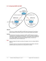
Figure 1 Porous high-density polyethylene (Medpor)
framework for a uricul ar reconstruction.
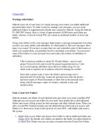
Figure 2 Surgical markings: the position of the intended
helix in blue, the ‘‘Y’’-shaped incision in black, the superficial
temporal artery in red, and the frontal branch of the facial
nerve in green.
122 FACIAL PLASTICS SURGERY/VOLUME 24, NUMBER 1 2008
Medpor has been described as a reconstructive
implant in various subsites within the head and neck. In
Frodel and Lee’s series, Medpor implants were used to
correct facial deformities after trauma, including orbital,
temporal fossa, frontocranial, maxillary, malar, calvarial
bone graft, and chin defects.
22
In two cases, microtic
ears were repaired. Similarly, Romano used Medpor in
140 patients with facial fractures, reporting a low com-
plication rate (one instance of implant infection requir-
ing removal and no implant migration or exposure).
23
Medpor has been used with success in the reconstruction
of auricular deformities in postburn and cryptotic pa-
tients.
24,25
Medpor, beneath a temporoparietal fascia (TPF)
flap and covered with full-thickness skin grafts, is an
expedient and useful alternative to costal cartilage re-
construction.
26
It eliminates the need for costal cartilage
harvest (with attendant chest morbidity), shortens the
surgical time, and standardizes results among patients
and surgeons. The Medpor prefabricated implant for
auricular reconstruction consists of both helical and base
components (Fig. 1). These two components are sutur ed
together, and shaped with a #10 scalpel, to form an
auricle customized in size and shape to the patient’s
aesthetic needs. This can be done with relative ease as
the basic shape of the auricle, with all of its component
structures, is already manufactured, allowing minute
changes to be made to patient specifications.
Medpor reconstruction is indicated for patients
with second- and third-degree microtic dysplasias, in
addition to failed microtia reconstructions with autoge-
nous rib grafts.
26
In preparation for the TPF flap, the
patient should be evaluated for a functioning superficial
temporal arterial and venous system.
27
A Doppler probe
is used to mark the position of the anterior and posterior
superficial branches of the superficial temporal artery.
Patients who have had previous surgery undergo mag-
netic resonance angiography imaging and, more recently,
computed tomogr aphic angiography. Angiography is
performed if a functioning artery is not visible.
The Medpor reconstruction takes place when the
child is 5 to 6 years old, when the opposite comparative
ear is 85% normal growth.
6,26
In contrast to rib
cartilage techniques, children at age 5 to 6 are candidates
for reconstruction, as there is no need to wait for chest
donor site growth. However, the child must be old
enough and psychologically prepared to deal with the
aftercare and restrictions of the surgery. The surgery
is performed in two stages: the first procedure places
the auric ular f ramework in t he temporal pocket, and in
the sec ond procedur e, 3 m onths later, the lobule is
transposed.
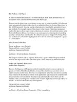
Figure 3 (A) Medpor framework inset into a temporal scalp pocket, temporoparietal fascial (TPF) flap harvested. (B) TPF flap
brought down to cover framework in its entirety.
AESTHETIC MICROTIA RECONSTRUCTION WITH MEDPOR/ROMO, REITZEN 123
SURGICAL TECHNIQUE
To establish the size of the new ear, an exposed radio-
graph template is traced to the borders of the normal or
contralateral ear.
6,26
If there is no opposi te ear, an ear of
a parent is used as a mo del. A second, scaled-down
Figure 5 Stage two microtia reconstruction: lobule transposition and tragal reconstruction. (A) Well-healed auricular
reconstruction after stage one. (B) Postoperative result after stage two. A scar revision of the scalp incisions was performed.
Figure 6 An example of the Romo-Guard
TM
(Medical
Concepts Inc., Murrieta, CA) to pro tect the ear from trauma
or compression.
Figure 4 Postoperative photograph at the completion of
stage one microtia reconstruction. The lower two-thirds of
the framework is covered by temporal skin, whereas the
upper one-third is covered by full-thickness skin graft.
124 FACIAL PLASTICS SURGERY/VOLUME 24, NUMBER 1 2008
version is then generated to account for the soft tissue
bulk that will surround the framework.
The auricular position of the scalp is determined
based on the aesthetic proportions discussed above.
6,26
Generally, the anterior helix sits 6 cm from the lateral
canthus and is positioned at a 20-degree angle from the
vertical. The position is marked with permanent black
marker. With a red marker, the anterior and posterior
branches of the superficial temporal artery are traced to a
height of the parietal scalp, using a handheld Doppler, in
preparation for the TPF flap. The frontal branch of the
facial nerve is marked in green, in a line midway between
the lateral eyebrow and the hair-bearing temporal skin
(Fig. 2).
A ‘‘Y’’-shaped incision line is planned with the
inferior aspect of the tail of the ‘‘Y’’ placed just superior
to the intended heli x.
6,26
The anterior extent of the
dissection is 10 cm superior to the intended helix, and
the posterior extent is 5 cm posterior to the helical rim.
The area is injected superiorly and posteriorly with 0.5%
lidocaine and 1:200,000 epinephrine. The full-thickness
skin graft donor site (postauricular or inguinal regions) is
also injected at this time. Using a #10 scalpel, an incision
is made just deep to the level of the dermal papillae of the
hair, taking care not to damage the subdermal plexus of
vessels, revealing the superficial aspect of the tempor-
oparietal fascia. Using a blunt-tipped Stevens scissors,
slow meticulous dissection with constant cutting in a
subfollicular plane develops the scalp flap to the anterior
extent of the dissection. After the temporal scalp is
elevated 5 cm posterior to the planned helical rim, the
area of non-hair-bearing skin including the vestige is
injected with 0.5% lidocaine and 1:200,000 epinephrine
in a subcutaneous plane. The dissection then continues
underneath the mastoid of the non-hair-bearing skin
with Metzenbaum scissors. Thorough dissection in this
plane will allow for sufficient contouring of a postaur-
icular sulcus. The vestige of cartilage is carefully exposed
Figure 7 Microtia reconstruction. (A) Preoperative photograph in a patient with grade two microtia. (B) Postoperative
photograph approximately 1 year after stage two. (C) The reconstructed ear has a natural-appearing, well-defined postauricular
sulcus. (D) Preoperative frontal view of the same patient. (E) Postoperative photograph. Note the symmetry of the bilateral
auricles on frontal view.
AESTHETIC MICROTIA RECONSTRUCTION WITH MEDPOR/ROMO, REITZEN 125
and removed. Hemostasis is achieved with bipolar elec-
trocautery.
The Medpor implant is washed with antibiotic
solution, sized with a #10 blade, sutured together with
3–0 Monocryl sutures, and placed into the inferior aspect
of the dissected temporal flap in the position as
marked.
6,26
A back cut is made along the incision, which
allows the framework to lateralize and project from
the temporal bone. A small Hemovac (Zimmer Inc.,
Roodeport, South Africa) drain at its helical aspect is
placed with the framework into the pocket; the frame-
work is sutured into place with 3–0 prolene suture, and
the drain sutured with 5–0 fast gut suture. The TPF flap
is then measured and harvested (Fig. 3). A 4–0 chromic
suture is tied to the distal end of the TPF flap, guiding the
flap as it is brought down to cover the framework in its
entirety. Care is taken not to kink the superficial temporal
vessels during this step. The Hemovac drain is brought
out of the skin opening at the inferior aspect of the wound
and sutured with 4–0 nylon suture. The scalp flaps are
closed over a 10-French barreled suction drain, which
is sutured to the skin with 3–0 prolene suture. Staples,
alternating with 4–0 prolene sutures, are used to close the
scalp flaps. The lower two-thirds of the flap is covered
with native temporal skin; the upper one-third of the
flap is covered with full-thickness skin grafts from either
the opposite postauricular or inguinal regions (Fig. 4).
The second stage consists of the lobul ar trans-
position, generally 3 months later.
6,26
The inferior, fatty
portion of the auricular vestige is incised and rotated
posteriorly on an inferior pedicle. The superior aspect of
the vestige is used to aid in tragal reconstruction. The
conchal bowl can also be deepened. In addition, some
patients may undergo revision of the scalp scar, in the
event of a widened scar. At this stage, patients may
undergo placement of a bone-anchored hearing aid
28
(Fig. 5).
POSTOPERATIVE CARE
The main activity restriction postoperatively is the
avoidance of compres sion of the framework, to protect
the newly positioned TP F flap.
26
A firm plastic cup is
placed over the reconstructed auricle for 24 hours per day
for 2 to 4 weeks (Fig. 6). The 10-French barreled suction
drain is removed in 7 to 10 days, and the Hemovac
removed in 10 to 14 days (Figs. 7–9).
COMPLICATIONS
As with any surgical procedure, infection is a possible
complication. In this case , infection can occur in a
localized, cellulitic form, requiring oral or intraven ous
antibiotics. Otherwise, infection can progress to flap
necrosis and potential loss of the implant.
6,26
Romo
et al have reported a complication rate of 4% over 250
cases.
26
Complete flap coverage over the auricular frame-
work is the main factor in avoiding this complication. It
Figure 8 Microtia reconstruction. (A) Preoperative photograph. (B) Postoperative photograph after stage two.
126 FACIAL PLASTICS SURGERY/VOLUME 24, NUMBER 1 2008
is imperative that the TPF flap drape over the framework
in its entirety. Compression ischemia can ensue when
the flap is compressed by either trauma or the plastic ear
cup itself or due to poor surgical planning of the TPF
flap placement. If the graft is lost, a second chance a
reconstruction is available with the use of a contralateral
TPF flap and new auricular Medpor framework. Only
two patients out of 250 have suffered a complete loss of
the framework. Due to the soft tissue integration of the
implant, defects less than 1 cm can generally be salvaged
via local advancement flap or additional full-thickness
skin grafts. Without tissue integration, flap loss is likely
to ensue.
SUMMARY
The complex architecture of the auricle makes it one of
the most challenging structures for the reconstructive
surgeon to re-crea te. Overlying the ear’s unique cartilage
framework are layers of varied soft tissues forming a
three-dimensional organ, which is distinctively posi-
tioned on the head. Arguably the most challenging
auricle to reconstruct is third-degree microtia due to a
near-total absence of native tissue and a need for lifelong
durability of the reconstruction. Many methods of re-
construction have been studied; autogenous costal carti-
lage reconstruction has been one of the more traditional
methods, with favorable long-term results reported by
several surgeons. However, this technique requires tre-
mendous artistic and technical skill on the part of the
surgeon-sculptor to construct a realistic-appearing ear.
At the same time, the possibility of chest donor site
morbidity is introduced. High-density porous polyethy-
lene (Medpor) is a stable, alloplastic implant that can
integrate with host tissues, is resistant to infection, and
has been successfully applied to reconstruction of many
areas within the head and neck. For auricular recon-
struction, Medpor —enveloped in a temporoparietal
fascial flap with full-thickness skin graft coverage—is
a durable and aesthetically gratifying alternative in
microtic patients. This alternative surgical technique
reduces surgical time and morbidity, standardizes results
among surgeons, and facilitates an aesthetic, natural-
appearing reconstruction of the auricle.
REFERENCES
1. Pham TV, Early SV, Park SS. Surgery of the auricle. Facial
Plast Surg 2003;19:53–74
2. Sclafani AP, Mashkevich G. Aesthetic reconstruction of the
auricle. Facial Plast Surg Clin North Am 2006;14:103–116
3. Tolleth H. A hierarchy of values in the design and
construction of the ear. Clin Plast Surg 1990;17:193–207
4. Quatela VC, Thompson SK, Goldman ND. Microtia
reconstruction. Facial Plast Surg Clin North Am 2006;14:
117–127
Figure 9 Microtia reconstruction. (A) Preoperative photograph. (B) Postoperative photograph after stage two.
AESTHETIC MICROTIA RECONSTRUCTION WITH MEDPOR/ROMO, REITZEN 127
5. Aguilar EF. Auricular reconstruction in congenital anomalies
of the ear. Facial Plast Surg Clin North Am 2001;9:159–169
6. Romo T III, Presti PM, Yalamanchili HR. Medpor
alternative for microtia repair. Facial Plast Surg Clin North
Am 2006;14:129–136
7. Jahrsdoerfer RA, Kesser BW. Issues on aural atresia for the
facial plastic surgeon. Facial Plast Surg 1995;11:274–277
8. Weerda H. Classification of congenital deformities of the
auricle. Facial Plast Surg 1988;5:385–388
9. Brent B. Technical advances in ear reconstruction with
autogenous rib cartilage grafts: personal experience with 1200
cases. Plast Reconstr Surg 1999;104:319–334
10. Brent B. Microtia repair with rib cartilage grafts: a review of
personal experience with 1000 cases. Clin Plast Surg 2002;
29:257–271
11. Tanzer RC. Microtia—a long-term follow-up of 44 recon-
structed auricles. Plast Reconstr Surg 1978;61:161–166
12. Leach JL Jr, Jordan JA, Brown KR, et al. Techniques for
improving ear definition in microtia reconstruction. Int J
Pediatr Otorhinolaryngol 1999;48:39–46
13. Avelar J. Importance of ear reconstruction for the aesthetic
balance of the facial contour. Aesthetic Plast Surg 1986;10:
147–156
14. Furnas DW. Complications of surgery of the external ear.
Clin Plast Surg 1990;17:305–318
15. Adams WP Jr, Rohrich RJ, Gunter JP, et al. The rate of
warping in irradiated and nonirradiated homograft rib
cartilage: a controlled comparison and clinical implications.
Plast Reconstr Surg 1999;103:265–270
16. Thomson HG, Kim TY, Ein SH. Residual problems in chest
donor sites after microtia reconstruction: a long-term study.
Plast Reconstr Surg 1995;95:961–968
17. Ohara K, Nakamura K, Ohta E. Chest wall deformities and
thoracic scoliosis after costal cartilage graft harvesting. Plast
Reconstr Surg 1997;99:1030–1036
18. Cronin TD, Ascough BM. Silastic ear construction. Clin
Plast Surg 1978;5:367–378
19. Sclafani AP, Romo T III, Silver L. Clinical and histologic
behavior of exposed porous high-density polyethylene
implants. Plast Reconstr Surg 1997;99:41–50
20. Shanbhag A, Friedman HI, Augustine J, et al. Evaluation of
porous polyethylene for external ear reconstruction. Ann
Plast Surg 1990;24:32–39
21. Williams J D, Romo T III, Sclafani AP, et al. Porous
high-density polyethylene implants in auricular reconstruc-
tion. Arch Otolaryngol Head Neck Surg 1997;123:578–
583
22. Frodel JL, Lee S. The use of high-density polyethylene
implants in facial deformities. Arch Otolaryngol Head Neck
Surg 1998;124:1219–1223
23. Romano JJ, Iliff NT, Manson PN. Use of Medpor porous
polyethylene implants in 140 patients with facial fractures.
J Craniofac Surg 1993;4:142–147
24. Kim DY, Cho KS, Lee SY, et al. Su rgical correction of
cryptotia using Medpor. Ann Plast Surg 1 999;42:693–
699
25. Wellisz T. Reconstruction of the burned external ear using a
Medpor porous polyethylene pivoting helix framework. Plast
Reconstr Surg 1993;91:811–818
26. Romo T III, Fozo MS, Sclafani AP. Microtia reconstruction
using a porous polyethylene framework. Facial Plast Surg
2000;16:15–22
27. Park C, Lew DH, Yoo WH. An analysis of 123
temporoparietal fascial flaps: anatomic and clinical consid-
erations in total auricular reconstruction. Plast Reconstr Surg
1999;104:1295–1306
28. Romo T III, Morris LGT, Kohan D, et al. Reconstruction of
congenital microtia-atresia: outcomes with the Medpor/
BAHA approach. Accepted for publication in Annals of
Plastic Surgery
128 FACIAL PLASTICS SURGERY/VOLUME 24, NUMBER 1 2008