self organising aggregates of zebrafish retinal cells for investigating mechanisms of neural lamination
Bạn đang xem bản rút gọn của tài liệu. Xem và tải ngay bản đầy đủ của tài liệu tại đây (7.16 MB, 42 trang )
Development Advance Online Articles. First posted online on 7 February 2017 as 10.1242/dev.142760
Access the most recent version at />
Self-organising aggregates of zebrafish retinal cells for
investigating mechanisms of neural lamination
Authors: Megan K. Eldred, Mark Charlton-Perkins, Leila Muresan, William A. Harris1
1Corresponding
Author, email
Affiliations: Department of Physiology, Development and Neuroscience
Cambridge University, UK
Key words: Müller cells, cell sorting, layer formation, organoid, reaggregation, SoFa.
Summary statement: Dissociated embryonic zebrafish retinal cells reaggregate and
laminate quickly in agarose microwells. We show that this self-organisation is partly
© 2017. Published by The Company of Biologists Ltd.
This is an Open Access article distributed under the terms of the Creative Commons Attribution License
( which permits unrestricted use, distribution and reproduction
in any medium provided that the original work is properly attributed.
Development • Advance article
dependant on Müller glia.
Abstract
To investigate the cell-cell interactions necessary for the formation of retinal layers, we
cultured dissociated zebrafish retinal progenitors in agarose microwells. Within these
wells, the cells re-aggregated within hours, forming tight retinal organoids. Using a
Spectrum of Fates zebrafish line, in which all different types of retinal neurons show
distinct fluorescent spectra, we found that by 48 hours in culture, the retinal organoids
acquire a distinct spatial organization, i.e. they became coarsely but clearly laminated.
Retinal pigment epithelium cells were in the centre, photoreceptors and bipolar cells
were next most central and amacrine cells and retinal ganglion cells were on the
outside. Image analysis allowed us to derive quantitative measures of lamination,
which we then used to find that Müller glia, but not RPE cells, are essential for this
Development • Advance article
process.
Introduction
The retina is a strikingly well-organised neural tissue, with each of the major cell types
sitting in its own specific layer. Such laminated cellular organisation, common in the
nervous system, may aid in wiring the brain efficiently during development. However,
the mechanisms involved in the development of lamination, are only beginning to be
understood. In the cerebral cortex, there is a well-known histogenetic organization,
with early born cells populating the deep layers and late born cells the superficial
layers, an “inside-out” order (McConnell 1995). But timing alone does not account for
this organisation, as is clearly shown in the example of reeler mutant mice, where the
neocortex, shows the opposite “outside-in” order of histogenesis even though the
different types of cortical cells are generated and migrate to the cortical plate at the
correct times (Caviness & Sidman 1973). The layering defect in reeler is due to the lack
of the glycoprotein (Reelin), which is secreted largely by a single transient cell type, the
Cajal Retzius cell; (D’Arcangelo & Curran 1998; Huang 2009) suggesting certain cells
and molecules play important roles in histogenesis.
Retinal cells, like cells of the cerebral cortex, show a histogenetic arrangement, with
early born retinal ganglion cells (RGCs) residing in the innermost retinal layer and late
born photoreceptors in the outermost (Cepko et al. 1996; Harris 1997). But again, the
mechanism here cannot simply be timing – i.e. cells piling up on top of each other
according to their birthdate. This is known because several studies have revealed that
the different retinal cell types are born with overlapping periods of birth, suggesting
that timing alone is insufficient (Holt et al. 1988). In zebrafish, live imaging studies have
revealed that sister cells born at exactly the same time may migrate to different but
appropriate layers (He et al. 2012), that late-born RGCs migrate through earlier born
postmitotic cells intermingle before they sort into their correct layers (Almeida et al.
2014; Chow et al. 2015). One question arising from these findings is whether these
behaviours arise from interactions between the different cell types, i.e. cell-cell
interactions, or from different cell types responding to common environmental cues
such as gradients of apicobasal cues. The latter possibility is consistent with in vivo
studies in which lamination is preserved even in the absence of specific cell types
(Green et al. 2003; Kay et al. 2004; Randlett et al. 2013). However, other studies suggest
Development • Advance article
amacrine cells (ACs) to reach the RGC layer, and that there is a period during which
that direct interactions between cell types are likely to be involved in normal layering
(Huberman et al. 2010; Chow et al. 2015). In addition, the involvement of cell-cell
interactions is indicated by the formation of rosettes in retinoblastoma (Johnson et al.
2007) and retinal dysplasias in which cell adhesion molecules such as N-cadherin are
compromised (Wei et al. 2006).
Aggregation cultures, used since the early 20th century have revealed the ability of
various cell types to re-aggregate and re-organise into histotypic tissues in the absence
of tissue scaffolds and extrinsic factors. This phenomenon was first seen in basic,
monotypic tissues, such as sponge and sea urchin (Herbst 1900; Wilson 1907), not only
revealing an innate ability of certain cell types to self-organise, but also providing a
platform on which we could begin to investigate the fundamental cell-cell interactions
involved in histogenesis. In the mid-century, Moscona and colleagues used aggregation
studies to investigate tissue formation in a variety of tissues including the chick retina
(Moscona & Moscona 1952; Moscona 1961), highlighting the ability of even complex,
multitypic tissues to self-organise. Later, Layer and colleagues were able to generate
fully stratified retinal aggregates, termed retinospheroids, from embryonic chick retinal
cells in rotary culture (Layer & Willbold 1993; Layer & Willbold 1994; Rothermel et al.
1997). The study of aggregation cultures has led to physical and theoretical
considerations of how tissues might self-organise including differential adhesion or
tension between cells (Steinberg 2007; Heisenberg & Bellaïche 2013).
In this paper we present the embryonic zebrafish retina as a model with which to
extend these investigations due to the increasing availability of genetic, molecular and
nanophysical tools with which to label and manipulate cells types and molecules of
cultures to examine the ability of zebrafish retinal cells to self-organise, and investigate
the importance of retinal pigment epithelial cells and Müller cells in retinal lamination.
Development • Advance article
interest. We use the transgenic SoFa fish, which labels all retinal cell types, in aggregate
Results
Dissection, dissociation and culture of zebrafish retinal cells
At 24 hours post fertilisation (hpf), the zebrafish retina is a pseudostratified epithelium
comprised of approximately 2000 progenitor cells, each stretching from the apical to
the basal surface. Over the next 48 hours, these progenitors divide several times to give
rise to a fully laminated retina of approximately 20,000 postmitotic neurons and glia of
all the major cell types (He et al. 2012). We dissected and dissociated retinas within this
time window in order to investigate the cell interactions at these times (Fig. 1A-B). To
assure ourselves that the dissociation protocol was satisfactory, we used a fluorescent
cell counter (see Materials and Methods) to assess several factors. Cell yield was
consistently high, between 2000 - 3000 cells per 24hpf retina (SFig. 1A); cluster
analysis showed that over 95% of these dissociated cells were counted as single cells
(SFig. 1B); and cell viability immediately after dissociation was over 96% as calculated
using the Acridine Orange/Propidium Iodide viability assay (SFig. 1C). With sufficient
cell yield and viability we began our reaggregation experiments in a basic L-15
supplemented with PSF, but found that the addition of Zebrafish Embryo Extract and
FBS promotes cell re-aggregation and growth (SFig. 1D-G). In agreement with previous
reports (Zolessi et al. 2006), we also found that N2 supplement supports RGC growth
and maturation in these cultures (data not shown).
To investigate the cell-cell interactions involved in layering, we wanted to reaggregate
the cells in a way that minimises interactions with the substrate, thus limiting all
interactions to those between the cells themselves. For this reason, we tried a
traditional hanging drop culture (Foty 2011). We seeded aliquots of the single cell
suspension in drops on the lids of culture dishes, which were then inverted (SFig. 1H).
clusters while others contained several smaller clusters (SFig. 1I). We obtained much
more consistent results when we plated the dissociated cells into agarose microwells
made using the 3D Petri Dish mould (Napolitano et al. 2007; Klopper et al.
2010)(Microtissues Ltd) (Fig 1C,D, S.Fig1 J,K). These agarose microwells provide a
confined, non-adhesive environment which minimises distance between cells. The
dissociated cells in these wells began to aggregate immediately after seeding. Within 3
Development • Advance article
After 48h, we found varied degrees of aggregation; some drops contained single large
hours most cells had aggregated (S.Mov.1), and by 15 hours the cells had undergone
compaction into similarly sized aggregates (Fig. 1E-J).
The ability of zebrafish retinal progenitors to reaggregate without the need for a
scaffold supports previous findings in chick from the Moscona laboratory (Moscona
1961; Sheffield & Moscona 1969). In those studies, they identified a cell reaggregationpromoting factor (Lilien & Moscona 1967), which was later cloned and identified as
retinal cognin (R-Cognin) (Hausman & Moscona 1976). To assess whether the same
factor was involved in the reaggregation of zebrafish retinal cells, we added PACMA31, a
small molecule inhibitor of the active site of R-Cognin, to our cultures. We found a dosedependent effect on aggregation; cells treated with 5μM of PACMA31 generated slightly
loose aggregates after 24 hours in culture (hic), whereas those treated with 50-200μM
were completely unable to aggregate (SFig. 2).
A Self-Organizing Retina: Identification of zebrafish retinal cells and
characterisation of organisation
The Spectrum of Fates (SoFa1) zebrafish transgenic line (Almeida et al. 2014) allows the
simultaneous identification of all 5 main retinal cell types based on 3 fluorophores, each
of which is expressed in particular combinations of retinal cell types (Fig. 2A-F). RGCs
express membrane-bound RFP (Fig. 2C); Amacrine and Horizontal cells (ACs and HCs)
express cytoplasmic GFP and membrane-bound RFP (Fig. 2D); Bipolar cells express
membrane-bound CFP (Fig. 2E); and Photoreceptors express membrane-bound CFP and
RFP (Fig. 2F). Whereas most studies of tissue organisation use techniques such as
immunohistochemistry or in situ hybridization to identify the different cell populations,
the use of SoFa1 line for the starting material for these studies allows immediate and
As was previously reported in the studies of chick retinal reaggregation assays
(Rothermel et al. 1997), we also found that the developmental stage of the cells when
they are dissociated and re-aggregated has an effect on their ability to organise.
Cultures from cells of younger stage embryos such as 24hpf are more capable of
organising than those from older stages, such as 72hpf (SFig.3) suggesting the
Development • Advance article
even live microscopic access to the process of lamination.
mechanisms responsible for retinal layering are active during the developmental stages
when these processes are normally occurring.
Using this strategy, we found that aggregates derived from 24hpf zebrafish retinal
progenitors are indeed capable of self-organising. Figure 2G-L shows the central sagittal
section of an aggregated retinal culture after 48 hours in culture. It can be seen quite
clearly that the Ptf1a:cytGFP expressing cells (ACs and HCs) organise in a distinct ring
near the outside of the aggregate (Fig. 2H), containing within them a cluster of
Crx:gapCFP expressing cells (PRs and BCs)(Fig. 2G). It is difficult to see the positioning
of RGCs in this preparation as Atoh7:gapRFP is expressed in many other cells types,
however a Zn5 antibody staining reveals RGCs positioned in the outer layer of the
aggregate, amongst the Ptf1a:cytGFP cells (SFig. 4). The organisation of these
aggregates appears to be “inside-out” with respect to the normal retina. Thus, while
situated near the basement membrane on the inner surface of the intact retina, RGCs in
our aggregates are found near the outer surface, and photoreceptors and bipolar cells,
which populate the outer layers of the intact retina, are found near the centres of our
aggregates. To assess whether this organisation was similar to that in the intact eye in
terms of cell numbers, we counted the relative proportions of cell types in our aggregate
cultures by counting the numbers in each fluorescent channel as a proportion of total
cells. We found the numbers of ACs, and HCs to be very similar to those in previously
published in vivo studies (Boije et al. 2015; He et al. 2012) whereas the numbers of BCs
and PRs were somewhat increased (Table 1.) The reason for this is unknown, but the
overall change in proportions is fairly modest. Therefore, perhaps it is not
unreasonable to find that the organisation seen in our aggregates resembles the
Quantification
This pattern of organisation clearly shows relative positions of cell types in our
aggregates as reflected in the fluorescence profiles, which are highly consistent within
and between experiments, making it a good platform from which to compare
experimental conditions. To begin to quantitate this pattern, we devised a Matlab
script, which generated an isocontour fluorescence profile for each aggregate. This fits a
mask to the aggregate (Fig. 2M) and isocontours from the periphery to the centre of the
Development • Advance article
situation in vivo.
aggregate (Fig. 2N) along which it gives a readout of the fluorescence distribution
across isocontours of the distance function from the outline of the aggregate for each
fluorescent protein (FP) (For further details, see Materials and Methods). Figure 2O
shows the fluorescence profile for the aggregate represented in G-L. The CFP expression
is high near the centre of the aggregate, tailing off towards the periphery, whereas the
GFP expression is low in the centre, but peaks near the periphery, corresponding
roughly with Crx:gapCFP and Ptf1a:cytGFP cell positions respectively. By plotting this
data as an empirical cumulative distribution function (ecdf) against radial position, we
are able to see how far these patterns of expression deviate from a random distribution
of expression, which would be a straight diagonal line from the bottom left to the top
right. Figure 2P shows that the distribution of Atoh7:gapRFP curve is close to such a
straight line (dotted line). This is due to the fact that Atoh7 is expressed in most of the
different cell types indicating an even patterning of that fluorescent marker across the
aggregate, consistent with a complete failure of patterning. The ecdf for Crx:gapCFP
expressing cells is clearly shifted to the left of this line, whereas distribution of
Ptf1a:cytGFP cells is shifted to the right. By measuring the areas between these curves
we can derive a measure of laminar organization in our organoids, and can easily
compare one experiment to another.
RPE is not required for self-organisation
With the experimental and analytical tools in hand, we moved our focus to the
mechanisms responsible for this organisation. One approach to investigate these is to
eliminate specific cell types to see if any particular cell type is required. Previous
studies in chick have pointed to the Retinal Pigment Epithelium (RPE) as being
important for retinal organization by providing polarity information. Chick retinal cells
layering, but when cultured in the presence of a monolayer of RPE, formed correctly
oriented, fully stratified retinospheroids (Rothermel et al. 1997).
We therefore made reaggregates with and without RPE. RPE cells were included (Fig.
3A-H) or excluded (Fig. 3I-P) either by gently removing the layer during dissection, or
by leaving the layer attached to the neural retina before dissociation. These
experiments were done using 32hpf embryos to allow us to identify RPE cells based on
Development • Advance article
cultured in the absence of RPE formed aggregates containing rosettes with inverted
pigment formation, yet retaining a similar level of organisation to those from 24hpf
(SFig. 3A-J). It is clear that the fluorescence profiles of cultures with RPE (Fig. 3G) and
without RPE (Fig. 3O) are in the same order, with the Crx:gapCFP profile peaking
towards the centre of the aggregate and the Ptf1a:cytGFP profile peaking towards the
periphery. This pattern is consistent across all aggregates analysed (Fig. 3Q,R). This is
also represented in the ecdf plots where for aggregates with RPE (Fig. 3H) and without
RPE (Fig. 3P) the Crx:gapCFP curve is shifted to the left of the Atoh7:gapRFP curve, and
the Ptf1a:cytGFP curve is shifted to the right. The somewhat different shapes of the
curves near the centre of the aggregate for the condition with RPE is due to the fact that
the pigment epithelial cells, which are themselves not fluorescent, are positioned more
to the centre of these aggregates. Areas measured between these curves for both
conditions show no significant difference (Fig 3 S-U). These results, together with the
fact that in both conditions, the aggregates show a similar degree of ordering in the
same relative patterns suggests that in these experiments, RPE cells may not have an
appreciable influence on the ability of developing retinal tissue to self-organise.
Müller glia are important for retinal cell organisation
We next tested whether Müller glia have a role in the lamination of our retinal
organoids. Importantly, we found that Müller cell numbers are similar in our aggregates
compared to those counted in vivo (Supplementary Table. 1). Müller glia cells were
eliminated by treatment with the Notch Inhibitor DAPT, which was applied to our
cultures from the time equivalent to 45-48hpf in the embryo, onwards. Treatment of
embryos at this time completely blocks the differentiation of Müller glia in vivo without
affecting the differentiation of any of the neural cell types (MacDonald et al. 2015). The
GFAP:GFP reporter line (Bernardos & Raymond 2006) was used to confirm the
show a high expression of GFAP:GFP, with Müller glia extending processes throughout
the aggregate (SFig. 5A), whereas aggregates treated with 25μM DAPT display vastly
reduced expression of GFAP:GFP and no process projections (SFig. 5C). We then
analysed the effect of removing Müller glia on the ability of all other cell types to
organise using the SoFa1 line. The morphology of the aggregates (Fig. 4 A-F) and
fluorescence profiles of DMSO treated aggregates (Fig. 4G) are similar to previous
control aggregates, with the Crx:gapCFP profile peaking towards the centre of the
Development • Advance article
presence or absence of Müller glia in our aggregates (SFig. 5). DMSO treated controls
aggregate and the Ptf1a:cytGFP profile peaking towards the periphery. This is
consistent across all aggregates analysed for this condition (Fig. 4Q). This is also
represented in the ecdf plot (Fig. 4H) where the Crx:gapCFP curve is shifted to the left of
the Atoh7:gapRFP curve, and the Ptf1a:cytGFP curve is shifted to the right. The DAPT
treated cultures show disorganised aggregates (Fig. 4I-N) and the correspondent
fluorescence profiles clearly differ from the controls (Fig. 4O), the lack of pattern seen in
all aggregates analysed for this condition (Fig. 4R). The Crx:gapCFP curve does not peak
in the centre of the aggregate, but rather shows more of a plateau, with two smaller
peaks; one nearer the centre and one nearer the periphery, while the Ptf1a:cytGFP
profile still peaks towards the periphery but the steepness is much reduced. These
trends are reflected in the ecdf plots for the DAPT treated culture, where it is clear that
both the Crx:gapCFP and the Ptf1a:cytGFP have both been shifted toward the
Atoh7:gapRFP curve (Fig. 4P), representing an almost complete failure of patterning.
Areas measured between these curves for both conditions show a significantly higher
order of organisation for the DMSO treated controls as compared to the DAPT treated
cultures (Fig. 4S-U). These results suggest that MG cells may play an important role in
the laminar organisation of retinal organoids.
To address the question of whether this phenotype may be due to effects of inhibiting
Notch during the later stages of organization, or due to an alternative effect of inhibiting
gamma secretase activity, we carried out further experiments where we applied DAPT
to our cultures at a later time point to allow some Müller Glia to differentiate, while
retaining exposure to DAPT at later stages of organoid development. Aggregates in
which DAPT was added at 63hpf, appear to organise better than those in which DAPT
was applied from 48hpf onwards (Fig. 5 G-M), indicating that the ability to organise
(Fig 5. A-F)).
Development • Advance article
correlates with the presence of Müller Glia in the cultures, (shown with GFAP staining
Discussion
Here, we present a novel model for analysing the cellular and molecular mechanisms
governing the cellular interactions that drive cellular lamination in the retina during
development. We show that dissociated zebrafish retinal progenitors, after
disaggregation, reaggregate quickly, and within only 48 hours in culture, are able to
organise themselves into layers. With the aid of the SoFa1 line, simple analysis of this
layering can be easily and reliably quantified. Using this model we have begun to
investigate the mechanisms of the cellular interactions that drive layer formation in this
system, and report here on the relative importance of RPE cells and Müller Glial cells in
this process.
Our aggregates organise with RPE in the centre, next to photoreceptors and bipolar
cells, next to horizontal and amacrine cells, and RGCs on the outside. This normal
progression of layers is apparently inverted with respect to the retina in situ, where the
RPE is the outer cell layer and the RGCs comprise the inner. Such inside-out
organisation was also seen in rosettes within the retinospheroids described by Layer
and colleagues (Layer et al. 2001; Layer et al. 2002), and such photoreceptor-centred
rosettes, surrounded by inner layer cells have frequently also been seen in vivo in
pathological conditions. This suggests that there is a natural tendency for retinal cells
to organise themselves in layers which does not rely on the polarity of the tissue, and
which can happen in vitro with disaggregated cells.
Layer and colleagues found that when Müller glia or RPE cells or even media
conditioned by these cell types are added to reaggregated chick retinospheroids, then
over the course of several days, the aggregates involute and show retinal-like polarity
Willbold et al. 2000). As we are most interested in the events that lead to the initial
laminar arrangements of cell types, we have not looked over these longer terms in our
culture system. In our reaggregation cultures, the lamination happens between 24hpf
and 72hpf, which is exactly when retinal layering normally occurs in vivo. Indeed, we
show that zebrafish retinal cells dissociated at 72hpf do not form organised aggregates,
suggesting that there is a restricted time window when this process needs to happen.
This finding is reminiscent of work in chick retinal reaggregates, which also showed
Development • Advance article
with photoreceptors on the outside and RGCs toward the centre (Rothermel et al. 1997;
that complete aggregation (Ben-Shaul et al. 1980) and good layering (Rothermel et al.
1997) in reaggregated cultures could only be achieved when starting with young cells.
It is nevertheless interesting that RPE cells find themselves in the centre of these
aggregates considering that these cells are normally found around the outside of the
retina in vivo. What is the explanation for this? One possibility is that RPE cells act as
seeds or pioneers in the lamination process. We found, however, that these aggregates
organise in the same manner in the presence or absence of RPE cells. We were only
able to eliminate RPE cells at 32hpf, leaving open the possibility that they may have an
organising influence between 24 and 32hpf. However, as fully disaggregated cells from
32hpf retinas organise into laminae, the simplest explanation is that RPE cells are
essential neither for the ability of the neural cells to organise into layers nor for the
central to peripheral order of these layers.
The differential adhesion hypothesis model of cellular organisation posits that cells in
an aggregate will laminate through cells minimising their interfacial free energies
(Steinberg 1970; Foty & Steinberg 2005; Steinberg 2007). Cells with the strongest
adhesions to each other in such aggregates move to the centre while cells with weaker
adhesions sit further out in the cultures. Recently, it has been shown that cell-cell
surface tensions rather than simple adhesion may also drive lamination in tissues
(Mtre et al. 2015). It would, we feel, be very interesting to investigate in our
aggregates how much of a role these physical factors play in retinal lamination. For
instance, the differential adhesion hypothesis would suggest the strongest adhesions
are between RPE cells and the next strongest between photoreceptors and/or bipolar
cells, which occupy the centre of the aggregates when the RPE is removed. These
tension (Puech et al. 2006) and adhesion (Mtre et al. 2012).
Previous work from this laboratory has shown that MG cells are among the last cell
generated during zebrafish retinogenesis and that the generation of MG are particularly
sensitive to blockers of the Notch pathway during this period (MacDonald et al. 2015).
The gamma-secretase inhibitor DAPT specifically inhibits the Notch pathway, and if
applied at 45-48hpf, completely blocks the formation of MG in vivo. Yet in the complete
Development • Advance article
possibilities can be tested using new advances in micro-physical measurements of
absence of MG in vivo in zebrafish, a normally organised retina forms (Randlett et al.
2013; MacDonald et al. 2015). The result reported in this paper, namely that lamination
is significantly impaired by the absence of MG in zebrafish organoids therefore suggests
that mechanisms operating in vivo but not in vitro, can compensate for the absence of
MG in zebrafish. The possibility that this phenotype is due to other effects of DAPT on
retinal lamination cannot be completely ruled out, but the strong correlation between
the effects on lamination and MG differentiation suggest that it is the MG themselves
that are critical. Interestingly, in this regard, mouse retinas treated with an antagonist
of BMP to block MG differentiation have disrupted lamination and formation of rosettes
(Ueki et al. 2015) suggesting that MG may also have a more critical role in retinal
lamination in mammals. The apicobasal polarity of the native neuroepithelium is badly
degraded, if not completely destroyed, in the disaggregation-reaggregation process. In
vivo in zebrafish, cells may be able to sense this gradient and organise themselves along
it. Indeed, the native optic cup is a pseudostratified epithelium in which all retinal
progenitor cells extend across the entire apicobasal axis and this structure may provide
a polarised and oriented substrate for cell migration. In the zebrafish organoids, as in
the mouse retina, MG might take on some important role in establishing neuroepithelial
conditions. Another possibility has to do with the fact that MG provide tensile strength
to the retina (MacDonald et al. 2015), which is lost when these cells are dissociated but
re-established as MG differentiate.
The work of Sasai and colleagues who generated well-laminated retinal structures
starting from mouse and human stem cells (Eiraku et al. 2011; Eiraku & Sasai 2012) has
been particularly exciting for the field of intrinsic tissue organization. Human organoids
of various tissues provide a model whereby one can study developmental mechanisms
organoids to study development in a model system like zebrafish where it is possible to
examine retinal lamination in vivo. While the retinal organoids from the Sasai
laboratory show that one doesn’t need a whole embryo to grow an organised tissue, it is
clear that these stem cell derived organoids laminate in the context of a great deal of
early pattern that develops in these complex systems, such as the apicobasal cues,
patterned extracellular matrix, and localised signalling molecules. Thus, the
mechanisms at play in these stem cell derived organoids may be almost as complex as
Development • Advance article
and diseases that are specific to humans, so one may wonder why it is useful to turn to
those in the tissues in vivo and so it is useful to work on a simplified system. In vivo
studies in zebrafish and mice have revealed that each cell type in the retina can be
eliminated and the remainder of the cells are able to laminate in the correct order
(Green et al. 2003; Randlett et al. 2013). In our reaggregated cultures, we provide
neither a substrate nor any extracellular matrix with which the cells can interact. This
means that the cells must interact with each other to sort out, the fact that they can sort
themselves into rough layers in the absence of exogenous pattern should help us to
identify the molecular and cellular mechanisms involved in the cell-cell interactions at
Development • Advance article
play during retinal lamination.
Materials and Methods
Animals and Transgenic Lines
All zebrafish lines were maintained and bred at 26.5°C. Embryos were raised at 28.5°C
or 32°C in Embryo Medium and staged in hours post fertilization (hpf) using
morphological features as described in (Kimmel et al. 1995). Some embryos were
treated with 0.003% phenylthiourea (PTU, Sigma) from 8hpf onwards to prevent
pigment formation. All embryos were anaesthetized with 0.04% MS-222 (Sigma) prior
to dissection. All animal work was approved by the Local Ethical Review Committee at
the University of Cambridge and performed under the UK Home Office license PPL
80/2198.
Transgenic lines Ptf1a:cytGFP (Godinho et al. 2005), Crx:gapCFP (Almeida et al. 2014),
GFAP:GFP (Bernardos & Raymond 2006), and the polytransgenic SoFa1 line
(Atoh7:gapRFP/Ptf1a:cytGFP/Crx:gapCFP) (Almeida et al. 2014) have all been
previously described. Ptf1a:cytGFP/Crx:gapCFP embryos were obtained by the crossing
of homozygous Ptf1a:cytGFP and Crx:gapCFP lines.
Dissection and Dissociation of Zebrafish Retinas
24hpf or 32hpf embryos were anaesthetised and transferred to calcium-free dissecting
medium (116.6 mM NaCl, 0.67 mM KCl, 4.62 mM Tris; 0.4 mM EDTA (pH 7.8)
supplemented with 100 μg/ml of heparin and 0.04% MS-222), for retinal dissection. 20
retinas per condition were collected in fresh dissecting medium in a glass well dish and
kept on ice. Retinas were allowed to come to room temperature and incubated with
0.25% Trypsin-EDTA (Sigma) for 12 minutes. After gentle removal of Trypsin-EDTA,
using a glass fire-polished Pasteur pipette, followed by more vigorous trituration with a
P200 pipette until a single cell suspension was achieved. Cells were collected in L-15
supplemented with 3% FBS and centrifuged at 300rcf for 7 minutes. After gentle
removal of 75% of the supernatant cells were washed once more with the same
conditions and re-suspended in the required volume for immediate seeding.
Development • Advance article
fresh dissecting medium was replaced and retinas dissociated by gentle trituration
Cell Counting, Viability and Cluster Analysis
Cells were counted and percentage viability and cluster sizes calculated using the
LUNA-FL™ Dual Fluorescence Cell Counter (Logos Biosystems). Cells in suspension
were mixed with an Acridine Orange / Propidium Iodide mix (Logos Biosystems) and
analysed in fluorescence mode.
Cell Culture
Agarose dishes were prepared and cast from 35-well or modified 15-well (cut to size)
PDMS moulds (Microtissues Ltd) as previously described (Napolitano et al. 2007) using
UltraPure LMP Agarose (Invitrogen) and equilibrated with L-15 supplemented with 1%
PSF (Thermo Fisher Scientific) within 4-well culture plates (Nunclon). Cells were
seeded in a drop-wise manner as a volume of 75ul into 35-well dishes or 35ul into
modified 15-well agarose dishes. Cells were allowed 15-20 minutes to settle before
750ul culture medium was added via the medium exchange ports. Culture medium
consisted of L-15 supplemented with 10% Embryo Extract (See ZFin for recipe), 3%
FBS (Thermo Fisher Scientific), 2% N2 (Thermo Fisher Scientific) 1% PTU (Sigma) and
1% PSF. Cells were incubated at 28.5°C for 48 hours before aggregates were harvested
for analysis.
Drug Application
For the Müller Glia experiments cells were incubated with 25μM DAPT (Sigma) or the
equivalent volume of DMSO starting from the equivalent time of 45-48hpf, or 63hpf.
For the R-Cognin experiments cells were incubated with 5, 50, 100, or 200μM PACMA31
(Sigma) or the highest equivalent volume of DMSO from the time of cell seeding
Harvesting of Aggregates, Fixation and Mounting
Aggregates were fixed with 4% PFA for 20 mins at room temperature followed by 3 x 5
min washes with PBS and collection by gentle downward flushing action using a P200
pipette. Aggregates were mounted in VectorShield mounting medium with DAPI (Vector
Laboratories) surrounded by a reinforcement ring between a microscope slide and a
13mm round coverslip.
Development • Advance article
onwards.
Immunostaining
Aggregates were fixed in 4% PFA for 15min at room temperature followed by a 10 min
wash with 0.1% PBT and then PBS. Aggregates were then incubated overnight at 4°C
with primary antibodies. Aggregates were washed for 10 min with 0.05% PBT and
incubated for 2 hours at room temperature with the secondary antibodies together with
DAPI (1:1000). Aggregates were washed for 10 min with PBT then 10 min with PBS
before mounting. Aggregates stained with Zn5 were additionally blocked for 20mins at
room temperature (10% HIGS, 1% BSA, 0.5% Triton in 1x PBS) before the primary
stain. Antibodies used: mouse anti-Zn5 (1:100; ZIRC), mouse anti-GFAP (1:100 zrf1;
ZIRC).
Confocal Image Acquisition and Analysis
Aggregates were imaged under an oil immersion 60x objective (NA = 1.30) using an
inverted laser-scanning confocal microscope (Olympus FV1000). Images were acquired
for 7 z-slices at the centre of each aggregate at 1μm optical sections using the same
settings throughout: 1024 x 1024 resolution, 12.5us/pixel scanning speed. Data was
acquired using Olympus FV1000 software and analysed using Volocity Software (Perkin
Elmer).
Analysis of Organisation by Isocontour Fluorescence Profiling
The central most section of each aggregate was analysed using custom made Matlab
scripts. The geometry of the aggregate was determined from the DAPI image via
automatic segmentation (active contour [Chan-Vese] and morphological operators
based) (Chan & Vese 2001) or manual segmentation. The manual method allowed the
aggregate, which no longer express fluorescent protein.
The fluorescence inside the aggregate was characterized by the intensity profile
obtained via averaging the pixel intensities in concentric bands of width w = 5 pixels.
We examined two ways to construct the fluorescent profiles: on one hand averaging
pixel intensities in circular crowns around the centroid of the aggregate, on the other
hand, averaging the intensities in bands of equal width starting from the periphery
Development • Advance article
user to correct for easily recognised artefacts such as dead cells on the outside of the
(outline of the aggregate) to the centre. Although the results are similar for both cases,
we adopted the latter method since it is more robust to variation of aggregate shape.
In order to be able to compare sets of profiles from different experiments, the
fluorescent intensity profile was normalised such that its integral is 1 and the distances
to the centre were rescaled between 0 and 100 radial units. Subsequently the
cumulative profiles were computed (ecdf plot), depicting how the distribution of
fluorescence for each FP differs from a random distribution. From this we used
trapezoidal numerical integration to find the area beneath each curve and then
subtracted that of the Ptf1a:cytGFP curve from the Crx:gapCFP curve to calculate the
area between the two curves.
Acknowledgements
The authors are very grateful to Alexandra D. Almeida for helpful discussions
throughout this project, to Ryan MacDonald and Mark Charlton-Perkins for discussions
on the Müller Glia experiments, to Afnan Azizi for help with the quantitation and Sara
Conde Berriozabal for assistance during the revisions.
Competing interests
No competing interests declared.
Author contributions
MKE did all the experimental work and wrote the manuscript. MC-P helped with the
Müller glia experiments and preparation of the manuscript. LM generated the Matlab
Funding
This work was funded by a Wellcome Trust Senior Investigator Award to WAH
(100329/Z/12/Z) and a BBSRC Studentship Award to MKE (BB/J014540/1).
Development • Advance article
script for the image analysis. WAH helped design and guide the project and the writing.
References
Almeida, A.D. et al., 2014. Spectrum of Fates: a new approach to the study of the
developing zebrafish retina. Development (Cambridge, England), 141(9), pp.1971–
80.
Ben-Shaul, Y., Hausman, R.E. & Moscona, A.A., 1980. Age-dependent differences in
cognin regeneration on embryonic retina cells: immunolabeling and SEM studies.
Developmental neuroscience, 3(2), pp.66–74.
Bernardos, R.L. & Raymond, P.A., 2006. GFAP transgenic zebrafish. Gene Expression
Patterns, 6(8), pp.1007–1013.
Boije, H. et al., 2015. The Independent Probabilistic Firing of Transcription Factors: A
Paradigm for Clonal Variability in the Zebrafish Retina. Developmental Cell, 34(5),
pp.532–543.
Caviness, V.S. & Sidman, R.L., 1973. Time of origin of corresponding cell classes in the
cerebral cortex of normal and reeler mutant mice: An autoradiographic analysis.
The Journal of Comparative Neurology, 148(2), pp.141–151.
Cepko, C.L. et al., 1996. Cell fate determination in the vertebrate retina. Proceedings of
the National Academy of Sciences of the United States of America, 93(2), pp.589–95.
Chan, T.F. & Vese, L.A., 2001. Active contours without edges. IEEE Transactions on Image
Processing, 10(2), pp.266–277.
Chow, R.W.-Y. et al., 2015. Inhibitory neuron migration and IPL formation in the
developing zebrafish retina. Development (Cambridge, England).
D’Arcangelo, G. & Curran, T., 1998. Reeler: new tales on an old mutant mouse.
BioEssays : news and reviews in molecular, cellular and developmental biology,
20(3), pp.235–44.
culture. Nature, 472(7341), pp.51–6.
Eiraku, M. & Sasai, Y., 2012. Mouse embryonic stem cell culture for generation of threedimensional retinal and cortical tissues. Nature protocols, 7(1), pp.69–79.
Foty, R., 2011. A simple hanging drop cell culture protocol for generation of 3D
spheroids. Journal of visualized experiments : JoVE, 20(51), pp.4–7.
Foty, R.A. & Steinberg, M.S., 2005. The differential adhesion hypothesis: a direct
evaluation. Developmental biology, 278(1), pp.255–63.
Development • Advance article
Eiraku, M. et al., 2011. Self-organizing optic-cup morphogenesis in three-dimensional
Godinho, L. et al., 2005. Targeting of amacrine cell neurites to appropriate synaptic
laminae in the developing zebrafish retina. Development (Cambridge, England),
132(22), pp.5069–79.
Green, E.S., Stubbs, J.L. & Levine, E.M., 2003. Genetic rescue of cell number in a mouse
model of microphthalmia: interactions between Chx10 and G1-phase cell cycle
regulators. Development (Cambridge, England), 130(3), pp.539–52.
Harris, W.A., 1997. Cellular diversification in the vertebrate retina. Current Opinion in
Genetics & Development, 7(5), pp.651–658.
Hausman, R.E. & Moscona, A.A., 1976. Isolation of retina-specific cell-aggregating factor
from membranes of embryonic neural retina tissue. Proceedings of the National
Academy of Sciences of the United States of America, 73(10), pp.3594–8.
He, J. et al., 2012. How variable clones build an invariant retina. Neuron, 75(5), pp.786–
98.
Heisenberg, C.-P. & Bellaïche, Y., 2013. Forces in Tissue Morphogenesis and Patterning.
Cell, 153(5), pp.948–962.
Herbst, C., 1900. über das Auseinandergehen von Furchungs- und Gewebezellen in
kalkfreiem Medium. Archiv für Entwicklungsmechanik der Organismen, 9(3),
pp.424–463.
Holt, C.E. et al., 1988. Cellular determination in the xenopus retina is independent of
lineage and birth date. Neuron, 1(1), pp.15–26.
Huang, Z., 2009. Molecular regulation of neuronal migration during neocortical
development. Molecular and Cellular Neuroscience, 42(1), pp.11–22.
Huberman, A.D., Clandinin, T.R. & Baier, H., 2010. Molecular and cellular mechanisms of
lamina-specific axon targeting. Cold Spring Harbor perspectives in biology, 2(3),
p.a001743.
retinoblastoma. Cancer research, 67(6), pp.2701–11.
Kay, J.N. et al., 2004. Transient requirement for ganglion cells during assembly of retinal
synaptic layers. Development (Cambridge, England), 131(6), pp.1331–42.
Kimmel, C.B. et al., 1995. Stages of embryonic development of the zebrafish.
Developmental dynamics : an official publication of the American Association of
Anatomists, 203(3), pp.253–310.
Klopper, A. V et al., 2010. Finite-size corrections to scaling behavior in sorted cell
Development • Advance article
Johnson, D.A. et al., 2007. Neuronal differentiation and synaptogenesis in
aggregates. The European physical journal. E, Soft matter, 33(2), pp.99–103.
Layer, P.G. et al., 2002. Of layers and spheres: the reaggregate approach in tissue
engineering. Trends in neurosciences, 25(3), pp.131–4.
Layer, P.G., Rothermel, A. & Willbold, E., 2001. From stem cells towards neural layers: a
lesson from re-aggregated embryonic retinal cells. Neuroreport, 12(7), pp.A39-46.
Layer, P.G. & Willbold, E., 1993. Histogenesis of the avian retina in reaggregation
culture: from dissociated cells to laminar neuronal networks. International review
of cytology, 146, pp.1–47.
Layer, P.G. & Willbold, E., 1994. Regeneration of the avian retina by retinospheroid
technology. Progress in Retinal and Eye Research, 13(1), pp.197–230.
Lilien, J.E. & Moscona, A.A., 1967. Cell Aggregation: Its Enhancement by a Supernatant
from Cultures of Homologous Cells. Science, 157(3784), pp.70–72.
MacDonald, R.B. et al., 2015. Müller glia provide essential tensile strength to the
developing retina. The Journal of Cell Biology, 210(7), pp.1075–1083.
Mtre, J.-L. et al., 2012. Adhesion functions in cell sorting by mechanically coupling the
cortices of adhering cells. Science (New York, N.Y.), 338(6104), pp.253–6.
Mtre, J.-L. et al., 2015. Pulsatile cell-autonomous contractility drives compaction in the
mouse embryo. Nature Cell Biology, 17(7), pp.849–855.
McConnell, S.K., 1995. Constructing the cerebral cortex: Neurogenesis and fate
determination. Neuron, 15(4), pp.761–768.
Moscona, A., 1961. Rotation-mediated histogenetic aggregation of dissociated cells.
Experimental Cell Research, 22, pp.455–475.
Moscona, A. & Moscona, H., 1952. The dissociation and aggregation of cells from organ
rudiments of the early chick embryo. Journal of anatomy, 86(3), pp.287–301.
Napolitano, A.P. et al., 2007. Scaffold-free three-dimensional cell culture utilizing
Puech, P.-H. et al., 2006. A new technical approach to quantify cell–cell adhesion forces
by AFM. Ultramicroscopy, 106(8), pp.637–644.
Randlett, O. et al., 2013. Cellular requirements for building a retinal neuropil. Cell
reports, 3(2), pp.282–90.
Rothermel, A. et al., 1997. Pigmented epithelium induces complete retinal reconstitution
from dispersed embryonic chick retinae in reaggregation culture. Proceedings.
Biological sciences / The Royal Society, 264(1386), pp.1293–302.
Development • Advance article
micromolded nonadhesive hydrogels. BioTechniques, 43(4), pp.494, 496–500.
Sheffield, J. & Moscona, A., 1969. Early stages in the reaggregation of embryonic chick
neural retina cells. Experimental Cell Research, 57(2–3), pp.462–466.
Steinberg, M.S., 2007. Differential adhesion in morphogenesis: a modern view. Current
Opinion in Genetics & Development, 17(4), pp.281–286.
Steinberg, M.S., 1970. Does differential adhesion govern self-assembly processes in
histogenesis? Equilibrium configurations and the emergence of a hierarchy among
populations of embryonic cells. The Journal of experimental zoology, 173(4),
pp.395–433.
Ueki, Y. et al., 2015. A transient wave of BMP signaling in the retina is necessary for
Müller glial differentiation. Development (Cambridge, England), 142(3), pp.533–43.
Wei, X. et al., 2006. Nok plays an essential role in maintaining the integrity of the outer
nuclear layer in the zebrafish retina. Experimental Eye Research, 83(1), pp.31–44.
Willbold, E. et al., 2000. Müller glia cells reorganize reaggregating chicken retinal cells
into correctly laminated in vitro retinae. Glia, 29(1), pp.45–57.
Wilson, H. V., 1907. On some phenomena of coalescence and regeneration in sponges.
Journal of Experimental Zoology, 5(2), pp.245–258.
Zolessi, F.R. et al., 2006. Polarization and orientation of retinal ganglion cells in vivo.
Development • Advance article
Neural development, 1(1), p.2.
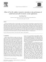
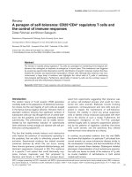
Fig 1. Dissociation, culture and re-aggregation of zebrafish retinal cells
(A-B) Schematic representing retinas dissected from 24hpf zebrafish (A), collected into
glass dishes and dissociated into single cells (B). (C) Agarose microwell dish cast from
the 3D Petri Dish PDMS Mould (adapted from ). (D)
Development • Advance article
Figures
Schematic representing the seeding chamber of the 3D Petri dish. After seeding, cells
settle into individual wells. (E-J) Time-lapse images of a single well from the 3D Petri
dish showing 24hpf cells re-aggregating. (H) Cells are almost fully reaggregated 3 hours
after seeding. (J) Cells have undergone compaction 15 hours after seeding. Time in
Development • Advance article
minutes and hours after seeding. Scale bar = 100 μm.
Development • Advance article