renal disease pathophysiology and treatment contributions from the rat
Bạn đang xem bản rút gọn của tài liệu. Xem và tải ngay bản đầy đủ của tài liệu tại đây (1.04 MB, 15 trang )
© 2016. Published by The Company of Biologists Ltd | Disease Models & Mechanisms (2016) 9, 1419-1433 doi:10.1242/dmm.027276
REVIEW
SPECIAL COLLECTION: TRANSLATIONAL IMPACT OF RAT
Renal disease pathophysiology and treatment: contributions from
the rat
ABSTRACT
The rat has classically been the species of choice for
pharmacological studies and disease modeling, providing a source
of high-quality physiological data on cardiovascular and renal
pathophysiology over many decades. Recent developments in
genome engineering now allow us to capitalize on the wealth of
knowledge acquired over the last century. Here, we review rat models
of hypertension, diabetic nephropathy, and acute and chronic kidney
disease. These models have made important contributions to our
understanding of renal diseases and have revealed key genes, such
as Ace and P2rx7, involved in renal pathogenic processes. By
targeting these genes of interest, researchers are gaining a better
understanding of the etiology of renal pathologies, with the promised
potential of slowing disease progression or even reversing the
damage caused. Some, but not all, of these target genes have proved
to be of clinical relevance. However, it is now possible to generate
more sophisticated and appropriate disease models in the rat, which
can recapitulate key aspects of human renal pathology. These
advances will ultimately be used to identify new treatments and
therapeutic targets of much greater clinical relevance.
KEY WORDS: Rat, Chronic kidney disease, Diabetic nephropathy,
Genetically modified rats, End-organ damage, Renal transplantation
Introduction
The prevalence of chronic kidney disease (CKD) is estimated to be
8-16% worldwide (Jha et al., 2013; Stevens et al., 2007). With an
aging population, and rising levels of hypertension, diabetes and
obesity, renal diseases pose an increasing burden on public
healthcare. Two million people worldwide are currently on renal
replacement therapy (RRT), dialysis or have a renal transplant.
However, this figure makes up only ∼10% of all individuals who
actually need RRT, with a greater number dying due to the
inadequate availability of therapies ( />kidneydisease/global-facts-about-kidney-disease#_ENREF_3) and
skewed treatment towards affluent countries with access to
healthcare (Jha et al., 2013). Furthermore, kidney disease
represents an independent risk factor for cardiovascular mortality
(Tonelli et al., 2006). Individuals often present with complex renal
pathologies resulting from numerous insults, both genetic and
environmental. The interactions of combined metabolic and
University of Edinburgh/British Heart Foundation Centre for Cardiovascular
Science, Queen’s Medical Research Institute, 47 Little France Crescent, Edinburgh
EH16 4TJ, UK.
*Author for correspondence ()
L.J.M., 0000-0002-6743-8707; J.J.M., 0000-0001-5745-5258
This is an Open Access article distributed under the terms of the Creative Commons Attribution
License ( which permits unrestricted use,
distribution and reproduction in any medium provided that the original work is properly attributed.
cardiovascular factors make it difficult to identify individuals who
will benefit most from available treatments to slow or prevent
disease progression (Jha et al., 2013). It is therefore imperative that
we develop new strategies to identify those at high risk of
progressive kidney disease and to discover new therapies to slow
the rate of disease progression in these individuals. Animal models
can provide insight into the pathophysiology of kidney disease and
can be used to test novel therapies. However, their utility is limited
by how well they recapitulate the key features and mechanisms of
progressive human disease. Although it can be argued that rodents
are poor replacements for humans in studies of kidney disease
(Becker and Hewitson, 2013), much valuable information about the
underlying etiology of renal disease has been revealed by studying
rat models.
The functional unit of the kidney is the nephron (see Glossary,
Box 1), which is closely integrated with the renal blood supply
(Fig. 1). The human kidney filters 180 liters of plasma through its
glomeruli, and produces 1 to 2 liters of urine daily. Approximately
99% of filtered sodium is retrieved as it passes through various
sections of the nephron before reaching the collecting duct.
Acute kidney injury (AKI) occurs when there is a rapid decline in
glomerular filtration rate (GFR; see Glossary, Box 1), usually
accompanied by impaired microcirculation, inflammation and/or
tubular injury or necrosis and reduced renal blood flow (Basile et al.,
2012). AKI is initiated by various clinical insults, including
hypotensive shock, sepsis, surgery or the administration of
nephrotoxic agents such as cisplatin (Tanaka et al., 2005) and
contrast agents (commonly used for medical imaging) (Mehran and
Nikolsky, 2006). Following mild kidney injury, an adaptive repair
response might ensue, leading to kidney regeneration. However,
with more severe injury, regeneration is incomplete and nephron
mass can be replaced by scar tissue, leading to CKD (Bucaloiu et al.,
2012; Chawla et al., 2011). There are limited treatment options
available for AKI, and its associated mortality remains high
(Ferenbach and Bonventre, 2015). AKI can be induced in rats by
performing ischemia-reperfusion surgery or by administering toxins
such as cisplatin. However, these single insults are unlikely to fully
recapitulate the multiple injurious processes that have typically
occurred in individuals with AKI.
CKD is an umbrella term for any renal disease that results in the
progressive loss of kidney function over time. The kidney possesses
only a limited capacity for regeneration, and repeated or sustained
injury to the kidney results in maladaptive responses (Ferenbach and
Bonventre, 2015), including the deposition of excess extracellular
matrix (ECM; see Glossary, Box 1), particularly collagen, in
the glomerulus and tubulointerstitium of the kidney (Fig. 2).
The pathological changes associated with CKD include
glomerulosclerosis and tubulointerstitial fibrosis (see Glossary,
Box 1), which result in the loss of normal renal architecture,
microvascular capillary rarefaction (see Glossary, Box 1), hypoxia
and tubular atrophy. These changes lead to the loss of renal filtrative
1419
Disease Models & Mechanisms
Linda J. Mullins*, Bryan R. Conway, Robert I. Menzies, Laura Denby and John J. Mullins
REVIEW
Disease Models & Mechanisms (2016) 9, 1419-1433 doi:10.1242/dmm.027276
Box 1. Glossary
Albuminuria: high levels of albumin ( protein) in the urine.
Arteriolar hyalinosis: the thickening of the arteriole wall with proteinaceous deposits of pink-staining hyaline material.
Capillary rarefaction: a reduction in capillary density.
Chronic allograft nephropathy (CAN): a leading cause of kidney transplant failure; it features a gradual decline in kidney function, often with an associated
increase in blood pressure.
Congenic: a rat strain that carries part of a chromosome from another, different rat strain.
Consomic: when two rat strains carry the same transgene inserted at the same place in the genome.
Cre recombinase/loxP: Cre recombinase enzymatically removes sequences that are flanked (floxed) by inserted loxP sequences.
CRISPR-Cas9: a genome-engineering technique. CRISPR stands for clustered regularly interspaced short palindromic repeats, which, together with transactivating guide RNAs, target the sequence-specific double-stranded breakage of DNA by the bacterial protein Cas9 endonuclease.
Diabetic nephropathy (DN): a progressive form of kidney disease in diabetics, characterized by albuminuria, a >50% decline in glomerular filtration rate
(GFR), increased glomerular basement-membrane thickness, arteriolar hyalinosis, mesangial sclerosis and tubulointerstitial fibrosis.
Embryonic stem cells (ES cells): pluripotent stem cells derived from the inner cell mass of a blastocyst, an early-stage preimplantation embryo.
End-organ damage: damage occurring in the major organs fed by the circulatory system.
Extracellular matrix (ECM): a proteinaceous matrix laid down outside the cell.
Focal segmental glomerulosclerosis: the deposition of excess ECM in a subset of glomeruli with only part of each glomerulus affected.
Glomerular filtration rate (GFR): the rate at which plasma is filtered through the glomerulus.
Glomerulosclerosis: the deposition of excess ECM in the glomerulus.
Hyperglycemia: abnormally increased sugar content in the blood.
Hyperkalemia: abnormally high potassium concentration in the blood.
Hypokalemia: abnormally low potassium concentration in the blood.
Ischemia-reperfusion injury (IRI): the tissue damage caused when blood supply returns to the tissue after a period of ischemia or lack of oxygen.
Malignant hypertension: a rapid and severe increase in blood pressure, leading to end-organ damage.
Mesangio-proliferative glomerulonephritis (MPGN): an autoimmune, inflammatory condition that damages the membrane supporting capillary loops of
the glomerulus.
Mineralocorticoid receptor (MR): a steroid-responsive nuclear receptor that controls fluid homeostasis in the kidney; it also has pro-inflammatory and proproteinuric effects.
Myofibroblast: a cell that combines the ultrastructural features of a fibroblast and a smooth-muscle cell.
Nephron: the functional unit of the kidney, consisting of the proximal tubule, the loop of Henle, and the distal convoluted tubule, each lined with specialized
tubular epithelial cells that express ion channels and transporters.
Nocturnal dipping: when systolic blood pressure falls by more than 10% at night compared to daytime levels.
Pericyte: contractile cell that wraps around the endothelial cells of capillaries and venules throughout the body.
Podocyte: a modified epithelial cell of the glomerulus that has foot-like processes, which contact the basal lamina of glomerular capillaries and allow blood
to filter through the slits.
Pressure-diuresis response: the increase in urine output for a given imposed increase in blood pressure.
Renin-angiotensin aldosterone system (RAAS): a hormone system involved in regulating sodium reabsorption from nephrons and blood pressure.
Tubulointerstitial fibrosis: the deposition of collagen in the interstitial region between tubules.
1420
features of human renal pathologies in vivo and how this model
organism has shed light on complex underlying mechanisms of
disease progression of therapeutic relevance – information that
might ultimately lead to the development of new drug treatments
and targets (Aitman et al., 2016, 2008).
Models of hypertensive renal damage
In up to 95% of individuals with hypertension, no specific
underlying genetic cause for the condition is identified despite
contributory factors such as smoking or obesity. However, in a small
proportion of cases, hypertension is secondary to endocrine or renal
disease. Sustained exposure to high blood pressure adversely affects
cardiac, brain, vascular and renal tissues, making hypertension a
major cause of end-organ damage (see Glossary; Box 1). Hence,
renal disease might be both a cause and consequence of
hypertension, forming a vicious circle whereby hypertension
causes kidney damage, which then exacerbates the high blood
pressure. Hypertensive nephrosclerosis is characterized by arterial
wall thickening, loss of renal autoregulation, glomerulosclerosis,
tubular atrophy and interstitial fibrosis (Hill, 2008). Arterial
stiffening due to increased pulse pressure affects autoregulation of
the preglomerular afferent arterioles, and leads to progressive
glomerular hypertrophy and damage with atrophy of the attached
tubule. Reduced glomerular filtration causes compensatory
Disease Models & Mechanisms
capacity and ultimately to end-stage renal disease. Many rodent
models mimic features of early CKD; however, only few exhibit
features of end-stage renal disease (ESRD).
The substantial wealth of physiological knowledge available for
the rat makes it the species of choice for modeling aspects of kidney
disease and for exploring therapeutic strategies in vivo. For several
decades, the mouse has been the pre-eminent mammalian organism
for disease modeling because of its genetic tractability. With recent
developments in genome engineering, the rat is rapidly catching up.
Genetic, congenic, transgenic, knockout, surgical or
pharmacological rat models have provided an opportunity to
investigate the molecular pathogenesis of renal disease, to
examine the disease in the context of live animals, and to assess
potential novel therapies. Table 1 lists the rat models (with key
genotypic and phenotypic features) discussed in this Review. The
interested reader is also directed to the Rat Genome Database (http://
rgd.mcw.edu/) for further information about these and additional
models (Shimoyama et al., 2016).
In this Review, we discuss how rat models have contributed to our
understanding of renal pathophysiology and hold promise for
developing improved treatments to halt the progression of CKD or
to repair kidney damage in humans. We consider aspects of
hypertensive renal damage, diabetic nephritis, AKI and CKD. We
emphasize the utility and limitations of the rat in recapitulating
REVIEW
Disease Models & Mechanisms (2016) 9, 1419-1433 doi:10.1242/dmm.027276
Absorption
Na+, Cl–, H2O, HCO3–
amino acids, glucose
Proximal
convoluted
tubule
Excretion
glucose
H Urea, NH3, K+
Distal
convoluted
tubule
+,
Absorption
NaCl, H2O, HCO3–
Excretion
H+, NH3, Urea, K+
Efferent
arteriole
Renal vein
Fig. 1. Schematic of a nephron. This
schematic shows a nephron, the functional
unit of the kidney. Blood is delivered to the
glomerulus, where plasma is filtered into
the lumen of the tubule. Various ions are
excreted and absorbed, and water is
retrieved, as plasma passes through the
different segments of the tubule, which are
intimately linked to peritubular capillaries.
Concentrated urine is formed by this
filtration process, which then passes
through the collecting duct to the renal
pelvis. The different components of a
nephron occupy distinct regions of the
kidney: the cortex and outer and inner
medulla, as shown.
Glomerulus
Peritubular
capillaries
Afferent
arteriole
Cortex
Thick
ascending
loop
NaCl
Renal artery
Thin
loop
Thin
descending
ascending
loop
Collecting
duct
H2O
Thin
ascending
loop
Outer medulla
NaCl
Inner medulla
H2O
H 2O
Urea
Loop of Henle
Absorption
Mg2+, Ca2+
hyperfiltration in other glomeruli, leading to glomerulosclerosis
(which also results from ischemic damage) and ultimately to tubular
damage and fibrotic lesions of the interstitial cells (Hill, 2008).
Classically, genetic animal models of high blood pressure, such
as the spontaneously hypertensive rat (SHR) and the related saltloaded stroke-prone (SHRSP) rat, generated by protracted rounds of
breeding and selection for high blood pressure (see also Table 1),
have been used to study the effects of chronic hypertension
(Okamoto and Aoki, 1963; Okamoto et al., 1964; Pravenec and
Křen, 2005). It has been proposed that the pathological progression
of hypertensive damage to kidney damage in this rat model mirrors
that seen in human hypertension (Hultström, 2012), with renal
damage resulting from altered pressure-dependent autoregulation of
renal blood flow.
The underlying mutations and their homeostatic sequelae, which
contribute to hypertension and to multi-end-organ damage in the
SHR, seem to be very complex. Renal microarray has identified
>200 genes that differ more than fourfold in their levels of
expression between adult SHRs or SHR substrains (Watanabe et al.,
2015) and Wistar Kyoto control rats. The availability of the entire
SHR genome sequence (Atanur et al., 2010) provides an
opportunity to identify potentially causative polymorphisms in
these genes. Undoubtedly, strains such as the SHR have helped to
confirm the involvement of multiple genes in hypertension and
kidney damage. However, identifying which mutations are primary
and which are secondary to the disease remains an unresolved
question for cardiovascular research.
Transgenesis allows researchers to investigate the biological
consequence(s) of a genetic perturbation. However, elucidating the
homeostatic effects of altered gene function is not always
straightforward, as exemplified by the mRen2 rat (Mullins et al.,
1990), which overexpresses the mouse renin (Ren2) gene, causing
severe hypertension (see Table 1). Renin is a key component of the
renin-angiotensin aldosterone system (RAAS; see Glossary, Box 1),
the activation of which increases levels of circulating angiotensin II
(AngII), and causes systemic vasoconstriction and sodium
resorption in the kidney in order to increase blood pressure. Both
kidney and plasma levels of renin are low in the mRen2 rat
(Bachmann et al., 1992) making this a low-renin hypertension
model. Hypertension was attenuated with captopril, which inhibits
the RAAS component angiotensin-converting enzyme (Ace),
indicating AngII dependence (Bader et al., 1992). High levels of
mouse-transgene-derived inactive renin, and low levels of active
renin, were produced in the adrenal gland, indicating that tissue
1421
Disease Models & Mechanisms
Urine passes to
renal pelvis,
ureter and bladder
REVIEW
Disease Models & Mechanisms (2016) 9, 1419-1433 doi:10.1242/dmm.027276
A
Normal healthy
cortical tubular
epithelium
Basement membrane
Peritubular capillary
B
Chronic injury
Hypertension
Diabetes
Glomerulonephritis
Flattened tubular
epithelium. Some cellcycle-arrested cells.
Atrophy of tubules
Hypoxia
Pro-fibrotic signals, e.g. TGFβ
Pro-inflammatory signals, e.g. IL-6
Tubulointerstitial fibrosis*
ECM production
Inflammatory cell infiltrate
Fibroblast activation
and recruitment
Injured activated endothelium
Increased apoptosis
Eventual capillary rarefaction
Increased hypoxia
Reduced glomerular filtration
Reduced renal perfusion
Loss of podocytes
Perivascular fibrosis
Glomerulosclerosis#
C
D
#
*
RAAS is responsible for hypertension in this model (Peters et al.,
1993). The crossing of the renin transgene onto a closely related
outbred Sprague Dawley strain generated animals that developed
malignant hypertension and end-organ damage by 8 weeks of age
(see Glossary, Box 1) (Whitworth et al., 1994). In particular, the
kidney exhibited glomerulosclerosis and interstitial fibrotic lesions.
When the mRen2 transgene was crossed onto the inbred Fischer
(F344) and Lewis rat strains, the resulting consomic strains (see
Glossary, Box 1) were susceptible and resistant to malignant
hypertension, respectively. Genome-wide screening and
quantitative trait analysis identified two modifier loci on
chromosomes 10 and 17, which contributed to malignant
hypertension susceptibility (Kantachuvesiri et al., 1999). The
mRen2 rat strains have been studied extensively for over 25 years,
under both hypertensive and hyperglycemic conditions.
In a more refined model, the Cyp1a1Ren2 rat (Kantachuvesiri
et al., 2001), expression of the mRen2 gene is under the control of an
inducible promoter in the inbred Fischer strain. This allows the
1422
researcher to control the degree of AngII-dependent hypertension
and consequent end-organ damage, its speed of attainment and,
also, to look at repair processes, once the inducer (indole-3carbinol; I-3-C) is withdrawn (see ‘Models of diabetic nephropathy’
below). The earliest hypertension-induced renal injury identified in
the Cyp1a1Ren2.Fischer strain is limited to the preglomerular
vasculature (Ashek et al., 2012). The later-onset hypertensive
kidney
damage
includes
arterial
wall
thickening,
glomerulosclerosis, interstitial fibrosis and tubular injury
(Kantachuvesiri et al., 2001) similar to the renal damage caused
by hypertension in humans. Increases in urinary albumin and
angiotensinogen were observed with malignant hypertension
(Milani et al., 2010), although the latter did not reflect changes in
angiotensinogen gene expression in the kidney cortex (Prieto et al.,
2011). Proteinuria was alleviated in this model by antagonism of
the mineralocorticoid receptor (MR; see Glossary, Box 1) with
spironolactone (Ortiz et al., 2007). After the transient induction
of hypertension, Cyp1a1Ren2 rats developed salt-sensitive
Disease Models & Mechanisms
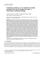
Fig. 2. The pathophysiological processes linked to kidney disease. (A) A normal, healthy kidney (left), and a magnified view of the structure of a tubule and its
associated vasculature (right). (B) A chronically diseased kidney, showing the processes that lead to tubulointerstitial fibrosis. (C,D) Histological sections of
an adult rat kidney, stained with Masson’s trichrome (20× magnification; scale bars: 50 µm). (C) The glomerular and tubular architecture of a normal adult rat
kidney, and (D) glomerulosclerosis (#) and tubulointerstitial fibrosis (*) in a 12-month-old hydroxysteroid dehydrogenase 2 (Hsd11b2)-knockout rat exhibiting
end-stage renal disease.
REVIEW
Disease Models & Mechanisms (2016) 9, 1419-1433 doi:10.1242/dmm.027276
Table 1. Rat models with renal pathophysiology
Strain
Type of model
Hypertensive kidney damage
Spontaneously
Inbred
Genetic: multiple
hypertensive
mutations
rat (SHR)
mRen2
Cyp1a1mRen2
(F344)
Sprague
Dawley/
Fischer
(F344)
Inbred
F344
Genetic: mouse Ren2
transgene
Phenotype
Strengths of model
Spontaneous
hypertension
Observe focal segmental
Complicated
Pravenec and
glomerulosclerosis
genetics and
Kren, 2005;
(FSGS) typical of human
phenotype
Okamoto et al.,
hypertensive
1964
nephrosclerosis
Mullins et al., 1990
Observe hyperplastic
Early mortality due
arteriosclerosis typical of
to MH (8human malignant
10 weeks)
hypertension (MH)
Control severity of
Genetic background Kantachuvesiri
hypertension; facilitates
must be
et al., 2001
study of renal or vascular
considered
repair
Fulminant (severe)
hypertension*; endorgan damage
Genetic: mouse Ren2 Inducible hypertension;
transgene under
susceptible to MH*
Cyp1a1 promoter;
inducible with indole3-carbanol (I-3-C)
Genetic: mouse Ren2 Inducible hypertension;
transgene under
resistant to MH
Cyp1a1 promoter;
inducible with I-3-C
Cyp1a1mRen2
(Lew)
Inbred
Lewis
(Lew)
Hsd2KO
Inbred
F344
Genetic: global
Hsd11b2 knockout
Syndrome of apparent
mineralocorticoid
excess (SAME); saltsensitive (SS)
hypertension*
Dahl saltsensitive (SS)
rat
Two-kidney, one
clip (2K1C)
model
Inbred
Genetic: multiple
mutations
SS hypertension
Various
Surgical
Hypertension;
nephropathy of
contralateral kidney
Diabetic nephropathy (DN)
mRen2/STZ
Sprague
Dawley
Cyp1a1mRen2
Inbred
F344
Genetic: mouse Ren2
transgene under
Cyp1a1 promoter;
inducible with I-3-C
and STZ
Inducible hypertension
and diabetes*
Pharmacological: e.g.
cisplatin or contrast
agent
Acute tubular necrosis
(ATN)
Various
Surgical
ATN
Various
Surgical
Inflammation and
fibrosis; obstructive
uropathy
Acute kidney injury (AKI)
Nephrotoxicity
Various
Ischemiareperfusion
injury (IRI)
Renal fibrosis
Unilateral
ureteral
obstruction
(UUO)
Genetic: mouse Ren2 Hypertension and
diabetes*
transgene;
pharmacological: DN
induced with STZ
Limitations of model References
As in cell above; facilitates
study of renal protection
Genetic background
must be
considered when
comparing with
F344 model
Hypertensive from young
SAME is a rare
age (∼5 weeks)
disease in
humans;
complicated
response to gene
knockout
Highly reproducible
Complicated
substrains: SS versus
genetics and
salt-resistant (SR) control
phenotype
Clipped kidney acts as
Variable phenotype
internal control, although
between labs
an untreated control
kidney should also be
included
Early mortality due
Some features of human
to MH (8DN, including
10 weeks); renal
glomerulosclerosis,
injury might be
tubulointerstitial fibrosis,
due to
arteriolar hyalinosis,
hypertension not
reduced glomerular
diabetes
filtration rate
Mimics pathology and renal No arteriolar
transcriptomic changes in
hyalinosis or
human DN
advanced kidney
failure
Liu et al., 2009
Mullins et al., 2015
Hu et al., 2014;
Dahl et al., 1962
Finne et al., 2014;
Goldblatt et al.,
1934; Okamura
et al., 1986
Kelly et al., 1998
Conway et al.,
2012; Conway
et al., 2014
Ease of induction of tubular Uncommon causes Mehran and
injury
of ATN in humans
Nikolsky, 2006;
Tanaka et al.,
2005
Straightforward surgery;
Human ATN usually Conger et al., 1991;
severity of tubular injury
multifactorial
Schrimpf et al.,
can be controlled by
2014; Kramann
altering duration of
and Humphreys,
ischemia
2014
Simple and rapid model of
fibrosis; mirrors features
of human congenital
UUO; useful as a
screening tool for antifibrotics
Adult human kidney
does not fibrose
as quickly during
obstruction
Terashima et al.,
2010
Continued
1423
Disease Models & Mechanisms
Rat model
REVIEW
Disease Models & Mechanisms (2016) 9, 1419-1433 doi:10.1242/dmm.027276
Table 1. Continued
Rat model
Strain
Type of model
Chronic kidney disease (CKD)
Human
Inbred
Genetic: human
diphtheria toxin
F344
diphtheria toxin
receptor
transgene
(hDTR)
AA-4E-BP1
Inbred
Genetic: AA-4E-BP1 ‡
F344
transgene driven by
podicin promoter
Sprague
Pharmacological: antiPassive
Dawley
Fx1A antibody
Heymann
nephritis
(PHN)
Phenotype
Strengths of model
Limitations of model References
Podocyte loss; focal
segmental
glomerulosclerosis
(FSGS)
Mechanical failure of
podocytes;
proteinuria; FSGS
PHN; membraneous
nephropathy
Develops nephrotic range
proteinuria, podocyte
loss, FSGS
Artificial mechanism Wharram et al.,
of injury: podocyte
2005
loss rapid and
simultaneous
Artificial mechanism Fukuda et al.,
of injury
2012a
Anti-Thy 1.1
Various
Pharmacological: IgA
nephropathy
Mesangio-proliferative
glomerulonephritis
(MPGN)
5/6th
nephrectomy
Various
Surgical
Reduced nephron
number; reduced
glomerular filtration
rate (GFR)
Pharmacological:
nephrotoxic globulin
Immune-complexmediated glomerular
nephritis; proteinuria;
P2RX7 increase
Nephrotoxic
Various
nephritis (NTN)
–
Develop immune deposits
and proteinuria
Antibody in human
Salant et al., 1979
disease is directed
against
phospholipase A2
receptor
Has several features of the Self-limiting disease Nazeer et al., 2009;
Denby et al.,
human clinical pathology,
course in rat,
2011
e.g. mesangial
limited tubular
proliferation, glomerular
involvement and
ECM deposition
minimal renal
functional change
Difficult surgery;
Gilbert et al., 2012
Can exhibit progressive
high mortality
decline in renal function
(strain specific) and
increase in blood
pressure
Develops proteinuria and
Batch-to-batch
Turner et al., 2007;
some histopathological
variation in
Taylor et al.,
changes that are
disease severity
2009
observed in human
disease
*UK Home Office regulations for animal research do not allow end-stage renal failure (ESRF) or malignant hypertension (MH) as end point of experiment.
AA-4E-BP1, eukaryotic translation initiation factor binding protein 1 (EIF4EBP1), a member of the mammalian target of rapamycin complex 1 pathway.
STZ, streptozotocin.
hypertension, which could be attenuated by the superoxide dismutase
mimetic tempol, implicating the superoxide anion in the development
of salt-sensitive hypertension (Howard et al., 2005).
The Cyp1a1Ren2 transgene is carried on the Y chromosome and,
by crossing the inducible Fischer male to a Lewis female, followed
by selective backcrossing of the F1 progeny to Lewis or Fischer
animals, congenic lines (see Glossary, Box 1) were derived. These
lines retain the transgene and either susceptibility or resistance to
end-organ damage, on an otherwise resistant or susceptible
background (Kantachuvesiri et al., 1999). Whole-renal,
microarray-based, gene-expression profiling studies of the
parental and congenic strains revealed genes in the congenic
region that were differentially expressed between the parental and
congenic strains (Liu et al., 2009). This strategy identified
angiotensin-converting enzyme Ace as a principal modifier of
hypertension-induced microvascular renal injury in the
Cyp1a1Ren2 rat model (Liu et al., 2009). The C-domain of Ace is
thought to mediate blood pressure control through its action on
angiotensin I. However, it is now recognized that Ace has other
effects, such as cleavage of the naturally occurring tetra-peptide
acetyl-N-Ser-Asp-Lys-Pro (AcSDKP) by the N-terminal domain of
Ace (Bernstein et al., 2011). AcSDKP has been shown to reverse
inflammation, cell proliferation and fibrosis in rat models of
hypertension (Liu et al., 2009; Zuo et al., 2013). As predicted,
AcSDKP was present at significantly lower levels in the kidneys of
the injury-susceptible Fischer rat than in the kidneys of the more
protected Lewis rat (Liu et al., 2009).
Microarray-based gene-expression profiling of the congenic
Fischer and Lewis kidneys was further used to identify previously
unknown candidate genes that might associate with a susceptibility
1424
to kidney injury (Menzies et al., 2013). A bioinformatic enrichment
analysis identified multiple candidate genes in addition to Ace. The
second- and third-ranked susceptibility genes were the purine
receptors P2X7 and P2X4 (Menzies et al., 2013). There are seven
P2X receptors in the rat, as in humans. These adenosine-5′triphosphate-activated cation channels are part of the larger
mammalian purine receptor family, which includes G-protein
coupled P2Y receptors and adenosine P1 receptors (Ralevic and
Burnstock, 1998). Both P2X and P2Y purine receptors have been
implicated in preclinical rodent models of hypertension (Menzies
et al., 2015b) and kidney disease (Menzies et al., 2016; Ralevic and
Burnstock, 1998). In humans, genetic variation that causes the
functional impairment of P2X7 is associated with a reduced risk of
stroke (Gidlöf et al., 2012). Conversely, P2X4 loss of function is
associated with increased pulse pressure (Stokes et al., 2011). The
renal pressure-diuresis response (see Glossary, Box 1) of Fischer,
but not of Lewis, rats was improved with combined P2X7 and P2X4
receptor antagonism using the dye, Brilliant Blue G (BBG)
(Menzies et al., 2013). Renal vascular resistance was unaffected
by BBG in Lewis rats, but both blood pressure and vascular
resistance decreased in Fischer rats, suggesting that P2X7 might
support tonic vasoconstriction in the susceptible strain. Specific
P2X7 receptor antagonism using the compound AZ11657312
caused rapid vasodilation. Acute antagonism of the receptor P2X7
in Fischer rats, chronically infused with AngII, significantly
improved renal perfusion and tissue oxygenation (Menzies et al.,
2015a). Recently, P2X7 receptor antagonism has also been shown
to attenuate renal injury in Dahl salt-sensitive rats (Ji et al., 2012).
P2X7 has been implicated in a wide range of neurological,
inflammatory and musculoskeletal disorders, in addition to its role
Disease Models & Mechanisms
‡
in hypertension and renal disease. Clinical trials of P2X7
antagonists in the treatment of inflammatory diseases have shown
limited therapeutic benefit to date (Bartlett et al., 2014). Given the
large number of splice variants (Cheewatrakoolpong et al., 2005)
and disease-related single-nucleotide polymorphisms (SNPs) (Jiang
et al., 2013) in the human P2RX7 gene, a productive future research
strategy could be the selective humanization of rats to develop
tissue-specific or disease-relevant therapeutic strategies.
In the two-kidney, one clip (2K1C) hypertensive system
(Goldblatt et al., 1934), which has been implemented in rats, a
clip on the left renal artery activates the RAAS system. Although
both kidneys are exposed to an equivalent increase in AngII, only
the non-clipped rat kidney shows hypertensive damage (Cervenka
et al., 1999). Recently, the non-clipped kidney was found to have
increased mRNA, protein and urinary levels of angiotensinogen,
suggesting that kidney damage occurs through increased AngII, and
that angiotensinogen could be used as an early biomarker of kidney
damage (Shao et al., 2016). Exposure of the non-clipped kidney to
increased AngII was ameliorated by nitric oxide (NO) release,
suggesting that this is a protective mechanism (Helle et al., 2008).
Additional early hypertension-induced changes in the renal tubules
were identified by micro-dissection of visibly undamaged
tubulointerstitial tissue from the non-clipped kidney. Proteomic
analysis using mass spectrometry revealed the differential
expression of over 300 proteins compared to control samples,
with profibrotic Rho-signaling proteins being the most highly
overrepresented (Finne et al., 2016). Such studies should help to
identify additional biomarkers of early tubule damage, which in
time could be used diagnostically. It should be noted, however, that
the clipped kidney is not physiologically equivalent to an untreated
(sham) control kidney; thus, the latter should always be included as
a control when comparing clipped and non-clipped kidneys (Palm
et al., 2008, 2010).
Despite complexities of the SHR, SHRSP and 2K1C hypertension
models, a recent gene-expression profiling study revealed a common
progression in hypertensive renal damage (Skogstrand et al., 2015).
Of the 88 genes similarly regulated in all three models, 40 were also
identified in gene-expression profiles from human fibrotic kidneys.
This suggests that pathogenic pathways underlying kidney damage
are conserved between rats and humans.
Hypertensive models generated by genetic modification
Gene-knockout technology has only recently become available for
the rat with the isolation of rat embryonic stem (ES) cells (see
Glossary, Box 1) (Buehr et al., 2008; Li et al., 2008), which can be
used as a tool for gene modification. The genetic tractability of the
rat has also been greatly facilitated by genome-engineering
technologies, such as zinc-finger nucleases (ZFNs) (Geurts et al.,
2009), transcription activator-like effector nucleases (TALENs)
(Tesson et al., 2011) and the CRISPR-Cas9 system (see Glossary,
Box 1) (Li et al., 2013). Genome endonuclease technologies
generate a sequence-specific DNA double-strand break, which is
repaired by error-prone, non-homologous end-joining. Any
insertions or deletions introduced at the target site cause missense
or nonsense mutations. The PhysGen knockout program (http://pga.
mcw.edu/) has utilized these technologies to generate a wide variety
of knockout rat models in genes associated with cardiovascular or
renal disease. One of the earliest ZFN-knockout rat models
generated with a clear renal phenotype was the hypotensive reninknockout rat (Moreno et al., 2011). Disruption of the renin gene
caused profound disruption to normal kidney development. The
inner renal medulla was morphologically rudimentary and there
Disease Models & Mechanisms (2016) 9, 1419-1433 doi:10.1242/dmm.027276
were signs of cortical interstitial fibrosis. These changes could be
related to the concomitant reduction in AngII production, and
support the assertion that the RAAS is essential for normal kidney
development in mammals (Guron and Friberg, 2000).
Another rat knockout model that exhibits reduced renin levels is
the Hsd2KO rat (Mullins et al., 2015). The enzyme 11-βhydroxysteroid dehydrogenase type 2 (Hsd11b2) protects the MR
from inappropriate activation by cortisol (corticosterone), in the
kidney principal cell, by inactivating it to cortisone (11dehydrocorticosterone). In this model, ZFN-induced knockout of
the Hsd11b2 gene causes inappropriate activation of the MR,
leading to salt-sensitive hypertension, suppression of renin
secretion, and hypokalemia (see Glossary, Box 1). This
phenotype closely models the human syndrome of apparent
mineralocorticoid excess (SAME). The rats exhibit severe renal
injury, including protein casts and atrophic tubules, segmental
glomerulosclerosis, tubule-interstitial fibrosis and proteinuria
(Mullins et al., 2015). These are all features associated with
chronic exposure to hypertension and with MR activation seen in
human kidney disease (Ueda and Nagase, 2014). Interestingly, the
Hsd2KO rat model demonstrates metabolic protection, including
increased insulin sensitivity and reduced mesenteric fat
accumulation, due to the depletion of the substrate for Hsd11b1 in
adipose tissue. This suggests that treatment with MR inhibitors
might reverse the adverse cardiovascular effects of SAME (which
include hypokalemia, hypertension, proteinuria and end-organ
damage), while promoting the beneficial metabolic effects of
Hsd11b2 inactivation (Mullins et al., 2015).
Salt-sensitive hypertension involves a complex feedback loop of
salt appetite and sodium retention. Hsd11b2 in the murine brain
triggers a central drive to consume salt (Evans et al., 2016). The rat
Hsd2KO model offers a more robust platform to investigate the
physiological mechanisms of central versus renal-centric salt
sensitivity than is feasible in the mouse. Decreasing dietary salt
consumption might reduce the burden of CKD in humans
(McMahon et al., 2013). Intriguingly, an alternative, albeit more
invasive, strategy to ameliorate salt-sensitive hypertension has been
recently demonstrated. Renal medullary dysfunction in saltsensitive Dahl rats (Dahl et al., 1962) was found to reflect a
reduction in adult (CD133+) mesenchymal stem cells (MSCs) in the
medulla. Injection of MSCs, but not of renal medullary interstitial
cells, into the renal medulla attenuated immune-cell infiltration and
sodium retention, and reduced systemic blood pressure (Hu et al.,
2014). The rationale for using MSCs stems from numerous animal
studies, which have demonstrated that these cells have protective
effects in acute and chronic kidney injury models (Fleig and
Humphreys, 2014; Wang et al., 2013).
The co-injection of single-strand oligonucleotides with ZFNs,
TALENs or CRISPR-Cas9 components can be used to introduce
targeted SNPs or to repair mutations, through homology-driven
repair (HDR). Rapid improvements in CRISPR-Cas9 technology,
using donor plasmids as HDR templates, have included the
introduction of fluorescent reporters (Ma et al., 2014a), the onestep generation of a floxed allele (loxP sites flanking an exon) (Ma
et al., 2014b) and conditional knockout using Cre-recombinase rat
strains (see Glossary, Box 1) (Ma et al., 2014a). Recently, WistarKyoto rats and SHRs that ubiquitously express GFP have been
produced, using the Sleeping Beauty transposon system. These
strains will prove useful for investigating cell fate and
transplantation in the hypertensive kidney (Garcia Diaz et al., 2016).
The identification of genes such as Ace, P2rx7 and Hsd11b2, or
specific genetic variants or splice variants of genes, that seem to
1425
Disease Models & Mechanisms
REVIEW
play key roles in moderating hypertensive damage, renal pathology
and salt-sensitivity has the potential to enable future identification
of individuals at risk of hypertensive kidney damage based on their
genetic profile. With the availability of humanized transgenic
models, Cre-loxP technology, reporter strains, gene knockouts and
knock-ins, and the ability to correct candidate genes in mutant rat
strains, many of the tools available to the mouse community are now
available in the rat. Although the inherent problem of off-target
events remain for genome-engineering technologies, targeting in rat
ES cells and screening for clones free of off-target events remains a
possibility. Thus, many more-refined and increasingly sophisticated
rat models, which more closely recapitulate human renal pathology
caused by hypertensive damage, can be expected in the future, and
might help to predict targeted therapeutic response more faithfully.
Models of diabetic nephropathy
Diabetic nephropathy (DN; see Glossary, Box 1) is the single most
common cause of end-stage kidney disease in the western world
(Saran et al., 2015). The use of reliable animal models of DN could
greatly facilitate research by providing mechanistic insights into this
disease to help identify novel therapeutic targets. These in turn
could provide a platform for preclinical testing of such novel
therapies. Unfortunately, one of the roadblocks to DN research is the
lack of preclinical models that recapitulate important functional,
structural and molecular pathological features of progressive human
diabetic kidney disease. Although several rodent models of type 1
diabetes [streptozotocin (STZ)-induced (Cooper et al., 1988)] and
type 2 diabetes [Zucker, Goto Kakizaki (Janssen et al., 2003)] have
been employed to study DN (see Glossary, Box 1), these models fail
to recapitulate all of the hallmarks of this disease as defined by
the Diabetic Complications Consortium (DiaComp; https://www.
diacomp.org/shared/validationcriteria.aspx). The inability of animal
models to fully replicate human DN might explain why many
therapies that have been beneficial in preclinical models of this
disease have proven to be ineffective in clinical trials. For example,
direct renin inhibitors were beneficial in reducing proteinuria in
rodent models (Kelly et al., 2007). However, the absence of
progressive renal failure in these models meant that the efficacy of
these inhibitors in reducing renal function could not be tested.
Human studies confirmed a beneficial effect of direct renin
inhibitors on reducing proteinuria (Parving et al., 2008) but,
importantly, they did not slow the rate of renal-function decline
(Parving et al., 2012). Furthermore, the increased risk of
hyperkalemia (see Glossary, Box 1) resulting from treatment with
direct renin inhibitors in patients with impaired renal function
(Parving et al., 2012) was not highlighted in the rodent models,
where blood potassium levels remained normal.
Although hyperglycemia (see Glossary, Box 1) is a pre-requisite
for the development of DN, hemodynamic factors play a substantial
role in the progression of this disease. Individuals with advanced
DN invariably have hypertension, and tight control of blood
pressure is as important as glycemic control in slowing disease
progression (Mogensen, 1998). Hypertension might not only be a
consequence of nephropathy but a key driver of kidney disease in
diabetes. Indeed, subtle abnormalities in blood pressure, such as
loss of nocturnal dipping (see Glossary, Box 1), precede the onset of
albuminuria (see Glossary, Box 1) in adolescents with type 1
diabetes (Lurbe et al., 2002). Furthermore, there are two case reports
regarding individuals with longstanding diabetes, hypertension and
unilateral renal artery stenosis (Berkman and Rifkin, 1973;
Béroniade et al., 1987) whose conditions mimic the 2K1C rat
model of hypertension. Autopsy findings in both cases revealed no
1426
Disease Models & Mechanisms (2016) 9, 1419-1433 doi:10.1242/dmm.027276
pathological evidence of nephropathy in the kidney downstream of
the arterial stenosis, despite severe nephropathy in the contralateral
kidney. The implications of these findings are that unilateral renal
artery stenosis might prevent the transmission of systemic
hypertension to the kidney parenchyma and the subsequent
development of nephropathy, even though both kidneys have been
exposed to an equivalent degree of hyperglycemia and to increased
AngII exposure. Thus, hyperglycemia or elevated angiotensin levels
alone are insufficient to promote advanced DN; the development of
hypertension is a prerequisite for disease progression. How
hypertension interacts with hyperglycemia to promote
nephropathy is unclear, but the application of cyclical stretch to
mesangial cells cultured in high-glucose media increases the
expression of pro-fibrotic genes, suggesting a role for increased
mechanical strain (Gruden et al., 2000). In rat mesangial cells grown
in high-glucose media, ATP and a P2X7 agonist dose-dependently
increased ECM deposition and levels of transforming growth factor
beta (TGFβ; a pro-fibrotic cytokine), whereas P2X7 inhibition
attenuated the response (Solini et al., 2005), indicating the
involvement of purinergic receptors.
Several approaches have been taken to recapitulate these
important hemodynamic factors in rodent models of DN. In the
1980s, the Brenner group determined that a high-protein diet
increased intra-glomerular pressure and promoted glomerular injury
in diabetic rats and that these features could be successfully
prevented by Ace inhibition (Zatz et al., 1986, 1985). These seminal
studies led directly to clinical trials of ACE inhibitors in patients
with DN, and they represent one of the best examples of how rodent
models can be utilized to provide important mechanistic insights
that subsequently lead to therapeutic advances. Indeed, ACE
inhibitors have since become the mainstay of preventing the
progression of renal disease in individuals with DN (Lewis et al.,
1993). Conversely, many therapies that have been effective in
animal models of DN that targeted hyperglycemia alone have
proven unsuccessful in clinical trials (B.R.C., personal
observation).
Rat models of DN
Genetic models of hypertension have also been utilized to model
progressive DN. The induction of diabetes with STZ leads to higher
levels of albuminuria in SHRs than in rat strains with diabetes or
hypertension alone (Cooper et al., 1988). Treatment with Ace
inhibitors abrogates the increase in albuminuria in SHR strains.
Activation of the RAAS plays a pre-eminent role in clinical DN.
Therefore, a logical approach was to induce diabetes in mRen2 rats
(Kelly et al., 1998). The renin-dependent hypertension in mRen2
rats accelerates the development of nephropathy, and this model has
been used to study not only the role of the RAAS in DN, but also
that of other pathways, including oxidative stress (Advani et al.,
2009). It has been shown that sustained hyperglycemia causes
increased tubular oxygen consumption due to mitochondrial
dysfunction and reduced electrolyte transport efficiency (reviewed
in Hansell et al., 2013). The onset of malignant hypertension in the
mRen2 model results in accelerated renal injury and in early
mortality, which is atypical of the slowly progressive course
observed in human diabetic kidney disease (Hartner et al., 2007).
This problem was overcome by using Cyp1a1mRen2 rats, where
adjustment of I-3-C concentration in the diet controls the timing and
severity of hypertension. Following induction of diabetes using
STZ, the addition of 0.125% I-3-C resulted in a gradual increase in
blood pressure, mimicking the evolution of hypertension in human
DN (Conway et al., 2012). The hyperglycemia and hypertension
Disease Models & Mechanisms
REVIEW
synergized to promote a 500-fold increase in albuminuria, and
caused moderate glomerulosclerosis and tubulointerstitial fibrosis –
all features of moderately advanced human DN. However, there was
no significant decline in renal function in this model, and some key
pathological features of DN, such as arteriolar hyalinosis (see
Glossary, Box 1), were not observed.
Microarray and RNA-sequencing technologies provide a nonbiased view of gene expression changes. Thus, comparing
transcriptomic changes in DN patients with rat models of the
disease might reveal common disease mechanisms, identify
relevant biomarkers and therapeutic targets, and enable the
rational selection of the rodent model that most closely
recapitulates changes seen in DN kidneys. Up to 50% of genes
that were differentially expressed in the tubulointerstitial
compartment of the kidney in human DN (Lindenmeyer et al.,
2007) were also similarly up- or downregulated in the renal cortex of
hyperglycemic and hypertensive Cyp1a1mRen2 rats (Conway et al.,
2012). For example, one downregulated gene in both the rat model
and in the kidneys of individuals with DN was epidermal growth
factor (EGF). Urinary EGF levels reflect renal EGF expression, and
subsequent studies confirmed that low levels of urinary EGF
excretion predict a poor renal outcome in individuals with DN and
with other CKDs (Betz et al., 2016; Ju et al., 2015). Hence, nonbiased transcriptomic approaches could be used to identify as-yetunknown prognostic biomarkers for therapeutic targets or to recruit
high-risk individuals for clinical trials. Such transcriptomic datasets
should be made freely available on databases such as Geodataset
( or Nephroseq (https://www.
nephroseq.org), as this will enable researchers to select the model
in which their pathway of interest is differentially activated in a
similar manner to human disease. Such ‘precision modeling’ could
improve the chances of translating findings made in rodent models
to the clinic.
Although the natural history of DN is one of inexorable
progression towards end-stage kidney disease, the tight control of
blood glucose and blood pressure can lead to the regression of
albuminuria in up to 50% of individuals with DN (Perkins et al.,
2003).
More
remarkably,
regression
of
established
glomerulosclerosis and tubulointerstitial fibrosis has been observed
in individuals with moderately advanced DN who achieve sustained
normoglycemia after receiving a pancreas transplant (Fioretto et al.,
1998, 2006), although this takes up to 10 years to become evident.
The pathways that promote regression remain poorly understood,
largely because serial biopsies are rarely performed in individuals
who are responding to treatment.
Rodent models provide insights into mechanisms of injury,
regeneration and repair. The Cyp1a1mRen2 rat model of DN is
particularly useful in this regard because hypertension can be
induced and then blood pressure normalized by adding and then
removing dietary I-3-C; inserting subcutaneous insulin implants can
also control STZ-induced hyperglycemia. In one study, 28 weeks of
hyperglycemia and hypertension (the injury phase) were followed
by tight glycemic and blood pressure control for an additional 8
weeks (the reversal phase), resulting in the partial regression of
albuminuria (Conway et al., 2014). Microarray analysis of the renal
transcriptome during both the injury and reversal phases revealed
∼650 genes that were upregulated during injury, almost 100 of
which reverted to control levels following reversal of
hyperglycemia and hypertension. This gene set was enriched for
genes that encoded ECM proteins, fibroblast markers and acutephase reactants, indicating that the tight control of glucose and
blood pressure might suffice to switch off the formation of new scar
Disease Models & Mechanisms (2016) 9, 1419-1433 doi:10.1242/dmm.027276
tissue. This was supported by the finding that there was no further
increase in the severity of glomerulosclerosis or tubulointerstitial
fibrosis during the 8-week reversal phase. In addition, many genes
of unknown function, which reverted to control levels during repair,
might be implicated in the fibrotic- or acute-phase response and
hence they merit further investigation. Conversely, almost 400
genes remained significantly upregulated despite the normalization
of blood glucose and blood pressure. This gene set was enriched for
genes that encoded proteins implicated in innate and adaptive
immunity, in particular pro-resolution macrophages and regulatory
T cells, suggesting that attempts at repair have been initiated.
Although glomerulosclerosis and tubulointerstitial fibrosis did not
reduce during the reversal phase, this was to be expected given the
protracted period required for regression of fibrosis following
pancreas transplantation in humans (Fioretto et al., 2006).
Permanent or long-term upregulation of some of these genes
might be responsible for the salt sensitivity observed in I-3-Cinduced rats (Howard et al., 2005).
Bilateral renal denervation has emerged as a potential treatment
for multiple-drug-resistant hypertension in individuals with bilateral
single renal arteries, but results from recent clinical trials have
questioned its efficacy for individuals with secondary (or accessory)
renal arteries (Bhatt et al., 2014; Hering et al., 2016; Khan et al.,
2014). When bilateral renal denervation was performed in the
mRen2/STZ rat model, it reduced signs of renal pathology,
albuminuria and the expression of fibrotic markers. This suggests
that renal denervation might attenuate renal injury in DN (Yao et al.,
2014), presumably with similar caveats regarding efficacy.
In summary, rat studies can mimic many of the features of human
DN, including progressive proteinuria, key pathological features
such as glomerulosclerosis and tubulointerstitial fibrosis, and the
activation of many pathways that are implicated in human DN.
However, none fully recapitulate human DN, with few exhibiting
arteriolar hyalinosis and a progressive decline in renal function. Rat
models have highlighted the benefits of Ace inhibitors and the
prognostic value of EGF in the treatment of DN. A comparison of
the results from microarray and RNA-sequencing technologies in
rodent models and human DN will continue to identify new
candidates for therapeutic interventions to prevent kidney damage
or to enhance repair and regeneration.
Models of acute and chronic kidney disease
AKI affects multiple cell types in the kidney, including endothelial
and tubular cells, which are adversely affected by hypoxia. It is not
clear whether hypoxia (the reduction of tissue oxygen supply to
below physiological levels) or re-oxygenation (increased exposure
to oxygen, as seen with reperfusion following ischemia) causes
AKI, but it is associated with altered intra-renal microcirculation
and oxygenation (Rosenberger et al., 2006). Ischemia-reperfusion
injury (IRI; see Glossary, Box 1) is extensively used as a model of
AKI, but hypoxic damage predominantly affects proximal tubule
segments in the outer stripe of the outer medulla and might not
recapitulate human AKI, which often includes medullary oxygen
insufficiency. Damage to the thick ascending limb is attenuated
following IRI, probably because the reduced solute transport leads
to improved oxygenation of the distal tubule (Rosenberger et al.,
2006). Following acute IRI, the vascular function of rats remains
impaired for several days (Conger et al., 1991). The pericyte (see
Glossary, Box 1) detaches from the endothelium under pathological
conditions, leading to microvascular rarefaction and hypoxia
(Schrimpf et al., 2014). Pericytes might contribute to the pool of
scar-forming myofibroblasts (see Glossary, Box 1) (Kramann and
1427
Disease Models & Mechanisms
REVIEW
Humphreys, 2014), making them key to both regeneration and the
development of fibrosis (Schrimpf and Duffield, 2011), although
myofibroblasts can also arise from other sources (Falke et al., 2015;
Micallef et al., 2012).
Agents affecting both cortical and medullary blood flow and
oxygen tension include radio-contrast agents (Heyman et al., 1991),
endotoxin [sepsis (Heyman et al., 2000)] and NO inhibitors (Brezis
et al., 1991). Together with non-steroidal anti-inflammatory drugs,
which cause a selective reduction in medullary blood flow and
tissue oxygenation, these could provide better models of AKI and
could enable investigation of hypoxia-inducible factors, adaptive
responses and potential therapies (Rosenberger et al., 2006). The
development of rat models should enhance our understanding of
AKI and help to design therapeutic strategies to block maladaptive
responses.
Pre-existing CKD affects the severity of AKI in humans and their
recovery (Liangos et al., 2006). This has been experimentally
modeled in rats using the renal-mass-reduction model of CKD with
an additional induced IRI. CKD develops in the 5/6th nephrectomy
rat model (in which the 5/6th of renal mass is surgically ablated; see
Table 1). When AKI is induced in this model via IRI, a
disproportionate number of regenerating tubules fail to redifferentiate. This is associated with significant loss of tubular
VEGF expression and with substantial capillary rarefaction.
Defective tubules also have pro-fibrotic properties that increase
tubulointerstitial fibrosis (Polichnowski et al., 2014). Further
investigation of this model will provide a greater understanding at
the molecular level of the AKI to CKD transition seen in humans.
Reporter rats should prove invaluable for mechanistic studies and
for the identification of the molecular pathways and cell lineages
involved in kidney disease (Garcia Diaz et al., 2016). The creation
of reporter transgenic rats has allowed the mapping of cells that
contribute to renal fibrosis and the testing of novel anti-fibrotic
agents on key pro-fibrotic pathways (Terashima et al., 2010). Using
transgenic rats carrying a luciferase reporter gene under the control
of rat α1(I) collagen and rat α2(II) collagen, the anti-fibrotic effects
of inhibiting TGFβ signaling (using a TGFβR1 inhibitor) and AngII
signaling [using an AngII-receptor blocker (ARB), olmesartan]
were examined (Terashima et al., 2010). This study revealed that
ARBs had an anti-fibrotic effect, independent of hemodynamic
effects, in the unilateral ureteral obstruction (UUO) model of rapid
renal fibrosis (see Table 1), which induces a marked change in renal
perfusion.
Rat models of AKI and CKD have been used as a platform to test
potential new therapies, including novel anti-fibrotic agents. FT011
is a derivative of the anti-allergy drug Tranilast (Miyazawa et al.,
1995), and it inhibits the proliferative actions of TGFβ and plateletderived growth factor (PDGF). FT011 stemmed the decline in GFR
in the 5/6th nephrectomy model of progressive CKD (see Table 1)
and reduced proteinuria and structural injury (Gilbert et al., 2012).
In the diabetic, hypertensive mRen2/STZ model, FT011 markedly
attenuated the development of proteinuria, as well as reducing
fibrosis in both the glomerulus and tubulointerstitium, and
interstitial macrophage infiltration, but GFR was unaffected
(Gilbert et al., 2012).
In a rat model of aristolochic-acid-induced nephropathy, the
neutralization of TGFβ with anti-TGFβ antibody improved renal
function and reduced acute tubular necrosis, interstitial
inflammation,
vascular
rarefaction
and
myofibroblast
accumulation (Pozdzik et al., 2016). The disruption of proximal
tubule organelle ultrastructure was also prevented. However, these
findings have not translated to the clinic; agents that block TGFβ and
1428
Disease Models & Mechanisms (2016) 9, 1419-1433 doi:10.1242/dmm.027276
retard CKD have failed to improve renal function despite the
promising preclinical results (Lee et al., 2015). These findings again
support the observation that animal models typically recapitulate
only part of the human condition – particularly CKD and its
progression to ESRD. Animal models such as the UUO rat, used as a
model of renal fibrosis, can be studied for a few weeks at most,
whereas, in humans, these conditions usually develop over many
years. Pathways that are important initially might not be as important
in the pathophysiology of later disease and could explain the lack of
translation of successful preclinical compounds.
Studies performed in various transgenic rat models have led to
new insights into glomerulosclerosis, and in particular into the role
of the podocyte (see Glossary, Box 1). A direct causative
relationship exists between the degree of podocyte depletion and
the development of proteinuria and glomerulosclerosis (Kim et al.,
2001; Wharram et al., 2005). However, the mechanisms by which
podocyte depletion can lead CKD to progress to end-stage kidney
disease are poorly understood.
To examine the effect of podocyte depletion, the human
diphtheria toxin receptor (hDTR) was specifically expressed in
podocytes, generating the hDTR Fischer rat model (see Table 1),
which has histopathological features commonly seen in the human
disease focal segmental glomerulosclerosis (FSGS; see Glossary,
Box 1), including mesangial expansion, segmental and global
sclerosis (Wharram et al., 2005). These features occur in proportion
to the degree of podocyte depletion. Although a return to normal
glomerular architecture over time did not occur, once the
glomerulus was destabilized by a critical degree of podocyte loss,
the continuous infusion of an ACE inhibitor (enalapril) and ARB
(losartan) was found sufficient to stabilize the glomeruli. The renoprotective effect of ARBs is not through blood pressure reduction
alone and seems to be due to a direct effect on the podocyte (Fukuda
et al., 2012b; Wharram et al., 2005).
Another transgenic Fischer rat model, this time expressing a
dominant-negative phosphorylation site mutant of AA-4E-BP1, the
eukaryotic translation initiation factor binding protein 1 (EIF4EBP1)
transgene (see Table 1), has been used to examine the effect of growth
on podocyte failure (Fukuda et al., 2012a). Driven by the podocin
promoter, the EIF4EBP1 transgene encodes a member of the
mammalian target of rapamycin complex 1 (mTORC1) pathway,
which is a key determinant of the cellular hypertrophic response,
driven by the podocin promoter. Transgenic AA-4E-BP1 rats have
normal kidney histology with no proteinuria below 100 g body weight,
but develop end-stage renal disease by 12 months. The observed
proteinuria and glomerulosclerosis were linearly related to body
weight increases and transgene dose. Histological observations
revealed bare areas of glomerular basement membrane, where
podocyte foot processes had pulled apart, and consequent adhesion
to the Bowman capsule. In the AA-4E-BP1 model, it seems that
proteinuria develops through mechanical failure of the podocyte
epithelial layer. This mechanism of podocyte depletion is different
from direct podocyte damage and death. It also provides a mechanistic
explanation for a separate group of diseases that lead to global
glomerulosclerosis or focal segmental glomerulosclerosis (see
Glossary, Box 1) in childhood and obesity (Fukuda et al., 2012a),
suggesting that limiting calorie intake could be beneficial in reducing
the severity of the human condition. With additional developments,
such as intravital imaging (Peti-Peterdi et al., 2016) and visualization
of calcium dynamics (Szebenyi et al., 2015) to observe podocyte
function/glomerular injury processes in real time, a deeper
understanding of the mechanisms that lead to the development of
renal pathology should identify novel therapeutic targets.
Disease Models & Mechanisms
REVIEW
Novel monogenic rat models of glomerulosclerosis have also been
generated, such as the TGR(hET-2)37 rat model, which expresses high
levels of human endothelin-2 (ET2) in the kidney (Hocher et al., 1996).
These rats develop blood-pressure-independent glomerulosclerosis,
which demonstrates that the human ET2 gene can have a bloodpressure-independent, growth-promoting effect on the rat glomerulus.
Apoptosis is a key feature of the progression of CKD. Recently,
ouabain, which is a cardiotonic steroid, has been found to have antiapoptotic actions. Chronic ouabain treatment of rats with passive
Heymann nephritis [PHN; a model of human membranous
nephropathy, a slow progressive proteinuric kidney disease
(Salant et al., 1979)] prevented the loss of podocytes, reduced the
level of apoptotic proximal tubule cells and reduced renal fibrosis
(Burlaka et al., 2016). Ouabain might represent a novel therapy that
could potentially protect against apoptosis and prevent the loss of
functional tissue in chronic proteinuric kidney disease.
The anti-Thy1.1 model of glomerulonephritis is an experimental
rat model that mimics human antigen-triggered, immune-induced
mesangio-proliferative glomerulonephritis (MPGN; see Glossary,
Box 1), such as IgA nephropathy. This well-characterized model of
glomerular injury has been used to investigate molecular
mechanisms of mesangial proliferation. Proteomic studies have
revealed several proteins that show altered expression in this model
(Nazeer et al., 2009), particularly the four and a half LIM domain
protein 2 (FHL2), which increases mesangial cell proliferation in
vitro (Lu et al., 2012) and could represent a new target for treating
MPGN. This model has proven to be useful in identifying key stressinduced microRNAs, such as miR-21 and miR-214 (Denby et al.,
2011), which are upregulated during renal injury. These
microRNAs have since been found to be differentially expressed
in human biopsies of individuals with IgA nephropathy, and their
upregulation correlates linearly with renal fibrosis (Hennino et al.,
2016), demonstrating the translational relevance of this model.
Other rat models of glomerulonephritis include the nephrotoxic
nephritis (NTN) model (see Table 1), which established that levels
of the P2X7 receptor protein are increased in the glomerulus. This
correlates with increased glomerular P2X7 in human biopsy
samples from patients with nephritis due to lupus (Turner et al.,
2007). In the rat NTN model, the P2X7 antagonist A-438079
prevented antibody-mediated glomerulonephritis through reduced
inflammatory damage due to a reduction in macrophage infiltration
into the glomerulus (Taylor et al., 2009).
Rat models have proved to be invaluable in the field of
regenerative cell therapy for renal disease. The potential of bonemarrow-derived MSCs to accelerate healing has been demonstrated
in several rat models of hypertension (as discussed above) and of
renal disease, including in the anti-Thy1.1 model (Li et al., 2006),
the 5/6th nephrectomy model of progressive CKD (Cavaglieri et al.,
2009; Choi et al., 2009) and in an AKI model induced by cisplatin
(Urt-Filho et al., 2016). MSCs might reverse AKI by a paracrine
mechanism rather than by MSC transdifferentiation. Intravenous
injection of microvesicles, released from cultured human MSCs,
inhibited tubular apoptosis and stimulated regeneration (Gatti et al.,
2011). The renoprotective effect was lost if microvesicles were pretreated with RNAse, or if the pro-angiogenic microRNAs, miR-126
and miR-296, were depleted. This suggests that the miRNAs,
delivered by microvesicles, are able to reprogram hypoxic resident
renal cells (Cantaluppi et al., 2012). Importantly, MSCs taken from
either the 5/6th nephrectomy model or the adenine-induced
nephropathy model and transplanted into the anti-Thy1.1 model
failed to induce healing. Both CKD and uremia adversely affected
transplanted MSCs, which exhibited cellular senescence
Disease Models & Mechanisms (2016) 9, 1419-1433 doi:10.1242/dmm.027276
(Klinkhammer et al., 2014). This result brings into question the
use of autologous MSCs for the treatment of CKD.
In summary, AKI and CKD share a spectrum of renal pathologies.
The identification of early biomarkers could allow the practitioner to
harness adaptive repair and regenerative mechanisms, and prevent the
maladaptive profibrotic pathways. A better understanding of the roles
of, and of the potential cross-talk between, pericytes, myofibroblasts,
tubular epithelium and podocytes is key to developing new therapies,
and the rat is well placed to deliver such advances.
Renal transplantation
Renal transplantation was first performed in the rat over 50 years
ago. Although the microsurgical techniques involved remain
challenging, they are more readily mastered in rats than in mice.
Several different combinations of inbred and outbred rat strains can
be used to model various complications of renal transplantation,
including IRI, acute rejection and chronic allograft nephropathy
(CAN; see Glossary, Box 1) (Shrestha and Haylor, 2014). Renal
transplantation from a Fischer donor to a Lewis recipient is the most
common model of CAN in rats (White et al., 1969). Fisher and
Lewis rat strains differ partially at the major histocompatibility loci
(MHC) I and II, and this weak histocompatible combination results
in CAN in the absence of immunosuppression (Hancock et al.,
1992; Paul et al., 1998). Ace inhibition can limit kidney damage in
this transplant model (Noris et al., 2003), which has also been used
to assess the development of alloimmunity (de Heer et al., 1994), the
efficacy of immunosuppressants (Chandraker et al., 1998), nonimmune therapies (Magee et al., 1999) and the development of
fibrosis in the graft (Jain et al., 2000). The small molecule BB3 is a
hepatocyte growth-factor mimetic, and studies in an IRI-induced rat
model of AKI revealed that BB3 protected the kidney from tubular
apoptosis and necrosis (Narayan et al., 2016). These data form the
basis of a clinical trial using BB3 in kidney-transplant recipients
who present with delayed graft function.
Allograft and isograft renal transplantation can also be used to
determine the relative importance of intrinsic renal cells versus
bone-marrow-derived cells in the pathogenesis of a wide range of
renal diseases. Ex vivo injection of MSCs into the kidney prior to
transplantation proved beneficial, whereas systemic injection of
MSCs failed to improve recipient survival (Iwai et al., 2014). Recent
improvements in the ability to genetically manipulate rats open up
an exciting new area of research for renal transplantation studies
(Doorschodt et al., 2014).
Conclusions and future perspectives
Disparities between animal models and human disease might have
resulted in promising preclinical therapies failing to be effective in
clinical trials. Recent developments in genome engineering and
transcriptomic profiling now allow the researcher to design and
refine models, to more closely interrogate specific aspects of renal
disease. The rat has and will continue to play a major role in the
identification of key genes that increase disease susceptibility, of
early biomarkers that highlight disease progression, and of genes,
pathways and cells that are fundamentally involved in kidney
regeneration or damage.
As highlighted in this Review, hypoxia, AngII, ACE and P2X7
play key roles in many aspects of kidney damage, placing them at
the forefront of therapeutic targets to be explored using rat models.
Given the complex nature of, for example, human P2X7 transcripts,
humanization of the rat could help to identify which isoforms are
disease-promoting, and could aid in the development of novel
treatment strategies.
1429
Disease Models & Mechanisms
REVIEW
Of particular interest is the application of MSC technology to the
treatment of AKI, CKD and renal transplantation. A number of
MSC-based clinical trials have been set up, despite safety concerns
raised by animal studies (Kunter et al., 2007). In a rat model of
glomerulonephritis, MSCs produced a short-term improvement, but
ultimately differentiated into intraglomerular adipocytes, resulting
in glomerulosclerosis (Kunter et al., 2007). Enhanced recruitment of
endogenous MSCs or the use of cell-free cocktails of secreted
factors might be preferable approaches (Kunter et al., 2011).
It is important to note that the ‘treatment’ of kidney disease might
not lead to repair of all aspects of organ damage. However, the
complexity of renal pathologies means that better design and use of
rat models as a resource could ultimately result in stratification of
diagnosis and tailored therapy.
This article is part of a special subject collection ‘Spotlight on Rat: Translational Impact’,
guest edited by Tim Aitman and Aron Geurts. See related articles in this collection at
/>Acknowledgements
Figures were adapted using the Servier Powerpoint image bank (vier.
com/Powerpoint-image-bank).
Competing interests
The authors declare no competing or financial interests.
Funding
The authors acknowledge support from the British Heart Foundation (BHF) Centre of
Research Excellence (RE/08/001/23904) and Kidney Research UK. R.I.M. is a BHF
Immediate Postdoctoral Basic Science Fellow (award number FS/15/60/31510).
L.D. is a Kidney Research UK Fellow (award number PD6/2012).
References
Advani, A., Gilbert, R. E., Thai, K., Gow, R. M., Langham, R. G., Cox, A. J.,
Connelly, K. A., Zhang, Y., Herzenberg, A. M., Christensen, P. K. et al. (2009).
Expression, localization, and function of the thioredoxin system in diabetic
nephropathy. J. Am. Soc. Nephrol. 20, 730-741.
Aitman, T. J., Critser, J. K., Cuppen, E., Dominiczak, A., Fernandez-Suarez,
X. M., Flint, J., Gauguier, D., Geurts, A. M., Gould, M., Harris, P. C. et al. (2008).
Progress and prospects in rat genetics: a community view. Nat. Genet. 40,
516-522.
Aitman, T., Dhillon, P. and Geurts, A. M. (2016). A RATional choice for
translational research? Dis. Model. Mech. 9, 1069-1072.
Ashek, A., Menzies, R. I., Mullins, L. J., Bellamy, C. O. C., Harmar, A. J., Kenyon,
C. J., Flatman, P. W., Mullins, J. J. and Bailey, M. A. (2012). Activation of
thiazide-sensitive co-transport by angiotensin II in the cyp1a1-Ren2 hypertensive
rat. PLoS ONE 7, e36311.
Atanur, S. S., Birol, I., Guryev, V., Hirst, M., Hummel, O., Morrissey, C.,
Behmoaras, J., Fernandez-Suarez, X. M., Johnson, M. D., McLaren, W. M.
et al. (2010). The genome sequence of the spontaneously hypertensive rat:
analysis and functional significance. Genome Res. 20, 791-803.
Bachmann, S., Peters, J., Engler, E., Ganten, D. and Mullins, J. (1992).
Transgenic rats carrying the mouse renin gene—morphological characterization
of a low-renin hypertension model. Kidney Int. 41, 24-36.
Bader, M., Zhao, Y., Sander, M., Lee, M. A., Bachmann, J., Bohm, M., Djavidani,
B., Peters, J., Mullins, J. J. and Ganten, D. (1992). Role of tissue renin in the
pathophysiology of hypertension in TGR(mREN2)27 rats. Hypertension 19,
681-686.
Bartlett, R., Stokes, L. and Sluyter, R. (2014). The P2X7 receptor channel: recent
developments and the use of P2X7 antagonists in models of disease. Pharmacol.
Rev. 66, 638-675.
Basile, D. P., Anderson, M. D. and Sutton, T. A. (2012). Pathophysiology of acute
kidney injury. Compr. Physiol. 2, 1303-1353.
Becker, G. J. and Hewitson, T. D. (2013). Animal models of chronic kidney disease:
useful but not perfect. Nephrol. Dial. Transplant. 28, 2432-2438.
Berkman, J. and Rifkin, H. (1973). Unilateral nodular diabetic glomerulosclerosis
(Kimmelstiel-Wilson): report of a case. Metabolism 22, 715-722.
Bernstein, K. E., Shen, X. Z., Gonzalez-Villalobos, R. A., Billet, S., OkwanDuodu, D., Ong, F. S. and Fuchs, S. (2011). Different in vivo functions of the two
catalytic domains of angiotensin-converting enzyme (ACE). Curr. Opin.
Pharmacol. 11, 105-111.
Bé roniade, V. C., Lefebvre, R. and Falardeau, P. (1987). Unilateral nodular
diabetic glomerulosclerosis: recurrence of an experiment of nature.
Am. J. Nephrol. 7, 55-59.
1430
Disease Models & Mechanisms (2016) 9, 1419-1433 doi:10.1242/dmm.027276
Betz, B. B., Jenks, S. J., Cronshaw, A. D., Lamont, D. J., Cairns, C., Manning,
J. R., Goddard, J., Webb, D. J., Mullins, J. J., Hughes, J. et al. (2016). Urinary
peptidomics in a rodent model of diabetic nephropathy highlights epidermal
growth factor as a biomarker for renal deterioration in patients with type 2 diabetes.
Kidney Int. 89, 1125-1135.
Bhatt, D. L., Kandzari, D. E., O’Neill, W. W., D’Agostino, R., Flack, J. M., Katzen,
B. T., Leon, M. B., Liu, M., Mauri, L., Negoita, M. et al. (2014). A controlled trial of
renal denervation for resistant hypertension. N. Engl. J. Med. 370, 1393-1401.
Brezis, M., Heyman, S. N., Dinour, D., Epstein, F. H. and Rosen, S. (1991). Role of
nitric oxide in renal medullary oxygenation. Studies in isolated and intact rat
kidneys. J. Clin. Invest. 88, 390-395.
Bucaloiu, I. D., Kirchner, H. L., Norfolk, E. R., Hartle, J. E., II and Perkins, R. M.
(2012). Increased risk of death and de novo chronic kidney disease following
reversible acute kidney injury. Kidney Int. 81, 477-485.
Buehr, M., Meek, S., Blair, K., Yang, J., Ure, J., Silva, J., McLay, R., Hall, J., Ying,
Q.-L. and Smith, A. (2008). Capture of authentic embryonic stem cells from rat
blastocysts. Cell 135, 1287-1298.
Burlaka, I., Nilsson, L. M., Scott, L., Holtbä ck, U., Eklö f, A.-C., Fogo, A. B.,
Brismar, H. and Aperia, A. (2016). Prevention of apoptosis averts glomerular
tubular disconnection and podocyte loss in proteinuric kidney disease. Kidney Int.
90, 135-148.
Cantaluppi, V., Gatti, S., Medica, D., Figliolini, F., Bruno, S., Deregibus, M. C.,
Sordi, A., Biancone, L., Tetta, C. and Camussi, G. (2012). Microvesicles
derived from endothelial progenitor cells protect the kidney from ischemiareperfusion injury by microRNA-dependent reprogramming of resident renal cells.
Kidney Int. 82, 412-427.
Cavaglieri, R. C., Martini, D., Sogayar, M. C. and Noronha, I. L. (2009).
Mesenchymal stem cells delivered at the subcapsule of the kidney ameliorate
renal disease in the rat remnant kidney model. Transplant. Proc. 41, 947-951.
Cervenka, L., Wang, C.-T., Mitchell, K. D. and Navar, L. G. (1999). Proximal
tubular angiotensin II levels and renal functional responses to AT1 receptor
blockade in nonclipped kidneys of Goldblatt hypertensive rats. Hypertension 33,
102-107.
Chandraker, A., Azuma, H., Nadeau, K., Carpenter, C. B., Tilney, N. L., Hancock,
W. W. and Sayegh, M. H. (1998). Late blockade of T cell costimulation interrupts
progression of experimental chronic allograft rejection. J. Clin. Invest. 101,
2309-2318.
Chawla, L. S., Amdur, R. L., Amodeo, S., Kimmel, P. L. and Palant, C. E. (2011).
The severity of acute kidney injury predicts progression to chronic kidney disease.
Kidney Int. 79, 1361-1369.
Cheewatrakoolpong, B., Gilchrest, H., Anthes, J. C. and Greenfeder, S. (2005).
Identification and characterization of splice variants of the human P2X7 ATP
channel. Biochem. Biophys. Res. Commun. 332, 17-27.
Choi, S., Park, M., Kim, J., Hwang, S., Park, S. and Lee, Y. (2009). The role of
mesenchymal stem cells in the functional improvement of chronic renal failure.
Stem Cells Dev. 18, 521-529.
Conger, J. D., Robinette, J. B. and Hammond, W. S. (1991). Differences in
vascular reactivity in models of ischemic acute renal failure. Kidney Int. 39,
1087-1097.
Conway, B. R., Rennie, J., Bailey, M. A., Dunbar, D. R., Manning, J. R., Bellamy,
C. O., Hughes, J. and Mullins, J. J. (2012). Hyperglycemia and renin-dependent
hypertension synergize to model diabetic nephropathy. J. Am. Soc. Nephrol. 23,
405-411.
Conway, B. R., Betz, B., Sheldrake, T. A., Manning, J. R., Dunbar, D. R., Dobyns,
A., Hughes, J. and Mullins, J. J. (2014). Tight blood glycaemic and blood
pressure control in experimental diabetic nephropathy reduces extracellular
matrix production without regression of fibrosis. Nephrology 19, 802-813.
Cooper, M. E., Allen, T. J., Macmillan, P., Bach, L., Jerums, G. and Doyle, A. E.
(1988). Genetic hypertension accelerates nephropathy in the streptozotocin
diabetic rat. Am. J. Hypertens. 1, 5-10.
Dahl, L. K., Heine, M. and Tassinari, L. (1962). Role of genetic factors in
susceptibility to experimental hypertension due to chronic excess salt ingestion.
Nature 194, 480-482.
de Heer, E., Davidoff, A., van der Wal, A., van Geest, M. and Paul, L. C. (1994).
Chronic renal allograft rejection in the rat. Transplantation-induced antibodies
against basement membrane antigens. Lab. Invest. 70, 494-502.
Denby, L., Ramdas, V., McBride, M. W., Wang, J., Robinson, H., McClure, J.,
Crawford, W., Lu, R., Hillyard, D. Z., Khanin, R. et al. (2011). miR-21 and miR214 are consistently modulated during renal injury in rodent models. Am. J. Pathol.
179, 661-672.
Doorschodt, B. M., Teubner, A., Kobayashi, E. and Tolba, R. H. (2014).
Promising future for the transgenic rat in transplantation research. Transplant.
Rev. 28, 155-162.
Evans, L. C., Ivy, J. R., Wyrwoll, C., McNairn, J. A., Menzies, R. I., Christensen,
T. H., Al-Dujaili, E. A. S., Kenyon, C. J., Mullins, J. J., Seckl, J. R. et al. (2016).
Conditional deletion of Hsd11b2 in the brain causes salt appetite and
hypertension. Circulation 133, 1360-1370.
Falke, L. L., Gholizadeh, S., Goldschmeding, R., Kok, R. J. and Nguyen, T. Q.
(2015). Diverse origins of the myofibroblast-implications for kidney fibrosis. Nat.
Rev. Nephrol. 11, 233-244.
Disease Models & Mechanisms
REVIEW
Ferenbach, D. A. and Bonventre, J. V. (2015). Mechanisms of maladaptive repair
after AKI leading to accelerated kidney ageing and CKD. Nat. Rev. Nephrol. 11,
264-276.
Finne, K., Vethe, H., Skogstrand, T., Leh, S., Dahl, T. D., Tenstad, O., Berven,
F. S., Reed, R. K. and Vikse, B. E. (2014). Proteomic analysis of formalin-fixed
paraffin-embedded glomeruli suggests depletion of glomerular filtration barrier
proteins in two-kidney, one-clip hypertensive rats. Nephrol. Dial. Transplant. 29,
2217-2227.
Finne, K., Marti, H.-P., Leh, S., Skogstrand, T., Vethe, H., Tenstad, O., Berven,
F. S., Scherer, A. and Vikse, B. E. (2016). Proteomic analysis of minimally
damaged renal tubular tissue from two-kidney-one-clip hypertensive rats
demonstrates extensive changes compared to tissue from controls. Nephron
132, 70-80.
Fioretto, P., Steffes, M. W., Sutherland, D. E. R., Goetz, F. C. and Mauer, M.
(1998). Reversal of lesions of diabetic nephropathy after pancreas transplantation.
N. Engl. J. Med. 339, 69-75.
Fioretto, P., Sutherland, D. E. R., Najafian, B. and Mauer, M. (2006). Remodeling
of renal interstitial and tubular lesions in pancreas transplant recipients. Kidney Int.
69, 907-912.
Fleig, S. V. and Humphreys, B. D. (2014). Rationale of mesenchymal stem cell
therapy in kidney injury. Nephron Clin. Pract. 127, 75-80.
Fukuda, A., Chowdhury, M. A., Venkatareddy, M. P., Wang, S. Q., Nishizono, R.,
Suzuki, T., Wickman, L. T., Wiggins, J. E., Muchayi, T., Fingar, D. et al.
(2012a). Growth-dependent podocyte failure causes glomerulosclerosis. J. Am.
Soc. Nephrol. 23, 1351-1363.
Fukuda, A., Wickman, L. T., Venkatareddy, M. P., Sato, Y., Chowdhury, M. A.,
Wang, S. Q., Shedden, K. A., Dysko, R. C., Wiggins, J. E. and Wiggins, R. C.
(2012b). Angiotensin II-dependent persistent podocyte loss from destabilized
glomeruli causes progression of end stage kidney disease. Kidney Int. 81, 40-55.
Garcia Diaz, A. I., Moyon, B., Coan, P. M., Alfazema, N., Venda, L., Woollard, K.
and Aitman, T. (2016). New Wistar Kyoto and spontaneously hypertensive rat
transgenic models with ubiquitous expression of green fluorescent protein. Dis.
Model. Mech. 9, 463-471.
Gatti, S., Bruno, S., Deregibus, M. C., Sordi, A., Cantaluppi, V., Tetta, C. and
Camussi, G. (2011). Microvesicles derived from human adult mesenchymal stem
cells protect against ischaemia-reperfusion-induced acute and chronic kidney
injury. Nephrol. Dial. Transplant. 26, 1474-1483.
Geurts, A. M., Cost, G. J., Freyvert, Y., Zeitler, B., Miller, J. C., Choi, V. M.,
Jenkins, S. S., Wood, A., Cui, X., Meng, X. et al. (2009). Knockout rats via
embryo microinjection of zinc-finger nucleases. Science 325, 433.
Gidlö f, O., Smith, J. G., Melander, O., Lö vkvist, H., Hedblad, B., Engströ m, G.,
Nilsson, P., Carlson, J., Berglund, G., Olsson, S. et al. (2012). A common
missense variant in the ATP receptor P2X7 is associated with reduced risk of
cardiovascular events. PLoS ONE 7, e37491.
Gilbert, R. E., Zhang, Y., Williams, S. J., Zammit, S. C., Stapleton, D. I., Cox,
A. J., Krum, H., Langham, R. and Kelly, D. J. (2012). A purpose-synthesised
anti-fibrotic agent attenuates experimental kidney diseases in the rat. PLoS ONE
7, e47160.
Goldblatt, H., Lynch, J., Hanzal, R. F. and Summerville, W. W. (1934). Studies on
experimental hypertension: I. The production of persistent elevation of systolic
blood pressure by means of renal ischemia. J. Exp. Med. 59, 347-379.
Gruden, G., Zonca, S., Hayward, A., Thomas, S., Maestrini, S., Gnudi, L. and
Viberti, G. C. (2000). Mechanical stretch-induced fibronectin and transforming
growth factor-beta1 production in human mesangial cells is p38 mitogen-activated
protein kinase-dependent. Diabetes 49, 655-661.
Guron, G. and Friberg, P. (2000). An intact renin-angiotensin system is a
prerequisite for normal renal development. J. Hypertens. 18, 123-137.
Hancock, W. W., Whitley, W. D., Baldwin, W. M., III and Tilney, N. L. (1992). Cells,
cytokines, adhesion molecules, and humoral responses in a rat model of chronic
renal allograft rejection. Transplant Proc. 24, 2315-2316.
Hansell, P., Welch, W. J., Blantz, R. C. and Palm, F. (2013). Determinants of
kidney oxygen consumption and their relationship to tissue oxygen tension in
diabetes and hypertension. Clin. Exp. Pharmacol. Physiol. 40, 123-137.
Hartner, A., Cordasic, N., Klanke, B., Wittmann, M., Veelken, R. and Hilgers,
K. F. (2007). Renal injury in streptozotocin-diabetic Ren2-transgenic rats is mainly
dependent on hypertension, not on diabetes. Am. J. Physiol. Renal. Physiol. 292,
F820-F827.
Helle, F., Hultstrom, M., Skogstrand, T., Palm, F. and Iversen, B. M. (2008).
Angiotensin II-induced contraction is attenuated by nitric oxide in afferent
arterioles from the nonclipped kidney in 2K1C. Am. J. Physiol. Renal Physiol.
296, F78-F86.
Hennino, M.-F., Buob, D., Van der Hauwaert, C., Gnemmi, V., Jomaa, Z., Pottier,
N., Savary, G., Drumez, E., Noë l, C., Cauffiez, C. et al. (2016). miR-21-5p renal
expression is associated with fibrosis and renal survival in patients with IgA
nephropathy. Sci. Rep. 6, 27209.
Hering, D., Marusic, P., Walton, A. S., Duval, J., Lee, R., Sata, Y., Krum, H.,
Lambert, E., Peter, K., Head, G. et al. (2016). Renal artery anatomy affects the
blood pressure response to renal denervation in patients with resistant
hypertension. Int. J. Cardiol. 202, 388-393.
Disease Models & Mechanisms (2016) 9, 1419-1433 doi:10.1242/dmm.027276
Heyman, S. N., Brezis, M., Epstein, F. H., Spokes, K., Silva, P. and Rosen, S.
(1991). Early renal medullary hypoxic injury from radiocontrast and indomethacin.
Kidney Int. 40, 632-642.
Heyman, S. N., Darmon, D., Goldfarb, M., Bitz, H., Shina, A., Rosen, S. and
Brezis, M. (2000). Endotoxin-induced renal failure. I. A role for altered renal
microcirculation. Exp. Nephrol. 8, 266-274.
Hill, G. S. (2008). Hypertensive nephrosclerosis. Curr. Opin. Nephrol. Hypertens.
17, 266-270.
Hocher, B., Liefeldt, L., Thone-Reineke, C., Orzechowski, H.-D., Distler, A.,
Bauer, C. and Paul, M. (1996). Characterization of the renal phenotype of
transgenic rats expressing the human endothelin-2 gene. Hypertension 28,
196-201.
Howard, L. L., Patterson, M. E., Mullins, J. J. and Mitchell, K. D. (2005). Saltsensitive hypertension develops after transient induction of ANG II-dependent
hypertension in Cyp1a1-Ren2 transgenic rats. Am. J. Physiol. Renal. Physiol.
288, F810-F815.
Hu, J., Zhu, Q., Xia, M., Guo, T. L., Wang, Z., Li, P.-L., Han, W.-Q., Yi, F. and Li, N.
(2014). Transplantation of mesenchymal stem cells into the renal medulla
attenuated salt-sensitive hypertension in Dahl S rat. J. Mol. Med. 92, 1139-1145.
Hultströ m, M. (2012). Development of structural kidney damage in spontaneously
hypertensive rats. J. Hypertens. 30, 1087-1091.
Iwai, S., Sakonju, I., Okano, S., Teratani, T., Kasahara, N., Yokote, S., Yokoo, T.
and Kobayash, E. (2014). Impact of ex vivo administration of mesenchymal stem
cells on the function of kidney grafts from cardiac death donors in rat. Transplant.
Proc. 46, 1578-1584.
Jain, S., Furness, P. N. and Nicholson, M. L. (2000). The role of transforming
growth factor beta in chronic renal allograft nephropathy. Transplantation 69,
1759-1766.
Janssen, U., Riley, S. G., Vassiliadou, A., Floege, J. and Phillips, A. O. (2003).
Hypertension superimposed on type II diabetes in Goto Kakizaki rats induces
progressive nephropathy. Kidney Int. 63, 2162-2170.
Jha, V., Garcia-Garcia, G., Iseki, K., Li, Z., Naicker, S., Plattner, B., Saran, R.,
Wang, A. Y.-M. and Yang, C.-W. (2013). Chronic kidney disease: global
dimension and perspectives. Lancet 382, 260-272.
Ji, X., Naito, Y., Hirokawa, G., Weng, H., Hiura, Y., Takahashi, R. and Iwai, N.
(2012). P2X(7) receptor antagonism attenuates the hypertension and renal injury
in Dahl salt-sensitive rats. Hypertens. Res. 35, 173-179.
Jiang, L.-H., Baldwin, J. M., Roger, S. and Baldwin, S. A. (2013). Insights into the
molecular mechanisms underlying mammalian P2X7 receptor functions and
contributions in diseases, revealed by structural modeling and single nucleotide
polymorphisms. Front. Pharmacol. 4, 55.
Ju, W., Nair, V., Smith, S., Zhu, L., Shedden, K., Song, P. X. K., Mariani, L. H.,
Eichinger, F. H., Berthier, C. C., Randolph, A. et al. (2015). Tissue
transcriptome-driven identification of epidermal growth factor as a chronic
kidney disease biomarker. Sci. Transl. Med. 7, 316ra193.
Kantachuvesiri, S., Haley, C. S., Fleming, S., Kurian, K., Whitworth, C. E.,
Wenham, P., Kotelevtsev, Y. and Mullins, J. J. (1999). Genetic mapping of
modifier loci affecting malignant hypertension in TGRmRen2 rats. Kidney Int. 56,
414-420.
Kantachuvesiri, S., Fleming, S., Peters, J., Peters, B., Brooker, G., Lammie,
A. G., McGrath, I., Kotelevtsev, Y. and Mullins, J. J. (2001). Controlled
hypertension, a transgenic toggle switch reveals differential mechanisms
underlying vascular disease. J. Biol. Chem. 276, 36727-36733.
Kelly, D. J., Wilkinson-Berka, J. L., Allen, T. J., Cooper, M. E. and Skinner, S. L.
(1998). A new model of diabetic nephropathy with progressive renal impairment in
the transgenic (mRen-2)27 rat (TGR). Kidney Int. 54, 343-352.
Kelly, D. J., Zhang, Y., Moe, G., Naik, G. and Gilbert, R. E. (2007). Aliskiren, a
novel renin inhibitor, is renoprotective in a model of advanced diabetic
nephropathy in rats. Diabetologia 50, 2398-2404.
Khan, N. A., Herman, R. J., Quinn, R. R., Rabkin, S. W., Ravani, P., Tobe, S. W.,
Feldman, R. D., Wijeysundera, H. C, Padwal, R. S. and Canadian
Hypertension Education Program. (2014). Renal denervation therapy for the
treatment of resistant hypertension: a position statement by the Canadian
Hypertension Education Program. Can. J. Cardiol. 30, 16-21.
Kim, Y. H., Goyal, M., Kurnit, D., Wharram, B., Wiggins, J., Holzman, L.,
Kershaw, D. and Wiggins, R. (2001). Podocyte depletion and glomerulosclerosis
have a direct relationship in the PAN-treated rat. Kidney Int. 60, 957-968.
Klinkhammer, B. M., Kramann, R., Mallau, M., Makowska, A., van Roeyen, C. R.,
Rong, S., Buecher, E. B., Boor, P., Kovacova, K., Zok, S. et al. (2014).
Mesenchymal stem cells from rats with chronic kidney disease exhibit premature
senescence and loss of regenerative potential. PLoS ONE 9, e92115.
Kramann, R. and Humphreys, B. D. (2014). Kidney pericytes: roles in regeneration
and fibrosis. Semin. Nephrol. 34, 374-383.
Kunter, U., Rong, S., Boor, P., Eitner, F., Muller-Newen, G., Djuric, Z., van
Roeyen, C. R., Konieczny, A., Ostendorf, T., Villa, L. et al. (2007).
Mesenchymal stem cells prevent progressive experimental renal failure but
maldifferentiate into glomerular adipocytes. J. Am. Soc. Nephrol. 18, 1754-1764.
Kunter, U., Rong, S., Moeller, M. J. and Floege, J. (2011). Mesenchymal stem
cells as a therapeutic approach to glomerular diseases: benefits and risks. Kidney
Int. Suppl. 1, 68-73.
1431
Disease Models & Mechanisms
REVIEW
Lee, S.-Y., Kim, S. I. and Choi, M. E. (2015). Therapeutic targets for treating fibrotic
kidney diseases. Transl. Res. 165, 512-530.
Lewis, E. J., Hunsicker, L. G., Bain, R. P. and Rohde, R. D. (1993). The effect of
angiotensin-converting-enzyme inhibition on diabetic nephropathy. The
Collaborative Study Group. N. Engl. J. Med. 329, 1456-1462.
Li, B., Morioka, T., Uchiyama, M. and Oite, T. (2006). Bone marrow cell infusion
ameliorates progressive glomerulosclerosis in an experimental rat model. Kidney
Int. 69, 323-330.
Li, P., Tong, C., Mehrian-Shai, R., Jia, L., Wu, N., Yan, Y., Maxson, R. E., Schulze,
E. N., Song, H., Hsieh, C.-L. et al. (2008). Germline competent embryonic stem
cells derived from rat blastocysts. Cell 135, 1299-1310.
Li, D., Qiu, Z., Shao, Y., Chen, Y., Guan, Y., Liu, M., Li, Y., Gao, N., Wang, L., Lu,
X. et al. (2013). Heritable gene targeting in the mouse and rat using a CRISPRCas system. Nat. Biotechnol. 31, 681-683.
Liangos, O., Wald, R., O’Bell, J. W., Price, L., Pereira, B. J. and Jaber, B. L.
(2006). Epidemiology and outcomes of acute renal failure in hospitalized patients:
a national survey. Clin. J. Am. Soc. Nephrol. 1, 43-51.
Lindenmeyer, M. T., Kretzler, M., Boucherot, A., Berra, S., Yasuda, Y., Henger,
A., Eichinger, F., Gaiser, S., Schmid, H., Rastaldi, M. P. et al. (2007). Interstitial
vascular rarefaction and reduced VEGF-A expression in human diabetic
nephropathy. J. Am. Soc. Nephrol. 18, 1765-1776.
Liu, X., Bellamy, C. O. C., Bailey, M. A., Mullins, L. J., Dunbar, D. R., Kenyon,
C. J., Brooker, G., Kantachuvesiri, S., Maratou, K., Ashek, A. et al. (2009).
Angiotensin-converting enzyme is a modifier of hypertensive end organ damage.
J. Biol. Chem. 284, 15564-15572.
Lu, Y., Liu, X., Shi, S., Su, H., Bai, X., Cai, G., Yang, F., Xie, Z., Zhu, Y., Zhang, Y.
et al. (2012). Bioinformatics analysis of proteomic profiles during the process of
anti-Thy1 nephritis. Mol. Cell. Proteomics 11, M111 008755.
Lurbe, E., Redon, J., Kesani, A., Pascual, J. M., Tacons, J., Alvarez, V. and
Batlle, D. (2002). Increase in nocturnal blood pressure and progression to
microalbuminuria in type 1 diabetes. N. Engl. J. Med. 347, 797-805.
Ma, Y., Ma, J., Zhang, X., Chen, W., Yu, L., Lu, Y., Bai, L., Shen, B., Huang, X. and
Zhang, L. (2014a). Generation of eGFP and Cre knockin rats by CRISPR/Cas9.
FEBS J. 281, 3779-3790.
Ma, Y., Zhang, X., Shen, B., Lu, Y., Chen, W., Ma, J., Bai, L., Huang, X. and
Zhang, L. (2014b). Generating rats with conditional alleles using CRISPR/Cas9.
Cell Res. 24, 122-125.
Magee, C. C., Azuma, H., Knoflach, A., Denton, M. D., Chandraker, A., Iyer, S.,
Buelow, R. and Sayegh, M. (1999). In vitro and in vivo immunomodulatory effects
of RDP1258, a novel synthetic peptide. J. Am. Soc. Nephrol. 10, 1997-2005.
McMahon, E. J., Bauer, J. D., Hawley, C. M., Isbel, N. M., Stowasser, M.,
Johnson, D. W. and Campbell, K. L. (2013). A randomized trial of dietary sodium
restriction in CKD. J. Am. Soc. Nephrol. 24, 2096-2103.
Mehran, R. and Nikolsky, E. (2006). Contrast-induced nephropathy: definition,
epidemiology, and patients at risk. Kidney Int., 69, S11-S15.
Menzies, R. I., Unwin, R. J., Dash, R. K., Beard, D. A., Cowley, A. W., Jr, Carlson,
B. E., Mullins, J. J. and Bailey, M. A. (2013). Effect of P2X4 and P2X7 receptor
antagonism on the pressure diuresis relationship in rats. Front. Physiol. 4, 305.
Menzies, R. I., Howarth, A. R., Unwin, R. J., Tam, F. W., Mullins, J. J. and Bailey,
M. A. (2015a). Inhibition of the purinergic P2X7 receptor improves renal perfusion
in angiotensin-II-infused rats. Kidney Int. 88, 1079-1087.
Menzies, R. I., Unwin, R. J. and Bailey, M. A. (2015b). Renal P2 receptors and
hypertension. Acta Physiol. 213, 232-241.
Menzies, R. I., Tam, F. W., Unwin, R. J. and Bailey, M. A. (2016). Purinergic
signaling in kidney disease. Kidney Int. [Epub ahead of print], doi:10.1016/j.
kint.2016.08.029.
Micallef, L., Vedrenne, N., Billet, F., Coulomb, B., Darby, I. A. and Desmouliere,
A. (2012). The myofibroblast, multiple origins for major roles in normal and
pathological tissue repair. Fibrogenesis Tissue Repair 5, S5.
Milani, C. J., Kobori, H., Mullins, J. J. and Mitchell, K. D. (2010). Enhanced
urinary angiotensinogen excretion in Cyp1a1-Ren2 transgenic rats with inducible
ANG II-dependent malignant hypertension. Am. J. Med. Sci. 340, 389-394.
Miyazawa, K., Kikuchi, S., Fukuyama, J., Hamano, S. and Ujiie, A. (1995).
Inhibition of PDGF- and TGF-beta 1-induced collagen synthesis, migration and
proliferation by tranilast in vascular smooth muscle cells from spontaneously
hypertensive rats. Atherosclerosis 118, 213-221.
Mogensen, C. E. (1998). Combined high blood pressure and glucose in type 2
diabetes: double jeopardy. British trial shows clear effects of treatment, especially
blood pressure reduction. BMJ 317, 693-694.
Moreno, C., Hoffman, M., Stodola, T. J., Didier, D. N., Lazar, J., Geurts, A. M.,
North, P. E., Jacob, H. J. and Greene, A. S. (2011). Creation and
characterization of a renin knockout rat. Hypertension 57, 614-619.
Mullins, J. J., Peters, J. and Ganten, D. (1990). Fulminant hypertension in
transgenic rats harbouring the mouse Ren-2 gene. Nature 344, 541-544.
Mullins, L. J., Kenyon, C. J., Bailey, M. A., Conway, B. R., Diaz, M. E. and
Mullins, J. J. (2015). Mineralocorticoid excess or glucocorticoid insufficiency:
renal and metabolic phenotypes in a rat Hsd11b2 knockout model. Hypertension
66, 667-673.
Narayan, P., Duan, B., Jiang, K., Li, J., Paka, L., Yamin, M., Friedman, S. L., Weir,
M. R. and Goldberg, I. D. (2016). Late intervention with the small molecule BB3
1432
Disease Models & Mechanisms (2016) 9, 1419-1433 doi:10.1242/dmm.027276
mitigates post-ischemic kidney injury. Am. J. Physiol. Renal Physiol. 311,
F352-F361.
Nazeer, K., Janech, M. G., Lin, J. J.-C., Ryan, K. J., Arthur, J. M. and
Budisavljevic, M. N. (2009). Changes in protein profiles during course of
experimental glomerulonephritis. Am. J. Physiol. Renal Physiol. 296, F186-F193.
Noris, M., Mister, M., Pezzotta, A., Azzollini, N., Cassis, P., Benigni, A.,
Gagliardini, E., Perico, N. and Remuzzi, G. (2003). ACE inhibition limits chronic
injury of kidney transplant even with treatment started when lesions are
established. Kidney Int. 64, 2253-2261.
Okamoto, K. and Aoki, K. (1963). Development of a strain of spontaneously
hypertensive rats. Jpn. Circ. J. 27, 282-293.
Okamoto, K., Aoki, K., Nosaka, S. and Fukushima, M. (1964). Cardiovascular
diseases in the spontaneously hypertensive rat. Jpn. Circ. J. 28, 943-952.
Okamura, T., Miyazaki, M., Inagami, T. and Toda, N. (1986). Vascular reninangiotensin system in two-kidney, one clip hypertensive rats. Hypertension 8,
560-565.
Ortiz, R. M., Graciano, M. L., Mullins, J. J. and Mitchell, K. D. (2007). Aldosterone
receptor antagonism alleviates proteinuria, but not malignant hypertension, in
Cyp1a1-Ren2 transgenic rats. Am. J. Physiol. Renal Physiol. 293, F1584-F1591.
Palm, F., Connors, S. G., Mendonca, M., Welch, W. J. and Wilcox, C. S. (2008).
Angiotensin II type 2 receptors and nitric oxide sustain oxygenation in the clipped
kidney of early Goldblatt hypertensive rats. Hypertension 51, 345-351.
Palm, F., Onozato, M., Welch, W. J. and Wilcox, C. S. (2010). Blood pressure,
blood flow, and oxygenation in the clipped kidney of chronic 2-kidney, 1-clip rats:
effects of tempol and Angiotensin blockade. Hypertension 55, 298-304.
Parving, H.-H., Persson, F., Lewis, J. B., Lewis, E. J., Hollenberg, N. K. and
Investigators, A. S. (2008). Aliskiren combined with losartan in type 2 diabetes
and nephropathy. N. Engl. J. Med. 358, 2433-2446.
Parving, H.-H., Brenner, B. M., McMurray, J. J. V., de Zeeuw, D., Haffner, S. M.,
Solomon, S. D., Chaturvedi, N., Persson, F., Desai, A. S., Nicolaides, M. et al.
(2012). Cardiorenal end points in a trial of aliskiren for type 2 diabetes.
N. Engl. J. Med. 367, 2204-2213.
Paul, L. C., Muralidharan, J., Muzaffar, S. A., Manting, E. H., Valentin, J. F., de
Heer, E. and Kashgarian, M. (1998). Antibodies against mesangial cells and their
secretory products in chronic renal allograft rejection in the rat. Am. J. Pathol. 152,
1209-1223.
Perkins, B. A., Ficociello, L. H., Silva, K. H., Finkelstein, D. M., Warram, J. H. and
Krolewski, A. S. (2003). Regression of microalbuminuria in type 1 diabetes.
N. Engl. J. Med. 348, 2285-2293.
Peters, J., Mü nter, K., Bader, M., Hackenthal, E., Mullins, J. J. and Ganten, D.
(1993). Increased adrenal renin in transgenic hypertensive rats, TGR(mREN2)27,
and its regulation by cAMP, angiotensin II, and calcium. J. Clin. Invest. 91,
742-747.
Peti-Peterdi, J., Kidokoro, K. and Riquier-Brison, A. (2016). Intravital imaging in
the kidney. Curr. Opin. Nephrol. Hypertens. 25, 168-173.
Polichnowski, A. J., Lan, R., Geng, H., Griffin, K. A., Venkatachalam, M. A. and
Bidani, A. K. (2014). Severe renal mass reduction impairs recovery and promotes
fibrosis after AKI. J. Am. Soc. Nephrol. 25, 1496-1507.
Pozdzik, A. A., Giordano, L., Li, G., Antoine, M.-H., Quellard, N., Godet, J., De
Prez, E., Husson, C., Declèves, A.-E., Arlt, V. M. et al. (2016). Blocking TGFbeta signaling pathway preserves mitochondrial proteostasis and reduces early
activation of PDGFRbeta+ pericytes in aristolochic acid induced acute kidney
injury in wistar male rats. PLoS ONE 11, e0157288.
Pravenec, M. and Kř en, V. (2005). Genetic analysis of complex cardiovascular
traits in the spontaneously hypertensive rat. Exp. Physiol. 90, 273-276.
Prieto, M. C., Williams, D. E., Liu, L., Kavanagh, K. L., Mullins, J. J. and Mitchell,
K. D. (2011). Enhancement of renin and prorenin receptor in collecting duct of
Cyp1a1-Ren2 rats may contribute to development and progression of malignant
hypertension. Am. J. Physiol. Renal. Physiol. 300, F581-F588.
Ralevic, V. and Burnstock, G. (1998). Receptors for purines and pyrimidines.
Pharmacol. Rev. 50, 413-492.
Rosenberger, C., Rosen, S. and Heyman, S. N. (2006). Renal parenchymal
oxygenation and hypoxia adaptation in acute kidney injury. Clin. Exp. Pharmacol.
Physiol. 33, 980-988.
Salant, D. J., Belok, S., Stilmant, M. M., Darby, C. and Couser, W. G. (1979).
Determinants of glomerular localization of subepithelial immune deposits: effects
of altered antigen to antibody ratio, steroids, vasoactive amine antagonists, and
aminonucleoside of puromycin on passive Heymann nephritis in rats. Lab. Invest.
41, 89-99.
Saran, R., Li, Y., Robinson, B., Ayanian, J., Balkrishnan, R., Bragg-Gresham, J.,
Chen, J. T. L., Cope, E., Gipson, D., He, K. et al. (2015). US Renal Data System
2014 annual data report: epidemiology of kidney disease in the United States.
Am. J. Kidney. Dis. 66, Svii, S1-305.
Schrimpf, C. and Duffield, J. S. (2011). Mechanisms of fibrosis: the role of the
pericyte. Curr. Opin. Nephrol. Hypertens. 20, 297-305.
Schrimpf, C., Teebken, O. E., Wilhelmi, M. and Duffield, J. S. (2014). The role of
pericyte detachment in vascular rarefaction. J. Vasc. Res. 51, 247-258.
Shao, W., Miyata, K., Katsurada, A., Satou, R., Seth, D. M., Rosales, C. B.,
Prieto, M. C., Mitchell, K. D. and Navar, L. G. (2016). Increased angiotensinogen
expression, urinary angiotensinogen excretion, and tissue injury in nonclipped
Disease Models & Mechanisms
REVIEW
kidneys of two-kidney, one-clip hypertensive rats. Am. J. Physiol. Renal Physiol.
311, F278-F290.
Shimoyama, M., Laulederkind, S. J. F., De Pons, J., Nigam, R., Smith, J. R.,
Tutaj, M., Petri, V., Hayman, G. T., Wang, S.-J., Ghiasvand, O. et al. (2016).
Exploring human disease using the Rat Genome Database. Dis. Model. Mech. 9,
1089-1095.
Shrestha, B. and Haylor, J. (2014). Experimental rat models of chronic allograft
nephropathy: a review. Int. J. Nephrol. Renovasc. Dis. 2014, 315-322.
Skogstrand, T., Leh, S., McClure, J., Dashti, M., Iversen, B. M., Graham, D.,
McBride, M. W. and Hultströ m, M. (2015). Identification of a common molecular
pathway in hypertensive renal damage: comparison of rat and human gene
expression profiles. J. Hypertens. 33, 584-596; discussion 596.
Solini, A., Iacobini, C., Ricci, C., Chiozzi, P., Amadio, L., Pricci, F., Di Mario, U.,
Di Virgilio, F. and Pugliese, G. (2005). Purinergic modulation of mesangial
extracellular matrix production: role in diabetic and other glomerular diseases.
Kidney Int. 67, 875-885.
Stevens, P. E., O’Donoghue, D. J., de Lusignan, S., Van Vlymen, J., Klebe, B.,
Middleton, R., Hague, N., New, J. and Farmer, C. K. T. (2007). Chronic kidney
disease management in the United Kingdom: NEOERICA project results. Kidney
Int. 72, 92-99.
Stokes, L., Scurrah, K., Ellis, J. A., Cromer, B. A., Skarratt, K. K., Gu, B. J.,
Harrap, S. B. and Wiley, J. S. (2011). A loss-of-function polymorphism in the
human P2X4 receptor is associated with increased pulse pressure. Hypertension
58, 1086-1092.
Szebenyi, K., Furedi, A., Kolacsek, O., Csohany, R., Prokai, A., Kis-Petik, K.,
Szabo, A., Bosze, Z., Bender, B., Tovari, J. et al. (2015). Visualization of
calcium dynamics in kidney proximal tubules. J. Am. Soc. Nephrol. 26,
2731-2740.
Tanaka, T., Kojima, I., Ohse, T., Inagi, R., Miyala, T., Ingelfinger, J. R., Fujita, T.
and Nangaku, M. (2005). Hypoxia-inducible factor modulates tubular cell survival
in cisplatin nephrotoxicity. Am. J. Physiol. Renal. Physiol. 289, F1123-F1133.
Taylor, S. R. J., Turner, C. M., Elliott, J. I., McDaid, J., Hewitt, R., Smith, J.,
Pickering, M. C., Whitehouse, D. L., Cook, H. T., Burnstock, G. et al. (2009).
P2X7 deficiency attenuates renal injury in experimental glomerulonephritis. J. Am.
Soc. Nephrol. 20, 1275-1281.
Terashima, H., Kato, M., Yasumo, H., Tsuchida, H., Mizuno, M. and Sada, T.
(2010). A sensitive short-term evaluation of antifibrotic effects using newly
established type I collagen reporter transgenic rats. Am. J. Physiol. Renal Physiol.
299, F792-F801.
Tesson, L., Usal, C., Mé noret, S., Leung, E., Niles, B. J., Remy, S., Santiago, Y.,
Vincent, A. I., Meng, X., Zhang, L. et al. (2011). Knockout rats generated by
embryo microinjection of TALENs. Nat. Biotechnol. 29, 695-696.
Tonelli, M., Wiebe, N., Culleton, B., House, A., Rabbat, C., Fok, M., McAlister, F.
and Garg, A. X. (2006). Chronic kidney disease and mortality risk: a systematic
review. J. Am. Soc. Nephrol. 17, 2034-2047.
Disease Models & Mechanisms (2016) 9, 1419-1433 doi:10.1242/dmm.027276
Turner, C. M., Tam, F. W. K., Lai, P.-C., Tarzi, R. M., Burnstock, G., Pusey, C. D.,
Cook, H. T. and Unwin, R. J. (2007). Increased expression of the pro-apoptotic
ATP-sensitive P2X7 receptor in experimental and human glomerulonephritis.
Nephrol. Dial. Transplant. 22, 386-395.
Ueda, K. and Nagase, M. (2014). Mineralocorticoid receptor activation as an
etiological factor in kidney diseases. Clin. Exp. Nephrol. 18, 16-23.
Urt-Filho, A. F., Oliveira, R. J., Hermeto, L. C., Pesarini, J. R., David, N., Cantero,
W. B., Falcao, G., Marks, G. and Antoniolli-Silva, A. C. (2016). Mesenchymal
stem cell therapy promotes the improvement and recovery of renal function in a
preclinical model. Genet. Mol. Biol. 39, 290-299.
Wang, Y., He, J., Pei, X. and Zhao, W. (2013). Systematic review and meta-analysis
of mesenchymal stem/stromal cells therapy for impaired renal function in small
animal models. Nephrology 18, 201-208.
Watanabe, Y., Yoshida, M., Yamanishi, K., Yamamoto, H., Okuzaki, D., Nojima,
H., Yasunaga, T., Okamura, H., Matsunaga, H. and Yamanishi, H. (2015).
Genetic analysis of genes causing hypertension and stroke in spontaneously
hypertensive rats: gene expression profiles in the kidneys. Int. J. Mol. Med. 36,
712-724.
Wharram, B. L., Goyal, M., Wiggins, J. E., Sanden, S. K., Hussain, S., Filipiak,
W. E., Saunders, T. L., Dysko, R. C., Kohno, K., Holzman, L. B. et al. (2005).
Podocyte depletion causes glomerulosclerosis: diphtheria toxin-induced
podocyte depletion in rats expressing human diphtheria toxin receptor
transgene. J. Am. Soc. Nephrol. 16, 2941-2952.
White, E., Hildemann, W. H. and Mullen, Y. (1969). Chronic kidney allograft
reactions in rats. Transplantation 8, 602-617.
Whitworth, C. E., Fleming, S., Cumming, A. D., Morton, J. J., Burns, N. J. T.,
Williams, B. C. and Mullins, J. J. (1994). Spontaneous development of
malignant phase hypertension in transgenic Ren-2 rats. Kidney Int. 46,
1528-1532.
Yao, Y., Fomison-Nurse, I. C., Harrison, J. C., Walker, R. J., Davis, G. and
Sammut, I. A. (2014). Chronic bilateral renal denervation attenuates renal injury in
a transgenic rat model of diabetic nephropathy. Am. J. Physiol. Renal. Physiol.
307, F251-F262.
Zatz, R., Meyer, T. W., Rennke, H. G. and Brenner, B. M. (1985). Predominance of
hemodynamic rather than metabolic factors in the pathogenesis of diabetic
glomerulopathy. Proc. Natl. Acad. Sci. USA 82, 5963-5967.
Zatz, R., Dunn, B. R., Meyer, T. W., Anderson, S., Rennke, H. G. and Brenner,
B. M. (1986). Prevention of diabetic glomerulopathy by pharmacological
amelioration of glomerular capillary hypertension. J. Clin. Invest. 77, 1925-1930.
Zuo, Y., Chun, B., Potthoff, S. A., Kazi, N., Brolin, T. J., Orhan, D., Yang, H.-C.,
Ma, L.-J., Kon, V., Myö hä nen, T. et al. (2013). Thymosin beta4 and its
degradation product, Ac-SDKP, are novel reparative factors in renal fibrosis.
Kidney Int. 84, 1166-1175.
Disease Models & Mechanisms
REVIEW
1433