quantification of tumor fluorescence during intraoperative optical cancer imaging
Bạn đang xem bản rút gọn của tài liệu. Xem và tải ngay bản đầy đủ của tài liệu tại đây (1.24 MB, 11 trang )
www.nature.com/scientificreports
OPEN
Quantification of tumor
fluorescence during intraoperative
optical cancer imaging
received: 22 May 2015
accepted: 29 September 2015
Published: 13 November 2015
Ryan P. Judy1, Jane J. Keating1, Elizabeth M. DeJesus1, Jack X. Jiang1,
Olugbenga T. Okusanya1, Shuming Nie2, David E. Holt3, Sean P. Arlauckas4, Phillip S. Low5,
E. James Delikatny4 & Sunil Singhal1
Intraoperative optical cancer imaging is an emerging technology in which surgeons employ
fluorophores to visualize tumors, identify tumor-positive margins and lymph nodes containing
metastases. This study compares instrumentation to measure tumor fluorescence. Three imaging
systems (Spectropen, Glomax, Flocam) measured and quantified fluorescent signal-to-background
ratios (SBR) in vitro, murine xenografts, tissue phantoms and clinically. Evaluation criteria included
the detection of small changes in fluorescence, sensitivity of signal detection at increasing depths
and practicality of use. In vitro, spectroscopy was superior in detecting incremental differences
in fluorescence than luminescence and digital imaging (Ln[SBR] = 6.8 ± 0.6, 2.4 ± 0.3, 2.6 ± 0.1,
p = 0.0001). In fluorescent tumor cells, digital imaging measured higher SBRs than luminescence
(6.1 ± 0.2 vs. 4.3 ± 0.4, p = 0.001). Spectroscopy was more sensitive than luminometry and
digital imaging in identifying murine tumor fluorescence (SBR = 41.7 ± 11.5, 5.1 ± 1.8, 4.1 ± 0.9,
p = 0.0001), and more sensitive than digital imaging at detecting fluorescence at increasing depths
(SBR = 7.0 ± 3.4 vs. 2.4 ± 0.5, p = 0.03). Lastly, digital imaging was the most practical and least timeconsuming. All methods detected incremental differences in fluorescence. Spectroscopy was the
most sensitive for small changes in fluorescence. Digital imaging was the most practical considering
its wide field of view, background noise filtering capability, and sensitivity to increasing depth.
Several groups have recently developed optical imaging strategies for clinical surgery using injectable fluorescent contrast agents to identify tumors intraoperatively1–3. Current fluorophores include
non-specific contrast agents, such as indocyanine green (ICG)2,4 and 5-aminolevulinic acid (5-ALA),
and receptor-targeted tumor-specific dyes, such as folate-fluorescein (EC-17)3. Intraoperative tumor fluorescence allows surgeons to identify small tumors2, margins1, lymph nodes5, and metastatic disease4. In
many clinical scenarios, however, tumors have low affinity for these tracers and autofluorescence from
normal tissue can cause difficulties in identifying small areas of cancer cells. Thus, one goal of optical
imaging instrumentation is to optimize the fluorescence data that is available in order to make intraoperative decisions.
The objective of this study was to perform a preclinical comparison of three technologies that can
be used for optical imaging: spectroscopy, luminometry, and digital imaging. Spectroscopy is based
on optical fibers that can sample tissues locally (typically 1 mm3 volume). Spectroscopic devices can
1
University of Pennsylvania Perelman School of Medicine, Department of Surgery, Philadelphia, 19104, United
States of America. 2Emory University, Departments of Biomedical Engineering and Chemistry, Atlanta, 30322,
United States of America. 3University of Pennsylvania School of Veterinary Medicine, Department of Clinical
Studies, Philadelphia, 19104, United States of America. 4Perelman School of Medicine at the University of
Pennsylvania, Department of Radiology, Philadelphia, 19104, United States of America. 5Purdue University,
Department of Chemistry, West Lafayette, 47907, United States of America. Correspondence and requests for
materials should be addressed to R.P.J. (email: )
Scientific Reports | 5:16208 | DOI: 10.1038/srep16208
1
www.nature.com/scientificreports/
be compact allowing light delivery and collection in close proximity. Several groups have previously
described spectroscopic devices to quantify fluorescent signal from solid tumors6–12. Luminometers are
photodiode detectors that can measure photoluminescence as a result of singlet–singlet electronic relaxation. Luminometers have the benefit of a wide range of detection wavelengths for measuring fluorescence. Digital imaging is based on intensified charge-coupled devices (CCD). Quantifying data from
digital imaging relies on region of interest (ROI) software that converts pixel counts from fluorescent
data into binary values and provides a ratio of tumor compared to background tissues13–18.
We judged each imaging modality based on three criteria. First, we identified the system providing
the best sensitivity for detection of small quantities of fluorescent tissue. One of the major goals of
intraoperative imaging is to locate residual tumor cells at the margins and wound bed after surgery so
we compared the ability of all three systems to detect minimal fluorescence. Second, many tumors are
located deep in solid organs. We hypothesized that although an imaging system may have excellent resolution, it may fail to detect fluorescence from tumors in deep tissues due to scattering and absorption.
Therefore, we considered the sensitivity for each system at increasing tissue depths using a tissue phantom. Third, we examined the practicality of each system. For patient safety concerns, it is not feasible to
prolong an operation excessively for the benefit of intraoperative imaging. Therefore, the ideal system for
surgical application is technically easy to handle, obtains real-time data and does not require significant
data processing.
Materials and Methods
Cell Lines. The murine lung cancer cell line, TC1, was derived from primary lung epithelial cells
from C57BL/6 mice and transformed with the c-Ha-ras oncogene19,20. It was kindly provided by Steven
Albelda, M.D., University of Pennsylvania. The murine Lewis Lung Carcinoma (LLC) is a non-small
cell lung cancer that was obtained from the American Type Culture Collection. The human cervical
carcinoma, KB, was established through HeLa cell contamination and has been previously described21.
It was a generous gift from Steven Albelda, M.D., University of Pennsylvania. The human renal clear cell
carcinoma, RCC10, has a mutated von Hippel-Lindau tumor suppressor gene and was kindly provided
by Celeste Simon Ph.D., University of Pennsylvania22. The human ovarian adenocarcinoma cell line,
IGROV-1, was isolated from a 47 year-old woman and is both drug resistant and hormone receptor
negative. It was kindly provided by Janos Tanyi, M.D., Ph.D., University of Pennsylvania23.
TC1, RCC10, KB and IGROV-1 cell lines were cultured and maintained in RPMI (RPMI 1640
Medium; Gibco) supplemented with 10% fetal bovine serum (FBS; Hyclone), 1% penicillin/1% streptomycin and 1% glutamine. The LLC cell line was cultured and maintained in Dulbecco’s Modified
Eagle Medium (DMEM; Gibco) supplemented with 10% FBS, 1% penicillin/1% streptomycin and 1%
glutamine. Cell lines were regularly tested and maintained negative for Mycoplasma spp. using the Lonza
MycoAlert Mycoplasma Detection Kit.
™
Mice. Female C57BL/6 mice were purchased from Jackson Laboratories and female NOD.Cg-Prkdcscid
Il2rgtm1Wjl/SzJ were bred at the CHOP Barrier at the Colket Translational Research Building at the
Children’s Hospital of Philadelphia. The mice were maintained in conditions approved by the Animal
Care and Use Committees of the Children’s Hospital of Philadelphia and the University of Pennsylvania
and in agreement with the Guide for the Care and Use of Laboratory Animals.
Reagents. Pharmaceutical grade indocyanine green (ICG) was purchased from Akorn, Inc.
(IC-GREEN, NDC 17478-701-25). Animals were injected intravenously with 5.0 mg/kg 24 hours before
imaging. Pharmaceutical grade EC-17 was kindly provided by On Target Laboratories, LLC (West
Lafayette, IN). Animals were injected i.v. with 0.1 mg/kg 4 hours before imaging. For in vitro studies,
serial dilutions were created in Dulbecco’s Phosphate-Buffered Saline (PBS) from Corning (21-031-CV).
Near-infrared and Fluorescence Imaging Platforms. The Spectropen is a home built handheld
NIR imaging system, which has previously been described in detail6. This fiber-optic spectroscopic system uses a Raman Probe detector connected to two fiber optic cables, one for laser excitation at 785 nm
and the other for light collection. This integrated spectrometer and sampling head has a wavelength
range 800–930 nm with 0.6 nm spectral resolution for fluorescent measurements.
The Glomax Multi Detection System (Promega , Madison, WI) was used in fluorimeter operation
mode to quantify EC-17 fluorescence from samples placed into 96-well microplates. Wavelength matched
LEDs provide the excitation light. The blue wavelength snap-in optical kit excites at 490 nm and has a
detection range of 510-570 nm. A PIN-photodiode top-reads the amount of emission. The SpectraMax
M5 Multi-Mode Microplate Reader (Molecular Devices, Sunnyvale, CA) was used to quantify NIR fluorescence. This fluorimeter uses a 50 watt xenon light source, and has a wavelength range from 250850 nm. A photomultiplier top-reads the emission intensity. The SpectraMax was used for all ICG
quantification experiments.
The “Flocam” is a home built digital imaging system based on a dual CCD camera system previously
described13 (BioVision Technologies Inc, Exeter, PA). The system uses two QIClick digital CCD cameras from QImaging (British Columbia, Canada), one for white brightfield and one for fluorescence overlay. The cameras have a 696 × 520 pixel resolution and have a fluorescence exposure time of 20–200 ms.
®
®
®
™
Scientific Reports | 5:16208 | DOI: 10.1038/srep16208
2
www.nature.com/scientificreports/
Each camera runs on 6 W supplied through a firewire interface. The light source is a Spectra X Light
Engine (Lumencor, Inc., Beavertown, OR). Six special-order NIR bandpass filters are employed to produce the excitation light. Using fluorescent images in ImageJ, the region-of-interest tool was used to
quantify pixel intensity of tumor, fluorescent tracer and a background signal. Muscle or PBS was used as
the background signal. SBR was generated by dividing the fluorescent signal by the background signal.
Each experiment was repeated in triplicate. Because Spectropen SBRs were often an order of magnitude
higher than luminometry or digital imaging SBRs, comparisons between these three were evaluated
using the natural log.
In vitro phantom models. For in vitro standard curve measurements, we used black 64-well plates
with serial dilutions: ICG and EC-17 ranged from 2.81 × 10⁻⁶ M to 7.26 × 10⁻⁶ M. For tissue depth penetration phantoms, rubber latex balloons were loaded with 3.23 μ M ICG until they reached 1.0 cm, 2.0 cm,
3.0 cm, 4.0 cm and 5.0 cm in diameter. To mimic human adipose tissue, the tumor phantom was placed
in a 1 L glass beaker and submerged in semi-solid butter at 5 mm increments between 0 and 3 cm. The
spectrometer and the digital capture software quantified fluorescence of the submerged tumor phantoms
at each depth. All measurements were taken at the top surface of the phantom. SBRs were generated for
each sized tumor phantom, and this information was plotted against the depth of penetration.
Murine Flank Tumor Model. Mice were injected subcutaneously in the flank with 1.2 × 10⁶ TC1 cells
(C57BL/6 mice), 1.0 × 10⁶ LLC cells (C57BL/6 mice), 2.0 × 106 KB cells (NOD.Cg-Prkdcscid Il2rgtm1Wjl/SzJ
mice) or 1.0 × 10⁶ IGROV-1 cells (NOD.Cg-Prkdcscid Il2rgtm1Wjl/SzJ mice). Tumor cells for subcutaneous
flank injections were suspended in 100 μ L of PBS. All mice were maintained in pathogen-free conditions
and used for experiments at ages 8 week or older. The Animal Care and Use Committees of the Children’s
Hospital of Philadelphia and the University of Pennsylvania approved all murine protocols in compliance
with the Guide for the Care and Use of Laboratory Animals (Protocol# 804894).
Once the tumors reached 500 mm3, mice were anesthetized with intramuscular ketamine (80 mg/kg)
and xylazine (10 mg/kg), shaved, and the surgical field was prepared for aseptic surgery. After surgical
resection was performed, both the tumor and tumor bed were fluorescently imaged according to a previously described model24. After imaging, the incision was closed using sterile silk 4-0 braided sutures
(Ethicon Inc., NJ).
Pilot Human Study. All research was approved by the Institutional Review Board (IRB) at the
University of Pennsylvania and patients gave informed consent for the procedure as previously published2. The study was carried out in accordance with all IRB approved guidelines. In conjunction with an
ongoing clinical trial (NCT02280954), patients with lung masses suspicious for pulmonary carcinomas
received ICG intravenously prior to lobectomy. After resection, portions of the tumor were placed in a
black 96-well plate and fluorescently imaged.
Data analysis. For experiments comparing differences between 2 groups, one-tailed Student t-tests
were used. One way Analysis of Variance (ANOVA) was used for experiments containing three sets of
data. All statistics were run with an alpha level α = 0.05. Data are presented as mean, and all values after
the mean are reported as standard deviations (STD). For purposes of consistency, we set data acquisition
times to 30 milliseconds on all imaging devices.
Results
Optical contrast agents can be used for intraoperative imaging. In order to model intraopera-
tive molecular imaging, a murine tumor model was developed to test two biocompatible fluorophores in
clinical use for cancer surgery: indocyanine green (ICG) and folate-fluorescein isothiocyanate (EC-17).
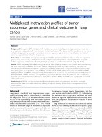
ICG is a water-soluble cyanine dye that was granted FDA approval in 1959 (Fig. 1a) and has multiple
applications for fluorescence-guided surgery in various specialties25–28. The molecular weight of ICG is
774.96 grams/mol. It is amphiphilic, and it has a peak excitation (λ ex) and peak emission (λ em) wavelength at 778 and 832 nm, respectively (Fig. 1b). EC-17 is a folate-fluorescein isothiocyanate (folateFITC) conjugate (Fig. 1d) with a molecular weight of 916.83 grams/mol. Its peak λ ex and λ em wavelengths
are 494 nm and 521 nm, respectively (Fig. 1e).
C57bl/6 mice (n = 15) were injected with either the TC1 (7 mice) or LLC (8 mice) cell line subcutaneously in the right flank. NOD.Cg-Prkdcscid Il2rgtm1Wjl/SzJ mice (n = 15) were injected with either
the KB cell line (7 mice) or IGROV cell line (8 mice). Once the tumors reached 500 mm3, the animals
were injected via tail vein with either 5 mg/kg of ICG (24 hours prior to surgery) or 0.1 mg/kg of EC-17
(4 hours prior to surgery). Once the flanks were exposed, two imaging devices (digital imaging and
spectroscopy) were utilized to assist in the surgical resection of the tumor as previously described1,6,13,24.
First, the surgeon reviewed optical images from the digital camera system and subjectively decided if
the tumor was fluorescent. In 15 animals that received ICG, the surgeon identified 15 out of 15 (100%)
animals to have fluorescent flank tumors (Fig. 1c). In 15 animals that received EC-17, the surgeon identified 15 out of 15 (100%) animals to have fluorescent flank tumors (Fig. 1f). When asked to subjectively
rank the tumors based on the degree of fluorescence, the surgeon could not identify any significant
difference.
Scientific Reports | 5:16208 | DOI: 10.1038/srep16208
3
www.nature.com/scientificreports/
Figure 1. (a) Chemical structure of ICG. (b) Absorbance spectrum of ICG. Y-axis is measured in arbitrary
units. (c) Fluorescence and bright field image of a C57BL/6 mouse bearing a LLC flank tumor injected
intravenously with with ICG. (d) Chemical structure of EC-17. (e) Absorbance spectrum of EC-17. Y-axis is
measured in arbitrary units. (f) Fluorescence and bright field image of an excised KB tumor from a C57BL/6
mouse that has been injected intravenously with EC-17.
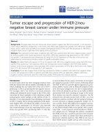
Figure 2. (a) Residual fluorescent tumor foci detected in the surgical bed by fluorescence imaging after
macroscopic tumor resection. (b) H&E staining was performed on the tumor margin and was confirmed by
a pathologist to contain tumor cells.
The surgeon then removed the tumors using standard-of-care palpation and gross visual inspection
to determine tumor margins. All tumors were imaged ex vivo using spectroscopy, luminometry and
digital imaging. In all cases, the surgeon felt the entire tumor had been removed. Then, spectroscopy
and digital imaging were used to confirm that the wound margins were tumor-free in vivo as previously
described1,2,24. Using both approaches, intraoperative imaging identified 3 out of 15 (20%) animals who
received ICG had residual disease, which required further resection (Fig. 2a). This residual tumor tissue
was removed, preserved in formalin and sectioned in paraffin blocks. It was stained with Hemotoxylin
and Eosin (H&E) (Fig. 2b). Similarly, this approach discovered 2 out of 15 (13%) mice who received
Scientific Reports | 5:16208 | DOI: 10.1038/srep16208
4
www.nature.com/scientificreports/
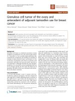
Figure 3. (a) Serial dilution concentrations of ICG and EC-17. (b) SBR vs ICG concentration. Y-axis
is measured in arbitrary units. Error bars are reported as standard deviations (STD). (c) SBR vs. EC-17
concentrations. Dilutions were prepared with phosphate buffered saline. Y-axis is measured in arbitrary
units. Error bars are reported as standard deviations (STD).
EC-17 to have positive margins that required further resections. All residual fluorescence was histologically confirmed to contain tumor cells by a pathologist. The wounds were surgically closed, and the
animals were monitored for recurrence for 6 weeks. None of the animals recurred. In total, intraoperative
imaging had 100% true positive and a 0% false positive rate for detecting fluorescence in non-cancerous
tissues.
Fine point discrimination between intraoperative imaging techniques. Although the surgeon
could not differentiate the degree of fluorescence by visual inspection, current imaging technologies can
provide quantitative measurements of tumor fluorescence. To identify the optimal method to quantitate signal-to-background (SBR) ratio of the tumor to the surrounding normal tissue, we compared the
three intraoperative imaging technologies: spectroscopy6, luminescence and optical imaging13. Our goal
was to determine which approach could provide superior sensitivity to small incremental differences in
fluorophore concentrations.
To standardize the quantity of ICG, ten serial dilutions (range: 7.26 × 10⁻⁶–2.81 × 10⁻⁶ M) were prepared in 96-well culture plates (Fig. 3a). The background signal was measured from a well with an equal
volume of PBS. Each method (spectroscopy, luminescence, digital imaging) was used to generate a SBR
ratio from each well (Fig. 3b).
From the digital images, the investigator subjectively identified fluorescence from 9 wells (range
2.81 × 10⁻⁶–7.26 × 10⁻⁶ M). Spectroscopy, luminescence and digital capture produced ln[SBR] ratios
ranging from 0.4–6.8, 0.0–2.4, and 0.2–2.6, respectively (n = 10). Intra-experiment variability between
replicates was small; the signal varied by 4.1% ± 4.0% in each experiment (n = 3). The SBR ratio was linear and strongly correlated with concentration for all three methods: spectroscopy, luminescence, digital
imaging (r2 = 0.92, 0.97 and 0.97, respectively).
Spectroscopy was the most sensitive at detecting small differences in the tracer concentrations. For
each 7.33 × 10⁻⁷ M increase in ICG concentration, the fluorescence quantified by spectroscopy changed
6,886.6 ± 924.1 arbitrary units (au). For the digital imaging system images, each 7.33 × 10⁻⁷ M increase in
ICG, the fluorescence increased by 6.7 ± 4.2 au. For the digital imaging, for each 7.33 × 10⁻⁷ M increase
in ICG, the fluorescence increased by 63.4 ± 2.3 au. Of note, the spectrometer generated a maximum
Ln[SBR] of 6.8 for ICG, whereas the luminometer and ROI software gave a maximum Ln[SBR] of less
than 3. The spectrometer’s fluorescent image sensor was saturated at the highest ICG concentration.
To evaluate EC-17, we created 10 dilutions over the same concentration range as ICG. Luminescence
and digital capture techniques produced Ln[SBR] ratios ranging from 2.4–4.5 and 1.8–3.3, respectively
in the 10 wells (Fig. 3c) (n = 10). Between the triplicate measurements in each technique, the signal was
Scientific Reports | 5:16208 | DOI: 10.1038/srep16208
5
www.nature.com/scientificreports/
Figure 4. (a) KB cells incubated with 18.4 μ M EC-17 under 200× magnification fluoresce upon excitation
by 490 nm light. (b) KB cells incubated with 18.4 μ M EC-17. Black wells containing increasing logarithmic
values of cells were imaged, and the signal was quantified using the luminometer and ROI software. The
pseudocolor map shows decreasing areas of detectable fluorescence, and some glare is present in all wells.
Y-axis is measured in arbitrary units. Error bars are reported as standard deviations (STD). (c) RCC10 cells
incubated 18.4 μ M EC-17. Black wells containing increasing logarithmic values of cells were imaged, and the
signal was quantified using the luminometer and ROI software. Y-axis is measured in arbitrary units. Error
bars are reported as standard deviations (STD).
within 6.1% ± 4.8% of the same value, thus there was strong data fidelity across experiments. The signal
was linearly correlated with EC-17 concentration using luminescence and digital imaging (r2 = 0.94 and
0.90, respectively).
In summary, digital imaging, luminescence and spectroscopy can quantify fluorescence. Signal intensity is directly proportional to tracer molarity. Spectroscopy appeared to be the most sensitive technique
for identifying small incremental changes in fluorophore concentrations.
Comparison of imaging techniques in vitro. In order to further evaluate each system, we
repeated our studies in vitro using tumor cells treated with EC-17. KB or RCC10 cells were incubated
with 18.4 μ M EC-17 for 45 minutes, washed and plated 1 cell/3.3 × 10−3m2 − 1 cell/3.3 × 10−9m2 in 96
well plates (3.3 × 10⁻³m2/well). The culture plates were then imaged using microscopy, digital imaging and the luminometer (Fig. 4a–c) SBRs were generated using background signal from wells with
non-fluorescent tumor cells. In a 96-well plate, the smallest quantity of cells the luminometer and optical
imaging could detect was similar and was between 104 and 105 cells. Both quantification systems detected
a more intense signal from KB cells (Fig. 4b) than RCC10 cells (Fig. 4c), reflecting greater uptake of
the probe in the folate-receptor positive KB cells. At 1 million KB tumor cells/well, optical imaging and
the luminometer had SBR ratios of 6.1 ± 0.2 and 4.3 ± 0.4 (p = 0.001), respectively (n = 3). At 1 million
RCC10 tumor cells/well ROI and the luminometer had SBR ratios of 4.3 ± 0.3 and 3.3 ± 0.1 (p = 0.004),
respectively (n = 3).
The digital ROI analysis was more sensitive than the luminometer for identifying trace levels of fluorescence from small quantities of fluorescent tumor cells. The digital capture software identified 104 cells/
well, whereas the luminometer was sensitive to 105 cells/well. Furthermore, when we compared the SBRs
for both devices at 106 cells/well, the fluorescent signal was higher with the digital ROI method. The SBR
generated for 106 KB cells/well co-cultured in EC-17 was larger when using ROI than when using the
luminometer (6.1 vs. 4.3) or the ROI software (4.3 vs. 3.3).
Quantification of tumor fluorescence in murine models. To test the quantification and sensitivity of each imaging technique in vivo, we established murine flank tumors and injected tumor-bearing
mice IV with either EC-17 or ICG. We tested non-small cell lung cancer (TC1, LLC), cervical carcinoma
(KB) and ovarian (IGROV) cell lines. NOD.Cg-Prkdcscid Il2rgtm1Wjl/SzJ mice were injected with KB or
IGROV cells into the right flank, and C57bl/6 mice were injected with TC1 or LLC cells into the right
flank. The tumors reached a mean volume of 500 mm3 after about 3 weeks. The mice were injected with
either 0.1 mg/kg of EC-17 or 5 mg/kg of ICG IV and the tumors were resected. The tumors were cut into
approximately 0.5 cm3 portions and placed in a black 96-well plate for fluorescence imaging (Fig. 5a).
Scientific Reports | 5:16208 | DOI: 10.1038/srep16208
6
www.nature.com/scientificreports/
Figure 5. (a) Brightfield, fluorescent, and pseudocolor images of LLC flank tumors from BL6 mice injected
with ICG. (b) Signal-to-background ratio of TC1 murine flank tumors imaged with ICG. Error bars
are reported as standard deviations (STD). (c) Signal-to-background ratio of LLC murine flank tumors
imaged with ICG. Error bars are reported as standard deviations (STD). (d) Signal-to-background ratio
of KB murine flank tumors imaged with EC17. Error bars are reported as standard deviations (STD).
(e) Signal-to-noise ratio of IGROV murine flank tumors imaged with EC17. Error bars are reported as
standard deviations (STD). (f) CT scan, bisected nodule of human adenocarcinoma patient, bright field, and
fluorescent,. (g) Signal-to-background ratio of human adenocarcinoma imaged with ICG. Error bars are
reported as standard deviations (STD).
Muscle adjacent to the tumor was used as the negative control (top row, right). Background fluorescent
readings were taken from these specimens. SBRs were generated and each measurement was repeated
five times.
The TC1 tumors generated the highest SBR reading (49.9 ± 8.7, n = 8) measured when ICG fluorescence was quantified with the spectrometer (Fig. 5b). The luminometer was more sensitive to small
amounts of tumor fluorescence when compared to ROI analysis but less sensitive than spectroscopy. The
LLC cell line was the one notable exception where digital imaging had a higher SBR than the luminometer (5.2 ± 1.5 vs. 3.0 ± 1.3, p = 0.003, n = 8) (Fig. 5c). The KB cell line (Fig. 5d) produced a tumor with
a SBR significantly brighter with the luminometer than with the digital analysis software (7.3 ± 3.9 vs.
3.5 ± 1.4, p = 0.01, n = 8). The ovarian flank tumor generated similar SBRs between the two quantification techniques (Fig. 5e). The average SBR of the spectrometer, luminometer and digital analysis was
41.7 ± 11.5, 5.1 ± 1.8 and 4.1 ± 0.9 (p = 0.0001), respectively (n = 3).
Together, these data suggest that it is possible to quantify the fluorescent signal from murine tumors
when the animals have been injected with EC-17 or ICG. Quantification of tumor fluorescence is highly
dependent on the imaging technology and must be done in concert with an ex vivo standard curve. This
high throughput murine model is reliable and was easy to use. Several cancer types were successfully
visualized with fluorescence imaging. It is an inexpensive strategy to investigate the quantification of
fluorescent tumors.
Quantification of tumor fluorescence in human clinical trial. Lastly, to test the ability of our
fluorescent technologies to detect optical contrast agents in humans, we investigated 3 human patients
who were injected with ICG 24 hours prior to the removal of a primary pulmonary adenocarcinoma as
part of an ongoing clinical trial2. A representative patient is shown in Fig. 5f.
The test patient was a 68 year old male with a 1.5 cm right upper lobe non-small cell lung cancer
(Fig. 5f). The patient had no evidence of systemic disease, thus he was scheduled for surgery with intraoperative imaging assistance. The patient was injected IV with 5 mg/kg of ICG 24 hours prior to surgery.
During surgery, the patient’s chest was opened and the nodule was resected (Fig. 5f). Intraoperatively,
the tumor was subjectively fluorescent on the digital imaging monitor when reviewed by the surgeon.
A piece of the tumor (3.6 grams, wells on left) and surrounding latissimus muscle (wells on right) were
harvested for more detailed quantification of the fluorescence. The latissimus muscle was used as a background signal. SBRs were generated and each measurement was repeated in triplicate. The tumor was
portioned and quantified using the luminometer, spectrometer and digital analysis (Fig. 5g).
The spectrometer generated the highest SBR (13.0 ± 4.6, n = 8) compared to the luminometer
(5.4 ± 1.6, n = 8) and digital analysis (3.7 ± 0.8, n = 8). Using the standard curve of ICG generated in
Fig. 3, we then attempted to quantify the concentration of the ICG in the tissue. According to our prior
Scientific Reports | 5:16208 | DOI: 10.1038/srep16208
7
www.nature.com/scientificreports/
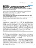
Figure 6. (a) Fluorescent images of 3 cm ICG balloon phantoms submerged under various depths of
liquefied butter. The Flocam ROI SBR data was gathered from these images. (b) Flocam ROI SBR vs. depth
of tumor. ICG phantom sizes range from 1–5 cm. (c) Spectropen SBR vs. depth of tumor. ICG phantom sizes
ranges from 1 cm–5 cm.
calculations, we estimate the [ICG] in the tumor tissue to be 5.5 × 10⁻⁶ M. These data indicate that the
optical contrast agents may accumulate at up to 13-fold higher concentrations in tumors than healthy
adjacent tissue. The spectrometer was the most sensitive at identifying fluorescence, however, the luminometer and digital analysis did produce distinguishable differences in SBR.
Signal detection as a function of depth. One of the shortcomings of intraoperative cancer detection is the limited ability of imaging techniques to locate tumors at increasing tissue depths. In order
to test the ability of each imaging technique to detect fluorescence at varying depths of penetration, we
imaged ICG phantoms in a semi-solid tissue model of adipose tissue. Balloons were filled with 3.23 μ M
ICG until they reached 1.0, 2.0, 3.0, 4.0, and 5.0 cm in diameter. They were submerged under semi-solid
butter from 0 to 3 cm at 0.5 cm intervals (Fig. 6a). The digital capture software and spectrometer were
used to quantify fluorescent signal from the phantoms.
Both digital imaging ROI and the spectrometer produced higher SBRs for un-submerged phantoms
compared to phantoms embedded in 3 cm of semi-solid butter (6.0 ± 0.5 vs. 2.4 ± 0.5 (p = 0.00001) and
261.5 ± 0.1 vs. 7.0 ± 3.4 (p = 0.00001), respectively (n = 3). The spectrometer generated a maximum SBR
value of 261.5 and the digital analysis software generated a maximum SBR of 6.7 (Fig. 6b,c). Likewise, the
5 cm phantom consistently had the highest SBR when measured with the spectrometer, and the different
sized phantoms had distinct differences. With the spectrometer, however, all phantoms smaller than
4.0 cm in diameter have similar SBRs (Fig. 6b). The 5.0 cm phantom generally had the highest signal.
There was a large decrease in the SBRs for each phantom when the depth of penetration increased from
2.5 cm to 3.0 cm. There was a larger gap between the signals from different-sized tumors when compared
to the spectrometer values.
Thus, we found that both the size of the phantom and depth below the surface had important effects
on the measured fluorescent signal. Using the model of adipose tissue, we found that although the spectroscopy signals were amplified in comparison to the digital capture system, they both maintained the
same trend.
Discussion
Non-specific and receptor targeted fluorophores are being used in combination with various fluorescent
imaging systems for intraoperative tumor visualization. This study compares three imaging modalities–
spectroscopy, luminometry and digital imaging – to obtain quantitative data from tumor fluorescence
during cancer surgery (Table 1). We found that all three imaging devices are clinically feasible and provide useful information. For detecting both small numbers of cancer cells and detecting tumors deeper
Scientific Reports | 5:16208 | DOI: 10.1038/srep16208
8
www.nature.com/scientificreports/
Imaging
Approach
Advantages
Disadvantages
Considerations
Suggested Uses
Spectroscopy
Most sensitive to small quantities of disease.
Can identify minimal disease up to 3 cm
depth of penetration.
Limited field of
view.
Provides 1800 data
points per reading,
thus data processing is
time consuming.
Identifying small
areas of residual
disease.
Luminometry
Precise reproducible measurements.
Cannot be used
in vivo.
Can only evaluate
small regions of tissue.
Not useful for
clinical application.
Digital imaging
Wide field of view.
Subject to user
bias.
Sensitivity depends
on quality of charge
coupled device.
Useful for broad
exploration of the
wound and body
cavity.
Table 1. Imaging modality comparison.
in tissues (up to 3 cm), spectroscopy is more sensitive and has superior resolution than luminometry and
digital imaging. With increasing residual disease > 106 cells), spectroscopy has no advantage over digital
imaging because the magnitude of fluorescence from the cancer cells overwhelms the resolution of spectroscopy, and this approach is slow and time-consuming. In these settings, digital imaging has substantial
advantages. It provides real-time, high-resolution images to the surgeon, which allows for intraoperative
decisions. Although it is not as sensitive for identifying small quantities of disease, it may be the most
useful approach for routine cancer operations. Ultimately, an approach using digital imaging to survey
a large region and then spectroscopy to verify targeted areas of interest may be the best combination.
In this study, we tested our 3 imaging systems using 2 commercially used fluorophores: ICG and
EC-17. EC-17’s extinction coefficient (7.5 × 10⁴ M−1cm−1) is two-fold that of ICG (4 × 10⁴ M−1cm−1),
thus it was useful to test each system with two different fluorophores. While EC-17 is a receptor-targeted
fluorophore, ICG does not specifically target cancer cells. ICG accumulates by the enhanced permeability
and retention (EPR) effect in solid cancers. When injected systemically, ICG passively accumulates in
tumors due to wide blood vessel fenestrations and defective endothelial cells29. The ICG is then retained
due to its molecular size, shape, differences in tumor oncotic pressure and poor lymphatic angiogenesis30–32. Spectroscopy, as predicted, is the most sensitive at detecting areas of minimal fluorescence. We
could detect 0.1 μ M ICG in vitro, whereas the lowest threshold for digital imaging was closer to 1 μ M.
Furthermore, spectroscopy was superior at fine-point discrimination of small increases in fluorescence.
Of note, in our murine model, we noticed that a standard curve was useful in estimating the relative
fluorescence of tumor tissues and normal background organs. Thus, in clinical situations where a surgeon
may be inspecting a close margin, spectroscopy provides the ideal approach for detailed interrogation to
detect residual cancer cells. Molecular imaging can detect up to 50% more residual tumor deposits than
traditional margin detection, generating a 50% increased recurrence-free survival rate33,34. Moreover,
multiple-organ recurrence is less likely to occur with fluorescence-guided surgery35. Our study confirms
the benefit of fluorescent-guided tumor resection over standard macroscopic resection, in which up to
85% residual tumor deposits are detected with fluorescent imaging versus only 9% for residual nodules
captured without fluorescence imaging24.
We also considered the ability of each imaging technology to identify small (1 cm) and large (5 cm)
ICG tumor phantoms located deeper in simulated tissue. With increasing tumor depth and tissue density,
scattering and absorption limit the fluorescence that can be measured at the surface. Again, we found
spectroscopy was capable of identifying low levels of fluorescence from 2 cm tumors as far as 5 cm from
the surface of the phantom. Digital imaging, on the other hand, could not measure detectable signal
beyond 2.5 cm below the phantom surface. For bigger tumors, however, both spectroscopy and digital
imaging could identify the location of the fluorescence. Clinically, spectroscopy may have greater value in
the localization of small tumors in solid organs (e.g. subcentimeter pulmonary nodules, hepatic colorectal metastases) that may be precarious to cut into due to bleeding or loss of tissue. For larger tumors that
have a high fluoroescence, digital imaging may be sufficient.
Finally, with regards to ease of use, the digital imaging approach was significantly better than spectroscopy or the luminometer. The ability of a surgeon to visualize the fluorescence in a wide field of view
provides rapid interrogation of an entire organ surface. We found we could inspect up to 100 mm2 of an
organ surface within an integration time of 1 second. Spectroscopy, on the other hand, only allowed us
to examine 1 mm2 per scan and this limits its practical application.
There are a number of factors that must be taken into account in the interpretation of the study.
First, we selected three representative imaging devices: spectroscopy, luminometer and digital imaging.
Each device has significantly different light sources (lasers versus light emitting diodes), illumination
methods, detectors and detection bandwidths. The spectroscopic device is a hand-held device that, to our
knowledge, is the only one available for clinical use. The luminometer is a mid-range device and more
expensive machines will have greater resolution power. Digital imaging charged coupled devices also
span a wide range of resolution. We constructed a device in the mid-price range, but the high-resolution
cameras that are 3 to 5 fold more expensive may begin to match spectroscopic devices.
Scientific Reports | 5:16208 | DOI: 10.1038/srep16208
9
www.nature.com/scientificreports/
The optical properties of the fluorophores must also be considered. Longer wavelength NIR fluorophores will be better observed in tissues as they are less subject to light scattering and absorption and
out of the range of tissue autofluorescence. The brightness of the probe, related to both the extinction
coefficient and the quantum yield is also important. Finally, we draw attention to the heterogeneity of
human tumors. Due to complex tumor heterogeneity and variable tumor perfusion and drainage, the
uptake of contrast agent is likely to be the major limiting factor in intraoperative imaging. Thus, it is
challenging to draw broad conclusions about devices without controlling the model, which is not possible
with human tumors. Further human trials will be needed to explore these issues.
Based on our observations, spectroscopy and digital imaging are likely to be the optimal approach
to intraoperative imaging, and more useful than luminometry. The tumors need to be imaged in a dark
environment under special conditions, which is often impractical in the confines of the operating room.
Future technologies that combine spectroscopy and digital imaging will have major advantages. This
approach will allow for detailed quantitative information about the fluorescence in and around the
tumor. It will also provide a wide field of view and practical information to the surgeon for ease of use
and clinical utility. The most useful strategy will be to use digital imaging to scan large regions of body
cavity and the wound. Then, for more detailed analysis of margins and lymph nodes, it will be necessary
to utilize the spectroscopic device.
References
1. Holt, D. et al. Intraoperative near-infrared imaging can distinguish cancer from normal tissue but not inflammation. PLoS One
9, e103342, doi: 10.1371/journal.pone.0103342 (2014).
2. Okusanya, O. T. et al. Intraoperative Near-Infrared Imaging Can Identify Pulmonary Nodules. Ann Thorac Surg. doi: 10.1016/j.
athoracsur.2014.05.026 (2014).
3. Van Dam, G. M. et al. Intraoperative tumor-specific fluorescence imaging in ovarian cancer by folate receptor-alpha targeting:
first in-human results. Nat Med. doi: 10.1038/nm.2472nm.2472 (2011).
4. Van der Vorst, J. R. et al. Near-infrared fluorescence-guided resection of colorectal liver metastases. Cancer 119, 3411–3418, doi:
10.1002/cncr.28203 (2013).
5. Hutteman, M. et al. Optimization of near-infrared fluorescent sentinel lymph node mapping for vulvar cancer. Am J Obstet
Gynecol 206, 89 e81–85, doi: 10.1016/j.ajog.2011.07.039 (2012).
6. Mohs, A. M. et al. Hand-held Spectroscopic Device for In Vivo and Intraoperative Tumor Detection: Contrast Enhancement,
Detection Sensitivity, and Tissue Penetration. Anal Chem, doi: 10.1021/ac102058k (2010).
7. Haka, A. S. et al. Diagnosing breast cancer by using Raman spectroscopy. Proc Natl Acad Sci USA 102, 12371–12376, doi:
10.1073/pnas.0501390102 (2005).
8. Haka, A. S. et al. In vivo margin assessment during partial mastectomy breast surgery using raman spectroscopy. Cancer Res 66,
3317–3322, doi: 10.1158/0008-5472.CAN-05-2815 (2006).
9. Haka, A. S. et al. Diagnosing breast cancer using Raman spectroscopy: prospective analysis. J Biomed Opt 14, 054023, doi:
10.1117/1.3247154 (2009).
10. Wagnieres, G. A., Star, W. M. & Wilson, B. C. In vivo fluorescence spectroscopy and imaging for oncological applications.
Photochem Photobiol 68, 603–632 (1998).
11. Zellweger, M. et al. In vivo autofluorescence spectroscopy of human bronchial tissue to optimize the detection and imaging of
early cancers. J Biomed Opt 6, 41–51 (2001).
12. Ntziachristos, V. Optical imaging of molecular signatures in pulmonary inflammation. Proc Am Thorac Soc43 6, 416–418, doi:
10.1513/pats.200901-003AW (2009).
13. Okusanya, O. T. et al. Small Portable Interchangeable Imager of Fluorescence for Fluorescence Guided Surgery and Research.
Technol Cancer Res Treat, doi: 10.7785/tcrt.2012.500400 (2013).
14. Gibbs-Strauss, S. L., Rosenberg, M., Clough, B. L., Troyan, S. L. & Frangioni, J. V. First-in-human clinical trials of imaging
devices: an example from optical imaging. Conf Proc IEEE Eng Med Biol Soc 2009, 2001–2004, doi: 10.1109/IEMBS.2009.5333429
(2009).
15. Troyan, S. L. et al. The FLARE() Intraoperative Near-Infrared Fluorescence Imaging System: A First-in-Human Clinical Trial in
Breast Cancer Sentinel Lymph Node Mapping. Ann Surg Oncol 16, 2943–2952, doi: 10.1245/s10434-009-0594-2 (2009).
16. Liu, Y. et al. Hands-free, wireless goggles for near-infrared fluorescence and real-time image-guided surgery. Surgery 149,
689–698, doi: S0039-6060(11)00063-810.1016/j.surg.2011.02.007 (2011).
17. Collins, T. J. ImageJ for microscopy. Biotechniques 43, 25–30, doi: 000112517 (2007).
18. Foo, J. L., Miyano, G., Lobe, T. & Winer, E. Tumor segmentation from computed tomography image data using a probabilistic
pixel selection approach. Comput Biol Med 41, 56–65, doi: 10.1016/j.compbiomed.2010.11.006S0010-4825(10)00163-0 (2011).
19. Predina, J. et al. Changes in the local tumor microenvironment in recurrent cancers may explain the failure of vaccines after
surgery. Proc Natl Acad Sci USA 110, E415–424, doi: 10.1073/pnas.1211850110 (2013).
20. Lin, K. Y. et al. Treatment of established tumors with a novel vaccine that enhances major histocompatibility class II presentation
of tumor antigen. Cancer Res 56, 21–26 (1996).
21. Eagle, H. Propagation in a fluid medium of a human epidermoid carcinoma, strain KB. Proc Soc Exp Biol Med 89, 362–364
(1955).
22. Krieg, M. et al. Up-regulation of hypoxia-inducible factors HIF-1alpha and HIF-2alpha under normoxic conditions in renal
carcinoma cells by von Hippel-Lindau tumor suppressor gene loss of function. Oncogene 19, 5435–5443, doi: 10.1038/sj.
onc.1203938 (2000).
23. Benard, J. et al. Characterization of a human ovarian adenocarcinoma line, IGROV1, in tissue culture and in nude mice. Cancer
Res 45, 4970–4979 (1985).
24. Madajewski, B. et al. Intraoperative near-infrared imaging of surgical wounds after tumor resections can detect residual disease.
Clin Cancer Res 18, 5741–5751, doi: 10.1158/1078-0432.CCR-12-11881078-0432.CCR-12-1188 (2012).
25. Aydogan, F. et al. Excision of Nonpalpable Breast Cancer with Indocyanine Green Fluorescence-Guided Occult Lesion
Localization (IFOLL). Breast Care (Basel) 7, 48–51, doi: 10.1159/000336497 (2012).
26. Metildi, C. A. et al. Fluorescence-guided surgery allows for more complete resection of pancreatic cancer, resulting in longer
disease-free survival compared with standard surgery in orthotopic mouse models. J Am Coll Surg 215, 126–135, doi: 10.1016/j.
jamcollsurg.2012.02.021S1072-7515(12)00242-6 (2012).
27. Rossi, E. C., Ivanova, A. & Boggess, J. F. Robotically assisted fluorescence-guided lymph node mapping with ICG for gynecologic
malignancies: a feasibility study. Gynecol Oncol 124, 78–82, doi: 10.1016/j.ygyno.2011.09.025S0090-8258(11)00795-5 (2012).
Scientific Reports | 5:16208 | DOI: 10.1038/srep16208
10
www.nature.com/scientificreports/
28. Tobis, S. et al. Near infrared fluorescence imaging after intravenous indocyanine green: initial clinical experience with open
partial nephrectomy for renal cortical tumors. Urology 79, 958–964, doi: 10.1016/j.urology.2011.10.016S0090-4295(11)02494-0
(2012).
29. Matsumara Y. & Maeda H. A new concept for macromolecular therapeutics in cancer chemotherapy: mechanism of tumoritropic
accumulation of proteins and the antitumor agent smancs. Cancer Res 46, 6387–6392 (1986).
30. Ishizawa, T. et al. Mechanistic background and clinical applications of indocyanine green fluorescence imaging of hepatocellular
carcinoma. Ann Surg Oncol 21, 440–8, doi: 10.1245/s10434-013-3360-4 (2014).
31. Shin, E. H. et al. Membrane potential mediates the cellular binding of nanoparticles. Nanoscale 7, 5879–86, doi: 10.1039/
c3nr01667f (2013).
32. Jiang, J. X. et al. Optimization of the enhanced permeability and retention effect for near-infrared imaging of solid tumors with
indocyanine green. Am J Nucl Med Mol Imaging 5, 390–400 (2015).
33. Keating, J. J. et al. Intraoperative molecular imaging of lung adenocarcinoma can identify residual tumor cells at the surgical
margins. Mol Imaging Biol, doi: 10.1007/s11307-015-0878-9 (2015).
34. Atallah, I. et al. Near-infrared fluorescence imaging-guided surgery improves recurrence-free survival rate in novel orthotopic
animal model of head and neck squamous cell carcinoma. Head Neck, doi: 10.1002/hed.23980 (2014).
35. Murakami, T. et al. Improved disease-free survival and overall survival after fluorescence-guided surgery of liver metastasis in
an orthotopic nude mouse model. J Surg Oncol, doi: 10.1002/jso.23986 (2015).
Acknowledgements
I would like to thank Pratik Bhojnagarwala, MS, for his contribution to this manuscript. This work was supported by a Transdisciplinary Awards Program in Translational Medicine and Therapeutics-Translational
Biomedical Imaging Core (TAPITMAT-TBIC) grant through UL1RR024134 (SS and EJD).
Author Contributions
Participated in data collection: R.P.J., J.X.J., E.M.D., J.J.K., O.T.O., S.S. and D.H. Helped with statistics:
R.P.J., S.S. and S.P.A. Designed the experiments: S.S. Performed the experiments: R.P.J., J.X.J., S.P.A. and
E.J.D. Analyzed the data: R.P.J., S.N., P.S.L. and S.S. Wrote the paper:R.P.J., J.J.K. and S.S. All authors
reviewed the manuscript.
Additional Information
Competing financial interests: S.N. is a consultant for Spectropath, Inc., a startup company to develop
advanced instrumentation and nanoparticle contrast agents.
How to cite this article: Judy, R. P. et al. Quantification of tumor fluorescence during intraoperative
optical cancer imaging. Sci. Rep. 5, 16208; doi: 10.1038/srep16208 (2015).
This work is licensed under a Creative Commons Attribution 4.0 International License. The
images or other third party material in this article are included in the article’s Creative Commons license, unless indicated otherwise in the credit line; if the material is not included under the
Creative Commons license, users will need to obtain permission from the license holder to reproduce
the material. To view a copy of this license, visit />
Scientific Reports | 5:16208 | DOI: 10.1038/srep16208
11