progress in prediction and interpretation of clinically relevant metabolic drug drug interactions a minireview illustrating recent developments and current opportunities
Bạn đang xem bản rút gọn của tài liệu. Xem và tải ngay bản đầy đủ của tài liệu tại đây (648.73 KB, 14 trang )
Curr Pharmacol Rep
DOI 10.1007/s40495-017-0082-5
MOLECULAR DRUG DISPOSITION (M HU, SECTION EDITOR)
Progress in Prediction and Interpretation of Clinically Relevant
Metabolic Drug-Drug Interactions: a Minireview Illustrating
Recent Developments and Current Opportunities
Stephen Fowler 1 & Peter N. Morcos 2 & Yumi Cleary 1 & Meret Martin-Facklam 1 &
Neil Parrott 1 & Michael Gertz 1 & Li Yu 2
# The Author(s) 2017. This article is published with open access at Springerlink.com
Abstract
Purpose of Review This review gives a perspective on the
current Bstate of the art^ in metabolic drug-drug interaction
(DDI) prediction. We highlight areas of successful prediction
and illustrate progress in areas where limits in scientific
knowledge or technologies prevent us from having full
confidence.
Recent Findings Several examples of success are highlighted.
Work done for bitopertin shows how in vitro and clinical data
can be integrated to give a model-based understanding of
pharmacokinetics and drug interactions. The use of interpolative predictions to derive explicit dosage recommendations for
untested DDIs is discussed using the example of ibrutinib, and
the use of DDI predictions in lieu of clinical studies in new
drug application packages is exemplified with eliglustat and
alectinib. Alectinib is also an interesting case where dose adjustment is unnecessary as the activity of a major metabolite
compensates sufficiently for changes in parent drug exposure.
This article is part of the Topical Collection on Molecular Drug
Disposition
Electronic supplementary material The online version of this article
(doi:10.1007/s40495-017-0082-5) contains supplementary material,
which is available to authorized users.
* Stephen Fowler
1
Pharmaceutical Research and Early Development, Roche Innovation
Centre Basel, F. Hoffmann-La Roche Ltd., Grenzacherstrasse 124,
CH-4070 Basel, Switzerland
2
Pharmaceutical Reseach and Early Development, Roche Innovation
Center New York, F. Hoffmann-La Roche Ltd., 430 East 29th Street,
New York City, NY, USA
Examples where Bunusual^ cytochrome P450 (CYP) and
non-CYP enzymes are responsible for metabolic clearance have
shown the importance of continuing to develop our repertoire
of in vitro regents and techniques. The time-dependent inhibition assay using human hepatocytes suspended in full plasma
allowed improved DDI predictions, illustrating the importance
of continued in vitro assay development and refinement.
Summary During the past 10 years, a highly mechanistic understanding has been developed in the area of CYP-mediated
metabolic DDIs enabling the prediction of clinical outcome
based on preclinical studies. The combination of good quality
in vitro data and physiologically based pharmacokinetic modeling may now be used to evaluate DDI risk prospectively and are
increasingly accepted in lieu of dedicated clinical studies.
Keywords Drug-drug interaction . Prediction .
Physiologically based pharmacokinetic model . Metabolism .
Regulatory submission . Cytochrome P450
Introduction
Quantification of a drug-drug interaction (DDI) effect in a
man is the basis for explicit dose recommendation in drug
labels to minimize the risk of adverse events or reduced
efficacy, thereby supporting appropriate use of the drug. It
is therefore essential that such quantitative DDI assessments are made with confidence. There has been a steady
development of in vitro assays and the reagents available
for the study of drug metabolism and metabolic enzyme
inhibition. This, combined with advances in our capability
to extrapolate in vitro data to in vivo, has brought us past
a Btipping point^ such that applying a model-based synthesis of the available data has become normal in drugdrug interaction assessments [1–5••]. Simple static
Curr Pharmacol Rep
models, built upon DDI studies reaching back to the
1970s [6•], still find utility in early drug discovery where
there are very limited data available for the drug candidate. However, the greatest DDI effects are observed
where the metabolism of an orally administered drug is
substantially inhibited in the first pass metabolism, potentially in both the intestine and liver. The combination of
increased drug reaching the systemic circulation as well as
reduced systemic clearance will result in a significantly
higher exposure (area under the plasma concentrationtime curve [AUC]) than when inhibition of systemic
clearance alone is considered. An example of this can be
seen when comparing the DDI effect of ketoconazole on
alprazolam and midazolam which are low and high clearance cytochrome P450 (CYP) 3A substrates, respectively.
In the recent study of Boulenc et al., peak concentration
(Cmax) for alprazolam and midazolam were increased by
1.18- and 4.21-fold, whereas AUC was increased by 2.63and 16.95-fold, respectively, when co-administered with
multiple once-daily doses of 400 mg ketoconazole [7].
Mechanistic static models have significantly extended
mathematical model usage, by incorporating additional
considerations such as intestinal metabolism, enzyme induction, and enzyme inactivation [8, 9]. Nevertheless,
these mathematical models cannot capture the full dynamic nature of drug metabolism in vivo since only a fixed
concentration of inhibitor is considered. For example,
DDI effects on simultaneous co-administration versus
staggered dosing situations may be different, especially
when the interacting drugs have short half-lives and high
first pass metabolism. Details of the different approaches
to DDI prediction were recently described in a
Pharmaceutical Industry Innovation and Quality working
group publication from Bohnert et al. and will not be
discussed further in this review [10•].
A more powerful approach to DDI prediction can be
taken using physiologically based pharmacokinetic
(PBPK) modeling, especially when human pharmacokinetic data are available. Validated PBPK models allow
high confidence in prospective DDI predictions [1]. This
application of modeling and simulation has been reflected
in the regular inclusion of PBPK model information into
new drug application (NDA) submissions [4, 5••] and recent use in final drug product labeling text with explicit
dosage recommendations (see examples below). Similarly,
the simulations may support selection of dose strengths to
be developed. In order to generate a well-validated PBPK
model for a drug, a large amount of data need to be collected. Such data include pharmacokinetics of drug substance and metabolites, drug solubility and permeability,
plasma protein binding, contributions of individual enzymes to hepatic and extrahepatic clearance, enzyme inhibition, inactivation and induction, clearance by non-
metabolic routes (e.g., urinary and biliary secretion information), and any existing clinical drug-drug interaction
information. Only when a good description of compound
pharmacokinetics and metabolism has been established
can drug-drug interaction predictions and the consequences for efficacy and safety be adequately addressed.
Improvements in in vitro technologies and the buildup of
system knowledge (enzyme abundance, physiological parameters, effect of disease, age, sex, and polymorphism status)
have allowed increasingly realistic computational models of
the human body to be developed [11]. Confidence in competitive CYP inhibition measurement and consequent DDI prediction is typically high. In contrast, although availability,
consistency, and sensitivity of time-dependent inhibition
(TDI) measurement have improved considerably [12], challenges still exist in the quantitative extrapolation of TDI data.
This is especially true in complex situations, for example,
where time-dependent inhibition is combined with active uptake or enzyme induction. The human immunodeficiency virus (HIV) drug ritonavir, used to boost the bioavailability of
antiviral agents such as saquinavir by inhibition of CYP3A4,
is an example of a complex case. As well as being a CYP3A4
substrate, ritonavir inhibits, inactivates, and induces CYP3A4
[13–15]. It also inhibits and induces other drug-metabolizing
enzymes [16].
The other facet of DDI assessment, that of victim DDIs, can
be made with confidence for drugs principally metabolized by
well-characterized metabolic enzymes (e.g., CYPs 1A2, 2C8,
2C9, 2C19, 2D6, 3A4). However, model validation is more difficult and prediction confidence is lower for enzymes such as
aldehyde oxidase (AO), flavin monooxygenases (FMOs) and
UGP-glucuronosyltransferases (UGTs) where human pharmacokinetic data for selective substrates and for in vivo interactions
with inhibitors are lacking.
This review draws on recent Roche experiences combined
with key literature examples to provide an overview of the
current state of the art in DDI prediction and ongoing developments in the field. The structures of the drugs featured in
this review, together with information relevant to their metabolic DDIs, can be found in Table 1.
The Recent Past: Mechanistic Understanding
of DDIs Through Retrospective Modeling
Bitopertin Case Study—Drug-Drug Interaction
with CYP3A4 Inhibitors
Bitopertin inhibits the glycine transporter type 1 (GlyT1),
which is expressed in the central nervous system and in peripheral tissues, mainly in erythroid cells [17, 18]. Bitopertin is
cleared slowly and almost exclusively by oxidative metabolism, primarily via CYP3A4 (fm(CYP3A enzymes) > 90% in vitro)
Curr Pharmacol Rep
Table 1
List of investigated drugs, their pharmacology and relevant metabolic DDI information
Drug
Pharmacological activity
(target disease)
Glyt-1 inhibitor
(clinical development for
schizophrenia)
Relevant DDI
information
Substrate of
CYP3A4/5
(fm (CYP3A) > 0.9)
BTK inhibitor
(oncology)
Substrate of
CYP3A4/5
fm(CYP3A4/5) > 0.9)
Glucosylceramide
synthase inhibitor
(Gaucher disease)
Substrate of
CYP2D6 and
CYP3A4
fm(CYP2D6) = 0.86
fm(CYP3A4) = 0.14
ALK inhibitor
(oncology)
Substrate of
CYP3A4/5
fm(CYP3A4/5)=0.4–0.5
Bitopertin
Ibrutinib
Eliglustat
Inhibitor of CYP2C8
Ki = 0.147
Alectinib
Prodrug of sphingosine 1 - Substrate of CYP4F
phosphate receptor agonist enzymes
(multiple sclerosis)
Fingolimod
SGLT2 inhibitor
(diabetes)
Substrate of
CYP2C18,
CYP4A11 and
CYP4F enzymes
TAAR1 agonist
(clinical development for
schizophrenia)
Sensitive substrate
of UGT2B10
CRTH2 inhibitor
(clinical development for
respiratory diseases)
Sensitive substrate
of UGT2B17
Tofogliflozin
RO5263397
MK-7246
Curr Pharmacol Rep
with less than 0.1% of the administered dose excreted in the
urine as unchanged drug [19•]. The half-life is approximately
2 days.
The pharmacokinetics of bitopertin was predicted prior to
clinical studies using a PBPK model developed on the basis of
non-clinical data [20]. After entry into the clinic, the modelpredicted pharmacokinetics were found to be in close agreement with observations and the model was refined [21] and
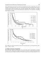
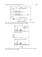
then applied to simulate the potential for drug-drug interactions. The clinical effect of CYP3A4 inhibition on bitopertin
exposure was assessed in two studies in healthy volunteers
with open-label, two-period, fixed-sequence designs [19•].
Ketoconazole, a strong CYP3A4 inhibitor, increased the
bitopertin AUC from 0 to 312 h (AUC0–312 h) 4.2-fold (90%
confidence interval [CI] 3.5–5.0) while erythromycin, a moderate CYP3A4 inhibitor, increased the AUC from time zero to
infinity (AUC0–inf) 2.1-fold (90% CI 1.9–2.3). The AUC0–inf
ratios predicted by PBPK modeling for these interactions were
in good agreement at 7.7 and 1.9, respectively (note that the
AUC0–312 h ratio underestimated the full DDI to some extent).
The effect on Cmax was minor, <25% for both inhibitors. This
was consistent with a high absolute bioavailability as simulated by PBPK for bitopertin with very limited first pass extraction in both the intestine and the liver. After discontinuation of
ketoconazole, the bitopertin elimination half-life decreased,
becoming similar to that observed in the absence of ketoconazole indicating the reversibility of the CYP3A4/5 inhibition
(Fig. 1). For bitopertin, therefore, an excellent picture of the
pharmacokinetics and a model describing the CYP3Amediated drug-drug interactions could be developed and retrospectively validated using emerging clinical data. Details of
the PBPK model can be found in Supplementary Table 1. DDI
study and simulation data are also available in the
Supplementary Materials.
Current State of the Art: Interpolation and Limited
Prospective DDI Prediction Gain Regulatory
Acceptance
PBPK models have initially found use in incorporating the
results of DDI studies into an overall description of the pharmacokinetics, then in interpolating results from DDI studies
with strong probe inhibitors/inducers for the enzyme of interest (Bmechanistic DDI study^) to DDIs with moderate and
mild inhibitors/inducers. In addition, PBPK models have been
applied to extrapolation of DDI results to subpopulations such
as organ failure, geriatrics, or certain phenotypes of the involved metabolic enzymes where it is often ethically and/or
practically challenging to investigate DDI [22, 23]. Such simulations have been used for guiding dose adjustment in drug
labels in lieu of actual clinical study results since 2009 [1].
Ibrutinib and eliglustat are two examples selected to illustrate
how a PBPK model was developed for drugs mainly metabolized by CYP3A and CYP2D6, respectively, and applied to
DDI assessment which were accepted in final product labels.
Ibrutinib Case Study—Model-Based Interpolation
of CYP3A4 Inhibition DDIs
Ibrutinib is a Bruton’s tyrosine kinase inhibitor developed for
treatment of leukemia. Ibrutinib is completely absorbed after
oral administration and extensively metabolized in the intestine and liver mostly by CYP3A4 and lesser extent by
Plasma Concentration (ng/mL)
100000
10000
Ketoconazole
1000
100
Bitopertin+ Ketoconazole
10
1
Bitopertin
0.1
0
48
96
144 192 240 288 336 384 432 480 528 576 624 672
Time (h)
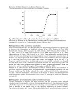
Fig. 1 Effect of ketoconazole on exposure of bitopertin. Symbols are
mean (±standard deviation) plasma concentration-time profiles after administration of 400 mg/day ketoconazole (filled black circles), bitopertin
10 mg alone (empty blue squares), or concurrently with ketoconazole
(filled red triangles). The lines are the plasma concentrations simulated
with a PBPK model in GastroPlus. Single dose of bitopertin alone
(dashed blue line), bitopertin with ketoconazole (dotted red line), ketoconazole 17 days (solid black line)
Curr Pharmacol Rep
CYP2D6 [24]. The absolute bioavailability of ibrutinib
560 mg (approved dose) was 3.9 and 8.4% in the fasted and
fed states, respectively [25]. The intestinal and hepatic bioavailability (Fg and Fh) evaluated after oral (140 mg) and
intravenous (100 μg, 13C6 labeled) administration in the fed
state were determined as 47.0 and 15.9%, respectively, in an
IV microdose study with grapefruit juice pretreatment [25]. A
PBPK model was developed by integrating available physicochemical properties, in vitro experiments, and clinical pharmacokinetic (PK) data [26]. The intrinsic clearance of
ibrutinib in human liver microsomes was inhibited 95.8% in
the presence of 1 μM of a strong CYP3A inhibitor, ketoconazole [27], and this information was incorporated into the
PBPK model for DDI simulations with various CYP3A4
modulators. Capability of the PBPK model to predict
CYP3A4 DDI for ibrutinib as substrate was examined by
predicting fold increase in Cmax and AUC of ibrutinib in the
presence of ketoconazole and compared to the observations in
the clinical study [28] (predicted vs. observed: 19- and 29fold for Cmax and 28- and 24-fold for AUC). Subsequently,
the PBPK model was verified by showing consistency between prospectively simulated fold decrease in Cmax and
AUC of ibrutinib in the presence of a CYP3A inducer, rifampicin, and the observations [28] (predicted vs. observed: 11and 13-fold for Cmax and 10- and 10-fold for AUC). The
verified PBPK model was then used to simulate unstudied
clinical DDIs with mild (fluvoxamine, azithromycin), moderate (diltiazem and erythromycin), and strong (voriconazole,
clarithromycin, itraconazole) CYP3A inhibitors to guide dose
reduction from 560 to 140 mg in concurrent administrations
with moderate CYP3A4 inhibitors.
The PBPK model simulations of DDI with moderate
(efavirenz) and strong (carbamazepine) CYP3A inducers supported ibrutinib dose of 560 mg in co-administrations with
moderate CYP3A inducers since predicted exposure was
within defined therapeutic exposure range [27]. The DDI risk
assessment and dose modification guidance for ibrutinib
based on the PBPK modeling and simulations were submitted
in new drug applications and approved in the USA [27, 29],
Canada [30], European Union [31], and Japan [32] and used in
drug labels.
Eliglustat Case Study—Model-Based Extrapolation
to Polymorphic CYP2D6 Phenotype Individuals
Eliglustat is an oral glucosylceramide synthase inhibitor and
indicated to treat symptoms of Gaucher disease type 1 [33].
This drug is extensively metabolized by CYP2D6
(f mCYP2D6 = 86%) and to a lesser extent by CYP3A4
(fmCYP3A4 = 14%). Clinical DDI investigations in CYP2D6
intermediate metabolizers (IMs) and extensive metabolizers
(EMs) showed increase in AUC of eliglustat by approximately
5-fold (IMs) to 10-fold (EMs) when co-administered with
paroxetine (strong CYP2D6 and weak CYP3A4 inhibitor)
and by 4-fold (in both IMs and EMs) when co-administered
with ketoconazole. Eliglustat is a time-dependent inhibitor of
CYP2D6 and multiple dose PK exhibited dose- and timedependent behavior. Multiple doses of eliglustat increased
AUC of metoprolol (CYP2D6 substrate) by approximately
2-fold in EMs and IMs. A PBPK model of eliglustat was
developed and its ability to describe PK in different
CYP2D6 phenotypes including poor metabolizers (PMs) and
to predict clinical DDIs was confirmed. The PBPK model was
then used for predicting DDIs with moderate inhibitors of
CYP2D6 (terbinafine) and CYP3A4 inhibitors (fluconazole)
in EMs and IMs. Moreover, DDIs with moderate to strong
CYP3A4 inhibitors (fluconazole and ketoconazole) in PMs
were predicted using the PBPK model since CYP3A4 inhibition effect on eliglustat has not been clinically investigated in
PMs. Predicted fold increase in AUC0–24 h of eliglustat in
concomitant administration with ketoconazole in PMs was
6.2 [34], higher than that in EMs and IMs, due to higher
dependency on elimination through CYP3A4 metabolism,
and concomitant use with strong CYP3A inhibitors is contraindicated in this population. The PBPK model enabled not
only interpolations from DDI with strong enzyme inhibitors
to the moderate inhibitors but also extrapolations of DDIs to
other CYP2D6 phenotypes which complemented DDI risk
assessments of eliglustat as a dual CYP2D6 and CYP3A4
substrate across CYP2D6 phenotypes. Dosage adjustment
guidance in the drug label approved by FDA [34] based on
these clinical studies and PBPK model predictions are summarized in Supplementary Table 2.
Repaglinide Case Study—Model-Based Prediction
of Insignificant DDI Effect to Support Appropriate
Dosing Recommendations
As a clinically relevant probe substrate, repaglinide is commonly used to assess the DDI risk for CYP2C8 inhibitors.
Repaglinide is an antidiabetic drug whose metabolism is mediated by CYP2C8, CYP3A4, and to a lesser extent UGT
enzymes [35, 36]. For assessing the DDI risk, therefore,
assigning the appropriate fm(CYP2C8) value for repaglinide is
of great importance given the sensitivity of predicted AUC
ratios to fm values [37]. CYP2C8 and CYP3A4 have been
reported to contribute equally to the in vitro metabolism of
repaglinide, ∼50% [36]. However, an alternative fm(CYP2C8)
value of 0.83 has been proposed based on meta-analysis of
in vivo data [38]. As these two fm> values would result in very
different maximal repaglinide DDI effects assuming complete
enzyme inhibition (2.4 and 5.9 for fm values of 0.59 and 0.83,
respectively, following oral administration), it was important
to consider both possibilities in the DDI assessment.
A number of clinically relevant DDIs with repaglinide have
been reported (Table 2). These DDIs include interactions with
Curr Pharmacol Rep
Table 2
Clinical drug-drug interactions with repaglinide as victim drug available in the University of Washington DDI database
AUC
change
(%)
Dose
(mg)
Gemfibrozil + Itraconazole
1830
Gemfibrozil
Gemfibrozil-glucuronide
443–726
Perpetrator
Ki (μmol/L)
CYP2C8a
Refs
[39]
CYP3A4a
OATP1B1/3b
600 + 100 Detailed below
Detailed below
Detailed below
171 (184–406)
n.r.
36 (13–68)
9.3–23
CYP2C8,
OATP1B1, and
CYP3A4
CYP2C8 (TDI)
and OATP1B1
KI = 26, kinact = 0.053/min
n.r.
4.0
11–34
CYP2C8 (TDI),
OATP1B1
3.2 (0.3–37)
0.019–0.032 (after
pre-incubation)
7.1
11–121
n.r.
8.26
OATP1B1,
(CYP3A4)
CYP2C8, (OATP)
CYP3A4
CYP2C8
CYP3A4 (TDI),
OATP
n.r.
n.r.
CYP3A4
Intestinal
metabolism
Cyclosporine
143
300–900 36 (9.3–87)
KI = 29,
kinact = 0.071/min
75–300 2.8–50
KI = 9.9,
kinact = 0.047
100
n.r.
Teriflunomide
Telithromycin
Trimethoprim
Clarithromycin
142
77
63
42
14–70
800
160
250
0.1
15
8.5
n.r.
Itraconazole
Grapefruit juice
41
21
100
n/a
31
n.r.
Clopidogrel
295–408
Clopidogrel-acyl-glucuronide
Mechanism
n.r.
87
n.r.
KI = 13.1 (0.85–37.4),
kinact = 0.058
(0.0192–0.14)
0.042 (0.0013–3.12)
TDI
[38, 93, 39,
94–96,
40]
[97]
[98]
[99]
[42]
[41]
[100]
[39]
[101]
Data in parenthesis represent the reported range
All data are available from [102]
n.r. not relevant, TDI time-dependent inhibition, n/a not applicable
a
Microsomal data
b
Data from HEK, or MDCK-transfected cell lines or human hepatocytes
inhibitors of CYP2C8, CYP3A4, and OATP1B1/3 as well as
compounds which interact via multiple mechanisms. The extent of clinical DDIs with repaglinide may be assessed as (1)
large extent (≥5-fold AUC change) due to inhibition of multiple processes or TDI of CYP2C8 [39, 40] and (2) a substantially lower risk can be anticipated for inhibition of a single
process, <2.5-fold AUC change for competitive CYP3A4 or
CYP2C8 inhibitors [41, 42].
Alectinib (Alecensa®) is a small molecule kinase inhibitor
which has received FDA accelerated approval for the treatment of patients with anaplastic lymphoma kinase (ALK)positive metastatic non-small cell lung cancer (NSCLC) who
have progressed on or are intolerant to crizotinib treatment
[43]. Alectinib has shown weak competitive and timedependent inhibition of CYP3A4 in vitro which has not translated in vivo [44]. Alectinib is also a competitive inhibitor of
CYP2C8 with an unbound in vitro Ki value of 0.147 μM [45].
DDI predictions with repaglinide were performed using a
PBPK modeling approach to evaluate the clinical relevance
of the CYP2C8 liability. The measured fu(plasma) and blood to
plasma concentration ratio used in the PBPK simulations were
0.003 and 2.64 (consequently the fu(blood) was 0.0011). In the
PBPK assessment of repaglinide, DDI potential fm(CYP2C8)
values of both 0.59 and 0.83 were used. In order to investigate
the sensitivity of the DDI simulations to the in vitro Ki value of
alectinib, the following scenarios were tested for both
repaglinide models: true in vivo Ki = 1×, 1/3×, 1/10×, and
1/30× of the in vitro Ki value [46].
Based on the simulations, no significant interaction (>25%
change of AUC) is anticipated regardless of the assumptions
around the in vivo fm(CYP2C8) value of repaglinide (0.59 or
0.83). A sensitivity analysis revealed that a risk for an AUC
change of greater than 25% can only be expected in case that
the in vivo inhibitory potency of alectinib is considerably
higher than anticipated from in vitro data and the in vivo
fm(CYP2C8) of repaglinide is 0.83. This model-based assessment for characterization of clinical DDI between alectinib
and CYP2C8 substrates was accepted in lieu of a clinical
DDI study with repaglinide and justified the product labeling
text BNo clinical meaningful effect on the exposure of …
repaglinide (sensitive CYP2C8 substrate) is expected following co-administration with ALESENSA^ [43].
Curr Pharmacol Rep
Interacting
Posaconazole
Cmax
M4
Cmax
Alectinib + M4
Cmax
(Strong CYP3A4
inhibitor)
Dose Recommendation for Alectinib
Fold Change and 90% Confidence Interval
Alectinib
AUCinf
AUCinf
No dose adjustment
AUCinf
Rifampicin
Alectinib
Cmax
M4
Cmax
Alectinib + M4
Cmax
(Strong CYP3A4
inducer)
AUCinf
AUCinf
No dose adjustment
AUCinf
0.0
0.5
1.0
1.5
2.0
Change Relative to Alectinib Alone
2.5
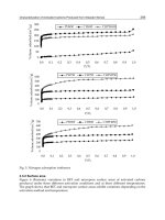
Fig. 2 Forrest plot of the drug-drug interaction potential between alectinib and the potent CYP3A inhibitor, posaconazole, or the potent CYP3A inducer,
rifampin [48]
Alectinib Efficacy Case Study—Translation of DDI
Effects Into Pharmacodynamic Effects: Relevance
and Contribution of a Major Active Metabolite
to Analysis and Interpretation of a Clinical DDI
Human metabolites are usually considered in terms of safety
when formed at greater than 10% of total drug-related systemic
exposure at steady state [47]. In terms of drug-drug interactions,
metabolites formed in vivo and reaching significant exposures
(e.g., ≥25% of parent drug exposure) have been recommended
to be characterized further in terms of metabolism, transport, and
for potential drug-drug interactions [48]. A metabolite may bind
to on- or off-target receptors and thus can be considered active
and contribute to intended and/or unintended effects [49–51].
Alectinib is metabolized by CYP3A4 and to a smaller extent by other enzymes to generate a number of metabolites
including a major metabolite M4 [52]. Population PK analysis
of the pivotal phase 2 studies showed that the geometric mean
M4 metabolite/parent (M/P) ratio in plasma was 0.4 with an
effective elimination half-life (t1/2) of approximately 33 and
31 h for alectinib and M4, respectively [53]. In vitro pharmacology studies demonstrated that both alectinib and M4 are
potent inhibitors of the target ALK with similar potency (IC50
of 1.9 and 1.2 nM, for alectinib and M4, respectively, in biochemical assays) and exhibit similar plasma protein binding
(>99% protein bound) [52].
As both alectinib and M4 are substrates of CYP3A, dedicated clinical pharmacology studies were undertaken to evaluate the effect of a strong CYP3A inhibitor (posaconazole)
and strong CYP3A inducer (rifampin) on the pharmacokinetics of alectinib and M4 [44]. Notably, the results from the
clinical DDI study with posaconazole showed that its coadministration increased alectinib exposure and decreased
M4 exposure while results from the rifampin DDI study
showed that its co-administration decreased alectinib exposure and increased M4 exposure [44] (Fig. 2). As both
alectinib and M4 are similarly active against ALK and exhibit
similar protein binding, it is expected that both substances
contribute to overall alectinib efficacy and safety. Therefore,
to support clinical dosing recommendations in the presence of
CYP3A inhibitors and inducers, changes in the combined molar exposure of alectinib and M4 (i.e., molar sum of alectinib +
M4) were evaluated (Fig. 2). The minor effects seen on the
combined exposure supported the statement Bno dosage adjustment required with co-administered CYP3A inhibitors or
inducers^ in US prescribing information for Alecensa® [43].
The alectinib case represents an approach to the understanding of drug-drug interaction potential by utilization of
integrated non-clinical and clinical data of a parent molecule
and its major active metabolite. The knowledge of clinical
pharmacology attributes of both the parent and metabolite
enabled dosing recommendations based on the changes occurring to both substances. To support this, characterization of
both alectinib and M4 was undertaken throughout the development process from preclinical safety and drug metabolism/
pharmacokinetic testing through to clinical exposure-response
evaluation of alectinib [54, 55]. Indeed, clinical exposureresponse analyses evaluated the relationship between key efficacy and safety endpoints emerging from alectinib pivotal
studies and the combined exposure of alectinib and M4 [53].
Thus, while the changes seen in the alectinib exposure when
co-administered with posaconazole or rifampin may have potentially warranted dosage adjustments, consideration of the
combined changes suggested that no dosage adjustments were
needed. This approach to consideration of parent and metabolite contributions to clinical DDI or exposure-response interpretation has been successfully applied previously for other
small molecules with active metabolites (e.g., regorafenib,
Curr Pharmacol Rep
ezetimibe, ruxolitinib, dabrafenib, and sunitinib) [56–61].
Cumulatively, the alectinib case illustrates the relevance and
contribution of a major active metabolite to clinical DDI analyses and interpretation.
Current Frontiers in DDI Prediction From In Vitro
Enhanced DDI Predictions From Time-Dependent
Inhibition Measurements Using Human Hepatocytes
Suspended in Full Plasma
Preclinical prediction of CYP inhibition-mediated DDIs has
been performed conventionally using the well-characterized
and intensively studied human liver microsomal (HLM) assay,
which shows high detection sensitivity and low likelihood of
false-negative predictions [62]. An in vitro assay using human
hepatocytes (hHEPs) suspended in whole human plasma
(plasma hHEPs) has been reported to give more accurate prediction of the extent of clinical relevant effect due to CYP
inhibition [63–66]. Advantages of assessing DDI in human
hepatocytes supplemented with 100% plasma include (1) inherent accounting for plasma protein and microsomal/
hepatocyte binding of a drug, (2) compound is available to
enzyme in its native environment within the cell, i.e., more a
physiologically relevant condition, (3) metabolism of the
compound by both CYP and non-CYP pathways is possible,
and (4) transporter-mediated uptake into hepatocytes may
occur.
An elegant study recently published by Mao et al. [67••]
compared side-by-side DDI prediction due to CYP3A inhibition from the plasma hHEP assay with three other assays: (a)
HLM, (b) plated hHEPs, and (c) hHEPs suspended in
Dulbecco’s modified Eagle’s medium (DMEM) for 12
marketed drugs (10 protein kinase inhibitors and 2 prototypical CYP3A time-dependent inhibitors). Kinetic parameters
were generated for the apparent reversible inhibition constant
(Ki,app) and/or TDI (KI,app and kinact) and directly used for
quantitative prediction of the fold-increase in midazolam
AUC0–inf (AUCR) following co-administration with CYP3A
inhibitors based on a static mechanistic model and the total
average systemic plasma concentration without correction for
free drug fraction (fu). The result from this study demonstrated
that the plasma hHEP assay offered a clear enhancement of
DDI prediction (95% accuracy) with no false-negative or
false-positive outcomes. The accuracies for the other three
assays were 58, 84, and 74% for HLM, plated hHEPs, and
DMEM hHEPs, respectively.
In this study [67••], a number of drugs were shown to give
both reversible inhibition and TDI for CYP3A in the HLM
assay (for example erlotinib, nilotinib, and pazopanib) but
interestingly, these drugs were not inhibitory in the plasma
HEP assay. While the clinical data confirmed low DDI due
to CYP3A inhibition for these drugs as predicted by the plasma HEP assay, a more complete mechanistic understanding
for the discrepancy between the two systems would be helpful
when considering the differential sensitivities of the test systems. The traditional HLM TDI assay is robust, sensitive, and
backed by a substantial body of published data [12, 68, 69]
which can be used to rank and to some extent to predict CYPmediated DDI during the discovery stage. It is however suggested to consider using the plasma hHEP TDI assay for an
enhanced assessment of the potential DDI during the candidate selection and early stages of drug development as a derisking approach for TDI-positive candidate compounds.
Challenges of DDI Prediction in Cases of Metabolism
by BUnusual^ CYP Enzymes or Non-CYP Enzymes
Despite advances in in vitro enzymology technologies, there
continues to be much to learn about enzymes which, while
unimportant in the metabolism of drugs in general, are key
contributors to the metabolism of particular drug compounds.
For example, the SGLT2 inhibitor tofogliflozin is metabolized
by CYPs 2C18, 4A11, and 4F3B [70], and the multiple sclerosis drug fingolimod is metabolized by CYP4F enzymes
[71]. These enzymes are usually regarded as Bminor^ CYP
isoforms and would not routinely be included in enzyme phenotyping screens [10•, 72]. This raises the question of how
one is to know that an important pathway is Bmissed^ in initial
in vitro assessments. Due to the availability of wellcharacterized and selective inhibitors for CYP isoforms, it
may be apparent should the activities of routinely tested
CYP enzymes not account for the majority of metabolism
in vitro. In such cases, additional in vitro work using
recombinantly expressed enzymes and (semi-)selective CYP
inhibitors may be performed to try to obtain more clarity on
enzyme contributions to metabolism, although this may prove
challenging. DDI risks could then be addressed through
screening of potential co-medicant substances either as inhibitors of the involved metabolic enzyme or, more empirically,
as inhibitors of turnover of the drug in development itself. Due
to a lack of system information (enzyme expression and activity levels, polymorphism status, effect of disease, ontogeny), it is unlikely that special population or polymorphism risk
assessments can be made at this time.
The situation is even more challenging when Bunusual^
non-CYP enzymes are involved. In one recent example, an
investigational trace amine-associated receptor antagonist
RO5263397 was found to be principally cleared by
UGT2B10 [73••]. At the time of compound selection,
UGT2B10 was not considered an important enzyme in drug
metabolism and was not commercially available for testing,
and no selective inhibitors were characterized [74–77]. Coadministration with potent UGT2B10 inhibitors could potentially mimic the UGT2B10 poor metabolizer phenotype which
Curr Pharmacol Rep
resulted in a 136-fold higher AUC for one individual after a
single 10 mg dose in a phase I clinical study [73••].
Such cases also provide substantial learning opportunities.
As a result of this observation, a new splice site polymorphism
was identified (prevalent in individuals of African origin but
almost absent in Caucasians). This is relevant for clearance of
other UGT2B10 substrates [78, 79]. In addition, increased understanding of the enzyme system and in vitro tools to assess
UGT2B10 contribution to metabolism have been developed
which can be rapidly employed in the future. In this way,
UGT2B10 illustrates the process by which an enzyme not
previously considered in drug metabolism testing progresses
from being an Bessentially uncharacterized^ to a Blargely
characterized^ metabolic enzyme system [80, 81]. A similar
experience had been reported by Wang et al. for a Merck development compound MK-7246 which is cleared by polymorphic UGT2B17 [82]. It is likely that such learning experiences
will be repeated as drug development continues to move into
areas of novel chemical space in pursuit of new drug targets
and further examples are discovered where previously little
studied enzymes are important for individual drug clearance.
Future Prospects for DDI Prediction
To date, most in vitro systems used in DDI prediction have
employed short timescale incubations to generate mechanistic
parameters which can then be used to build up long-term
model predictions of DDIs in vivo. Short timescale incubations cannot however address issues such as enzyme inactivation by highly metabolically stable compounds or the interplay of enzyme inactivation and induction which will drive
the effective steady-state change in metabolic enzyme capacity. Although the sensitivity of short-term plated human hepatocytes to inhibition and induction has been demonstrated
[83], such systems are unlikely to reflect steady-state conditions due to the transient nature of the cell cultures used. The
advent of long-term hepatocyte culture systems may allow
effective in vitro pharmacokinetic assessments to be made
which will better reflect the clinical situation. The potential
of long-term hepatocyte cultures has initially been demonstrated for clearance assessment of metabolically stable compounds [84–87••]. Their application to more sophisticated
ADME assessments, such as induction [88], the effect of active uptake on apparent induction potency [89•], metabolism
profiling/cross-species comparison [90], and to a limited extent for drug-drug interactions [85] have also been demonstrated. New long-term hepatocyte systems may therefore offer a completely new opportunity to simultaneously study
multiple processes involved in drug-drug interactions which
were not previously possible in vitro, especially for highly
metabolically stable compounds. The development of longterm hepatocyte systems may also be seen as a first step in
the direction of functional in vitro test systems with cells from
multiple organs such as the liver, intestine, kidney, skin, and
brain [91, 92], within a single test system (Bchip^). When
validated, data from the new experimental systems will quickly be incorporated into PBPK-based modeling tools further
enhancing prediction of clinical DDIs.
Conclusions
This review has drawn upon personal experiences and recent
literature reports to highlight achievements and ongoing challenges in the rapidly developing areas of metabolic DDI assessment, prediction, interpretation, and drug product labeling. Examples have been shown of how a model-based approach to understanding DDIs has progressed from data integration (bitopertin) to being accepted for interpolative
(ibrutinib) and increasingly extrapolative DDI predictions
(eliglustat and alectinib). Scientific confidence in and regulatory acceptance of PBPK modeling have increased with growing knowledge of DDIs, availability and robustness of in vitro
test systems, and experience in DDI prediction. Predictions
from well-executed analyses using validated models have enabled explicit dosing recommendations in product labels for
clinical DDIs based on PBPK modeling in lieu of dedicated
clinical DDI studies. Modeling approaches may indeed offer
the only way to explore some potential DDIs where clinical
investigation is unfeasible due to ethical considerations or the
inability to recruit suitable study subjects.
The impact of characterizing major active metabolites during drug development has also been exemplified in the case of
alectinib. This has been shown to be critical in the interpretation of clinical DDIs where exposure changes occur to both
the parent and an active metabolite and are relevant to clinical
efficacy and safety. Understanding the pharmacological, pharmacokinetic, and disposition properties of a metabolite using
in vitro and in vivo studies can allow for estimation of its
contribution in clinical DDI interpretation and subsequently
its potential impact on clinical efficacy and safety in support of
appropriate dosing recommendations.
A sometimes underemphasized factor affecting DDI predictions is the availability of good quality clinical DDI data
with which can be used for validation purposes. This is especially the case for drugs predominantly metabolized by
Bunusual^ CYP enzymes or non-CYP enzymes. Examples
where CYP4F enzymes or UGT2B10 catalyze drug clearance
have been discussed. We can expect that the continued development of experimental techniques and the increases in
knowledge of enzymology and DDI will be reflected in increased DDI prediction confidence for such drugs.
Use of a whole plasma human hepatocyte TDI assay has
shown to improve in vitro-in vivo extrapolation. Further TDI
assay developments are anticipated using long-term
Curr Pharmacol Rep
hepatocyte culture systems and other Borgans on a chip^ technologies. These offer the promise of in vitro systems where an
integrated assessment of enzyme inhibition, inactivation, and
induction can be made. The ability to use the same modeling
approaches to understand such Bin vitro pharmacokinetics/
DDI^ experiments and then directly transfer this understanding to the human DDI situation may allow a further step forward in DDI prediction to be made in the near future.
Acknowledgments The authors thank Franz Schuler and Christoph
Funk (Roche, Basel) for helpful suggestions and Alexander Nürnberg
(Roche, Basel) for his diligent assistance in preparation of the manuscript.
Compliance with Ethical Standards
6.•
7.
8.
9.
Conflict of Interest All authors are employees of F. Hoffmann-La
Roche Ltd. There are no conflicts of interest to declare.
Human and Animal Rights and Informed Consent This article does
not contain previously unpublished studies with human or animal subjects performed by any of the authors.
Open Access This article is distributed under the terms of the Creative
Commons Attribution 4.0 International License (http://
creativecommons.org/licenses/by/4.0/), which permits unrestricted use,
distribution, and reproduction in any medium, provided you give
appropriate credit to the original author(s) and the source, provide a link
to the Creative Commons license, and indicate if changes were made.
10.•
11.
12.
References
Papers of particular interest, published recently, have been
highlighted as:
• Of importance
•• Of major importance
1.
Jamei M. Recent advances in development and application of
physiologically-based pharmacokinetic (PBPK) models: a transition from academic curiosity to regulatory acceptance. Curr
Pharmacol Rep. 2016;2:161–9. doi:10.1007/s40495-016-0059-9.
2. Yu J, Ritchie TK, Mulgaonkar A, Ragueneau-Majlessi I. Drug
disposition and drug-drug interaction data in 2013 FDA new drug
applications: a systematic review. Drug Metab Dispos.
2014;42(12):1991–2001. doi:10.1124/dmd.114.060392.
3. Yu J, Ritchie TK, Zhou Z, Ragueneau-Majlessi I. Key findings
from preclinical and clinical drug interaction studies presented in
new drug and biological license applications approved by the
Food and Drug Administration in 2014. Drug Metab Dispos.
2016;44(1):83–101. doi:10.1124/dmd.115.066720.
4. Sager JE, Yu J, Ragueneau-Majlessi I, Isoherranen N.
Physiologically based pharmacokinetic (PBPK) modeling and
simulation approaches: a systematic review of published models,
applications, and model verification. Drug Metab Dispos.
2015;43(11):1823–37. doi:10.1124/dmd.115.065920.
5.•• Yu J, Zhou Z, Owens KH, Ritchie TK, Ragueneau-Majlessi I.
What can be learned from recent new drug applications? A systematic review of drug interaction data for drugs approved by the
US FDA in 2015. Drug Metab Dispos. 2017;45(1):86–108.
doi:10.1124/dmd.116.073411. Highly informative overview of
13.
14.
15.
16.
17.
18.
19.•
NDA information from 2015, covering many data which are
not published as journal articles.
Rowland M, Matin SB. Kinetics of drug-drug interactions. J
Pharmacokinet Biopharm. 1973;1(6):553–67. A classic paper
and relevant reminder of where the static mechanistic modelling came from and its original basis in half-life change
estimation.
Boulenc X, Nicolas O, Hermabessiere S, Zobouyan I, Martin V,
Donazzolo Y, et al. CYP3A4-based drug-drug interaction:
CYP3A4 substrates’ pharmacokinetic properties and ketoconazole dose regimen effect. Eur J Drug Metab Pharmacokinet.
2016;41(1):45–54. doi:10.1007/s13318-014-0235-4.
Obach RS. Predicting drug-drug interactions from in vitro drug
metabolism data: challenges and recent advances. Curr Opin Drug
Discov Devel. 2009;12(1):81–9.
Fahmi OA, Hurst S, Plowchalk D, Cook J, Guo F, Youdim K, et al.
Comparison of different algorithms for predicting clinical drugdrug interactions, based on the use of CYP3A4 in vitro data:
predictions of compounds as precipitants of interaction. Drug
M e t a b D i s p o s . 2 0 0 9 ; 3 7 ( 8 ) : 1 6 5 8 – 6 6 . d o i : 1 0 . 11 2 4
/dmd.108.026252.
Bohnert T, Patel A, Templeton I, Chen Y, Lu C, Lai G, et al.
Evaluation of a new molecular entity as a victim of metabolic
drug-drug interactions—an industry perspective. Drug Metab
Dispos. 2016;44(8):1399–423. doi:10.1124/dmd.115.069096.
An excellent overview of current practices for metabolic
enzyme phenotyping work.
Rowland M, Peck C, Tucker G. Physiologically-based pharmacokinetics in drug development and regulatory science. Annu Rev
Pharmacol Toxicol. 2011;51:45–73. doi:10.1146/annurevpharmtox-010510-100540.
Grimm SW, Einolf HJ, Hall SD, He K, Lim HK, Ling KH, et al.
The conduct of in vitro studies to address time-dependent inhibition of drug-metabolizing enzymes: a perspective of the pharmaceutical research and manufacturers of America. Drug Metab
Dispos. 2009;37(7):1355–70. doi:10.1124/dmd.109.026716.
Kumar GN, Rodrigues AD, Buko AM, Denissen JF. Cytochrome
P450-mediated metabolism of the HIV-1 protease inhibitor ritonavir (ABT-538) in human liver microsomes. J Pharmacol Exp Ther.
1996;277(1):423–31.
Eagling VA, Back DJ, Barry MG. Differential inhibition of cytochrome P450 isoforms by the protease inhibitors, ritonavir, saquinavir and indinavir. Br J Clin Pharmacol. 1997;44(2):190–4.
Koudriakova T, Iatsimirskaia E, Utkin I, Gangl E, Vouros P,
Storozhuk E, et al. Metabolism of the human immunodeficiency
virus protease inhibitors indinavir and ritonavir by human intestinal microsomes and expressed cytochrome P4503A4/3A5:
mechanism-based inactivation of cytochrome P4503A by ritonavir. Drug Metab Dispos. 1998;26(6):552–61.
Kharasch ED, Mitchell D, Coles R, Blanco R. Rapid clinical induction of hepatic cytochrome P4502B6 activity by ritonavir.
Antimicrob Agents Chemother. 2008;52(5):1663–9. doi:10.1128
/AAC.01600-07.
Alberati D, Moreau JL, Lengyel J, Hauser N, Mory R, Borroni E,
et al. Glycine reuptake inhibitor RG1678: a pharmacologic characterization of an investigational agent for the treatment of schizophrenia. Neuropharmacology. 2012;62(2):1152–61. doi:10.1016
/j.neuropharm.2011.11.008.
Winter M, Funk J, Korner A, Alberati D, Christen F, Schmitt G,
et al. Effects of GlyT1 inhibition on erythropoiesis and iron homeostasis in rats. Exp Hematol. 2016;44(10):964–74. e4
doi:10.1016/j.exphem.2016.07.003.
Boetsch C, Parrott N, Fowler S, Poirier A, Hainzl D, Banken L,
et al. Effects of cytochrome P450 3A4 inhibitors-ketoconazole
and erythromycin-on bitopertin pharmacokinetics and comparison
with physiologically based modelling predictions. Clin
Curr Pharmacol Rep
20.
21.
22.
23.
24.
25.
26.
27.
28.
29.
30.
31.
32.
33.
Pharmacokinet. 2016;55(2):237–47. doi:10.1007/s40262-0150312-0. The publication detailing bitopertin DDIs for readers
wanting to simulate a classical DDI using the data given in the
Supplementary information.
Parrott N, Hainzl D, Alberati D, Hofmann C, Robson R,
Boutouyrie B, et al. Physiologically based pharmacokinetic
modelling to predict single- and multiple-dose human pharmacokinetics of bitopertin. Clin Pharmacokinet. 2013;52(8):673–83.
doi:10.1007/s40262-013-0061-x.
Parrott N, Hainzl D, Scheubel E, Krimmer S, Boetsch C, Guerini
E, et al. Physiologically based absorption modelling to predict the
impact of drug properties on pharmacokinetics of bitopertin.
AAPS J. 2014;16(5):1077–84. doi:10.1208/s12248-014-9639-y.
Grillo JA, Zhao P, Bullock J, Booth BP, Lu M, Robie-Suh K, et al.
Utility of a physiologically-based pharmacokinetic (PBPK)
modeling approach to quantitatively predict a complex drugdrug-disease interaction scenario for rivaroxaban during the drug
review process: implications for clinical practice. Biopharm Drug
Dispos. 2012;33(2):99–110. doi:10.1002/bdd.1771.
Huang SM, Abernethy DR, Wang Y, Zhao P, Zineh I. The utility of
modeling and simulation in drug development and regulatory review. J Pharm Sci. 2013;102(9):2912–23. doi:10.1002/jps.23570.
Scheers E, Leclercq L, de Jong J, Bode N, Bockx M, Laenen A,
et al. Absorption, metabolism, and excretion of oral (1)(4)C
radiolabeled ibrutinib: an open-label, phase I, single-dose study
in healthy men. Drug Metab Dispos. 2015;43(2):289–97.
doi:10.1124/dmd.114.060061.
de Vries R, Smit JW, Hellemans P, Jiao J, Murphy J, Skee D, et al.
Stable isotope-labelled intravenous microdose for absolute bioavailability and effect of grapefruit juice on ibrutinib in healthy adults. Br J
Clin Pharmacol. 2016;81(2):235–45. doi:10.1111/bcp.12787.
de Zwart L, Snoeys J, De Jong J, Sukbuntherng J, Mannaert E,
Monshouwer M. Ibrutinib dosing strategies based on interaction
potential of CYP3A4 perpetrators using physiologically based
pharmacokinetic modeling. Clin Pharmacol Ther. 2016;100(5):
548–57. doi:10.1002/cpt.419.
NDA 205552 (Ibrutinib): Clinical pharmacology and
biopharmaceutics review(s). U. S. Food and Drug Administration,
Center for Drug Evaluation and Research. 2014. Available from:
/>Orig2s000ClinPharmR.pdf. Accessed: 28 Nov 2016
de Jong J, Skee D, Murphy J, Sukbuntherng J, Hellemans P, Smit
J, et al. Effect of CYP3A perpetrators on ibrutinib exposure in
healthy participants. Pharmacol Res Perspect. 2015;3(4):e00156.
doi:10.1002/prp2.156.
IMBRUVICA™: Prescribing information. U. S. Food and Drug
Administration. 2014. Available from: .
gov/drugsatfda_docs/label/2014/205552Orig2lbl.pdf. Accessed:
28 Nov 2016
IMBRUVICA™: Summary basis of decision. Health Canada.
2 01 5. Ava i l a bl e f r om : ht tp:// www.hc-s c.gc.ca /dhpmps/prodpharma/sbd-smd/drug-med/sbd_smd_2015_
imbruvica_174029-eng.php. Accessed: 28 Nov 2016
IMBRUVICA™: EPAR—product information. European Medicines
Agency. 2014. Available from: />GB/document_library/EPAR_-_Product_Information/human/003791
/WC500177775.pdf. Accessed: 28 Nov 2016
IMBRUVICA™: Regulatory decision [in Japanese]. Ministry of
Health, Labour and Welfare, Pharmaceuticals and Medical
Devices Agency. 2016. Available from: .
jp/drugs/2016/P20160404001/800155000_22800AMX00387_
A100_1.pdf Accessed: 28 Nov 2016
NDA 205494 (CERDELGA®): Clinical pharmacology and
biopharmaceutics review(s). U. S. Food and Drug Administration,
Center for Drug Evaluation and Research. 2014. Available from:
34.
35.
36.
37.
38.
39.
40.
41.
42.
43.
44.
45.
46.
47.
/>Orig1s000ClinPharmR.pdf. Accessed: 28 Nov 2016
CERDELGA™: Prescribing information. U. S. Food and Drug
Administration. 2014. Available from: essdata.
fda.gov/drugsatfda_docs/label/2014/205494Orig1s000lbl.pdf.
Accessed: 28 Nov 2016
Gan J, Chen W, Shen H, Gao L, Hong Y, Tian Y, et al.
Repaglinide-gemfibrozil drug interaction: inhibition of
repaglinide glucuronidation as a potential additional contributing
mechanism. Br J Clin Pharmacol. 2010;70(6):870–80.
doi:10.1111/j.1365-2125.2010.03772.x.
Sall C, Houston JB, Galetin A. A comprehensive assessment of
repaglinide metabolic pathways: impact of choice of in vitro system and relative enzyme contribution to in vitro clearance. Drug
M e t a b D i s p o s . 2 0 1 2 ; 4 0 ( 7 ) : 1 2 7 9 – 8 9 . d o i : 1 0 . 11 2 4
/dmd.112.045286.
Ito K, Hallifax D, Obach RS, Houston JB. Impact of parallel
pathways of drug elimination and multiple cytochrome P450 involvement on drug-drug interactions: CYP2D6 paradigm. Drug
Metab Disp os. 200 5;33 (6):83 7 –4 4. doi:1 0.1124
/dmd.104.003715.
Honkalammi J, Niemi M, Neuvonen PJ, Backman JT. Dosedependent interaction between gemfibrozil and repaglinide in
humans: strong inhibition of CYP2C8 with subtherapeutic gemfibrozil doses. Drug Metab Dispos. 2011;39(10):1977–86.
doi:10.1124/dmd.111.040931.
Niemi M, Backman JT, Neuvonen M, Neuvonen PJ. Effects of
gemfibrozil, itraconazole, and their combination on the pharmacokinetics and pharmacodynamics of repaglinide: potentially hazardous interaction between gemfibrozil and repaglinide.
Diabetologia. 2003;46(3):347–51. doi:10.1007/s00125-0031034-7.
Tornio A, Niemi M, Neuvonen M, Laitila J, Kalliokoski A,
Neuvonen PJ, et al. The effect of gemfibrozil on repaglinide pharmacokinetics persists for at least 12 h after the dose: evidence for
mechanism-based inhibition of CYP2C8 in vivo. Clin Pharmacol
Ther. 2008;84(3):403–11. doi:10.1038/clpt.2008.34.
Niemi M, Kajosaari LI, Neuvonen M, Backman JT, Neuvonen PJ.
The CYP2C8 inhibitor trimethoprim increases the plasma concentrations of repaglinide in healthy subjects. Br J Clin Pharmacol.
2004;57(4):441–7. doi:10.1046/j.1365-2125.2003.02027.x.
Kajosaari LI, Niemi M, Backman JT, Neuvonen PJ.
Telithromycin, but not montelukast, increases the plasma concentrations and effects of the cytochrome P450 3A4 and 2C8 substrate repaglinide. Clin Pharmacol Ther. 2006;79(3):231–42.
doi:10.1016/j.clpt.2005.11.002.
ALECENSA®: Prescribing information. U. S. Food and Drug
Administration. 2015. Available from: essdata.
fda.gov/drugsatfda_docs/label/2015/208434s000lbl.pdf.
Accessed: 28 Nov 2016
Morcos PN, Cleary Y, Guerini E, Dall G, Bogman K, De Petris L,
et al. Clinical drug-drug interactions through cytochrome P450 3A
(CYP3A) for the selective ALK inhibitor alectinib. Clin
Pharmacol Drug Dev. 2016; doi:10.1002/cpdd.298.
Sekiguchi N, Nagao S, Takanashi K, Kato M, Kaneko A, Morita
K, et al. Preclinical evaluation of the potential for cytochrome
P450 inhibition and induction of the selective ALK inhibitor,
alectinib. Xenobiotica. 2016:1–10. doi:10.1080
/00498254.2016.1261308.
Cleary Y, Gertz M, Morcos P, Youdim K, Fowler S, Yu L et al.
Physiologically-based pharmacokinetic (PBPK) modelling to
evaluate drug-drug interaction (DDI) risk of alectinib. American
Society for Clinical Pharmacology and Therapeutics - 118th
Annual Meeting 2017.
Guidance for Industry: Safety testing of drug metabolites. Revision 1.
U. S. Food and Drug Administration, Center for Drug Evaluation
Curr Pharmacol Rep
48.
49.
50.
51.
52.
53.
54.
55.
56.
57.
58.
and Research. 2016. Available from: .
gov/downloads/drugs/guidancecomplianceregulatoryinformation/
guidances/ucm079266.pdf. Accessed: 06 Jan 2017
Guidance for Industry: Drug interaction studies—study design, data
analysis, implications for dosing, and labeling recommendations [draft
guidance]. U. S. Food and Drug Administration, Center for Drug
Evaluation and Research. 2012. Available from: .
gov/downloads/drugs/guidancecomplianceregulatoryinformation/
guidances/ucm292362.pdf. Accessed: 28 Nov 2016
Guidance for Industry: Safety testing of drug metabolites U. S.
Food and Drug Administration, Center for Drug Evaluation and
R e s e a r c h . 2 0 0 8 . Av a i l a b l e f r o m : h t t p : / / w w w. f d a .
gov/OHRMS/DOCKETS/98fr/FDA-2008-D-0065-GDL.pdf.
Accessed: 28 Nov 2016
Guidance for Industry M3(R2): Nonclinical safety studies for the
conduct of human clinical trials and marketing authorization for
pharmaceuticals. Revision 1. U. S. Food and Drug
Administration, Center for Drug Evaluation and Research. 2010.
Available from: />GuidanceComplianceRegulatoryInformation/
Guidances/UCM073246.pdf. Accessed: 28 Nov 2016
Guidance for Industry M3(R2): Nonclinical safety studies for the
conduct of human clinical trials and marketing authorization for
pharmaceuticals. Questions and Answers (R2). Revision 1. U. S.
Food and Drug Administration, Center for Drug Evaluation and
Research. 2013. Available from: />downloads/Drugs/GuidanceCompliance
RegulatoryInformation/Guidances/UCM292340.pdf. Access
Ibrutinibed: 28 Nov 2016
Nakagawa T, Takanashi K, Hoshino-Yoshino A, Yamauchi T,
Kawashima K, Tachibana Y et al. In vitro metabolism of alectinib,
a novel highly potent ALK inhibitor: contribution of CYP3A enzymes in microsomes and hepatocytes. International Society for
the Study of Xenobiotics (ISSX). 2016; Busan, South Korea 2016.
Hsu J, Carnac R, Henschel V, Bogman K, Martin-Facklam M,
Guerini E et al. Population pharmacokinetics (popPK) and
exposure-response (ER) analyses to confirm alectinib 600 mg
BID dose selection in a crizotinib-progressed or intolerant population. J Clin Oncol 2016;34:(suppl; abstr e20598).
NDA 208434 (ALECENSA ®): Clinical pharmacology and
b i op h a r m a ce u t ic s r ev i e w( s ) . U . S . F o o d a n d D r u g
Administration, Center for Drug Evaluation and Research. 2015.
Available from: />docs/nda/2015/208434Orig1s000ClinPharmR.pdf. Accessed: 28
Nov 2016
NDA 208434 (ALECENSA®): Pharmacology review(s). U. S.
Food and Drug Administration, Center for Drug Evaluation and
Research. 2015. Available from: .
gov/drugsatfda_docs/nda/2015/208434Orig1s000PharmR.pdf.
Accessed: 28 Nov 2016
NDA 203085 (STIVARGA ®): Clinical pharmacology and
b i o p h a r m a c e u t i c s r e v i e w ( s ) . U . S . F oo d a n d D r u g
Administration, Center for Drug Evaluation and Research. 2012.
Available from: />docs/nda/2012/203085Orig1s000ClinPharmR.pdf. Accessed: 28
Nov 2016
NDA 202806 (TAFINLAR ® ): Clinical pharmacology and
b i op h a r m a c e u t i c s r e v i e w ( s ) . U . S. F o o d a n d D r u g
Administration, Center for Drug Evaluation and Research. 2013.
Available from: />docs/nda/2013/202806Orig1s000ClinPharmR.pdf. Accessed: 28
Nov 2016
NDA 21-938/21-968 (SUTENTTM): Clinical pharmacology and
biopharmaceutics review(s). U. S. Food and Drug Administration,
Center for Drug Evaluation and Research. 2006. Available from:
/>/021938_S000_Sutent_BioPharmR.pdf. Accessed: 28 Nov 2016
59. Houk BE, Bello CL, Poland B, Rosen LS, Demetri GD, Motzer
RJ. Relationship between exposure to sunitinib and efficacy and
tolerability endpoints in patients with cancer: results of a
pharmacokinetic/pharmacodynamic meta-analysis. Cancer
Chemother Pharmacol. 2010;66(2):357–71. doi:10.1007/s00280009-1170-y.
60. Shi JG, Chen X, Emm T, Scherle PA, McGee RF, Lo Y, et al. The
effect of CYP3A4 inhibition or induction on the pharmacokinetics
and pharmacodynamics of orally administered ruxolitinib
(INCB018424 phosphate) in healthy volunteers. J Clin
Pharmacol. 2012;52(6):809–18. doi:10.1177
/0091270011405663.
61. Kosoglou T, Statkevich P, Johnson-Levonas AO, Paolini JF,
Bergman AJ, Alton KB. Ezetimibe: a review of its metabolism,
pharmacokinetics and drug interactions. Clin Pharmacokinet.
2005;44(5):467–94.
62. Fowler S, Zhang H. In vitro evaluation of reversible and irreversible cytochrome P450 inhibition: current status on methodologies
and their utility for predicting drug-drug interactions. AAPS J.
2008;10(2):410–24. doi:10.1208/s12248-008-9042-7.
63. Mao J, Johnson TR, Shen Z, Yamazaki S. Prediction of crizotinibmidazolam interaction using the Simcyp population-based simulator: comparison of CYP3A time-dependent inhibition between
human liver microsomes versus hepatocytes. Drug Metab Dispos.
2013;41(2):343–52. doi:10.1124/dmd.112.049114.
64. Mao J, Mohutsky MA, Harrelson JP, Wrighton SA, Hall SD.
Prediction of CYP3A-mediated drug-drug interactions using human hepatocytes suspended in human plasma. Drug Metab
Dispos. 2011;39(4):591–602. doi:10.1124/dmd.110.036400.
65. Mao J, Mohutsky MA, Harrelson JP, Wrighton SA, Hall SD.
Predictions of cytochrome P450-mediated drug-drug interactions
using cryopreserved human hepatocytes: comparison of plasma
and protein-free media incubation conditions. Drug Metab
Dispos. 2012;40(4):706–16. doi:10.1124/dmd.111.043158.
66. Lu C, Miwa GT, Prakash SR, Gan LS, Balani SK. A novel model
for the prediction of drug-drug interactions in humans based on
in vitro cytochrome p450 phenotypic data. Drug Metab Dispos.
2007;35(1):79–85. doi:10.1124/dmd.106.011346.
67.•• Mao J, Tay S, Khojasteh CS, Chen Y, Hop CE, Kenny JR.
Evaluation of time dependent inhibition assays for marketed oncology drugs: comparison of human hepatocytes and liver microsomes in the presence and absence of human plasma. Pharm Res.
2016;33(5):1204–19. doi:10.1007/s11095-016-1865-9.
Development of good in vitro–in vivo correlations using the
Bfull plasma^ short-term suspension culture CYP3A4 TDI
assay system.
68. Zimmerlin A, Trunzer M, Faller B. CYP3A time-dependent inhibition risk assessment validated with 400 reference drugs. Drug
M e t a b D i s p o s . 2 0 11 ; 3 9 ( 6 ) : 1 0 3 9 – 4 6 . d o i : 1 0 . 11 2 4
/dmd.110.037911.
69. Orr ST, Ripp SL, Ballard TE, Henderson JL, Scott DO, Obach RS,
et al. Mechanism-based inactivation (MBI) of cytochrome P450
enzymes: structure-activity relationships and discovery strategies
to mitigate drug-drug interaction risks. J Med Chem. 2012;55(11):
4896–933. doi:10.1021/jm300065h.
70. Yamane M, Kawashima K, Yamaguchi K, Nagao S, Sato M,
Suzuki M, et al. In vitro profiling of the metabolism and drugdrug interaction of tofogliflozin, a potent and highly specific
sodium-glucose co-transporter 2 inhibitor, using human liver microsomes, human hepatocytes, and recombinant human CYP.
Xenobiotica. 2015;45(3):230–8. doi:10.3109
/00498254.2014.976296.
71. Jin Y, Zollinger M, Borell H, Zimmerlin A, Patten CJ. CYP4F
enzymes are responsible for the elimination of fingolimod
Curr Pharmacol Rep
72.
73.••
74.
75.
76.
77.
78.
79.
80.
81.
82.
83.
84.
(FTY720), a novel treatment of relapsing multiple sclerosis. Drug
Metab Dispos. 2011;39(2):191–8. doi:10.1124/dmd.110.035378.
Zientek MA, Youdim K. Reaction phenotyping: advances in the
experimental strategies used to characterize the contribution of
drug-metabolizing enzymes. Drug Metab Dispos. 2015;43(1):
163–81. doi:10.1124/dmd.114.058750.
Fowler S, Kletzl H, Finel M, Manevski N, Schmid P, Tuerck D,
et al. A UGT2B10 splicing polymorphism common in African
populations may greatly increase drug exposure. J Pharmacol
Exp Ther. 2015;352(2):358–67. doi:10.1124/jpet.114.220194.
The largest so far reported clinical effect of a UGT
polymorphism on exposure. Also highlights high ethnic
variation in polymorphism frequencies and the need to
consider this in clinical trials.
Kaivosaari S, Toivonen P, Aitio O, Sipila J, Koskinen M, Salonen
JS, et al. Regio- and stereospecific N-glucuronidation of
medetomidine: the differences between UDP glucuronosyltransferase (UGT) 1A4 and UGT2B10 account for the complex kinetics of human liver microsomes. Drug Metab Dispos. 2008;36(8):
1529–37. doi:10.1124/dmd.108.021709.
Kaivosaari S, Toivonen P, Hesse LM, Koskinen M, Court MH,
Finel M. Nicotine glucuronidation and the human UDPglucuronosyltransferase UGT2B10. Mol Pharmacol. 2007;72(3):
761–8. doi:10.1124/mol.107.037093.
Kazmi F, Yerino P, Barbara JE, Parkinson A. Further characterization of the metabolism of desloratadine and its cytochrome P450
and UDP-glucuronosyltransferase inhibition potential: identification of desloratadine as a relatively selective UGT2B10 inhibitor.
Drug Metab Dispos. 2015;43(9):1294–302. doi:10.1124
/dmd.115.065011.
Pattanawongsa A, Nair PC, Rowland A, Miners JO. Human UDPglucuronosyltransferase (UGT) 2B10: validation of cotinine as a
selective probe substrate, inhibition by UGT enzyme-selective inhibitors and antidepressant and antipsychotic drugs, and structural
determinants of enzyme inhibition. Drug Metab Dispos.
2016;44(3):378–88. doi:10.1124/dmd.115.068213.
Kaivosaari S, Finel M, Koskinen M. N-glucuronidation of drugs
a n d o t h e r x e n ob i o t i c s by hu m a n a n d ani m a l U D P glucuronosyltransferases. Xenobiotica. 2011;41(8):652–69.
doi:10.3109/00498254.2011.563327.
Kato Y, Izukawa T, Oda S, Fukami T, Finel M, Yokoi T, et al.
Human UDP-glucuronosyltransferase (UGT) 2B10 in drug Nglucuronidation: substrate screening and comparison with
UGT1A3 and UGT1A4. Drug Metab Dispos. 2013;41(7):1389–
97. doi:10.1124/dmd.113.051565.
Kazmi F, Barbara JE, Yerino P, Parkinson A. A long-standing
mystery solved: the formation of 3-hydroxydesloratadine is catalyzed by CYP2C8 but prior glucuronidation of desloratadine by
UDP-glucuronosyltransferase 2B10 is an obligatory requirement.
Drug Metab Dispos. 2015;43(4):523–33. doi:10.1124
/dmd.114.062620.
Gradinaru J, Romand S, Geiser L, Carrupt PA, Spaggiari D, Rudaz
S. Inhibition screening method of microsomal UGTs using the
cocktail approach. Eur J Pharm Sci. 2015;71:35–45. doi:10.1016
/j.ejps.2015.02.001.
Wang YH, Trucksis M, McElwee JJ, Wong PH, Maciolek C,
Thompson CD, et al. UGT2B17 genetic polymorphisms dramatically affect the pharmacokinetics of MK-7246 in healthy subjects
in a first-in-human study. Clin Pharmacol Ther. 2012;92(1):96–
102. doi:10.1038/clpt.2012.20.
Beumer JH, Pillai VC, Parise RA, Christner SM, Kiesel BF,
Rudek MA, et al. Human hepatocyte assessment of imatinib
drug-drug interactions—complexities in clinical translation. Br J
Clin Pharmacol. 2015;80(5):1097–108. doi:10.1111/bcp.12723.
Bonn B, Svanberg P, Janefeldt A, Hultman I, Grime K.
Determination of human hepatocyte intrinsic clearance for slowly
metabolized compounds: comparison of a primary hepatocyte/
stromal cell co-culture with plated primary hepatocytes and
HepaRG. Drug Metab Dispos. 2016;44(4):527–33. doi:10.1124
/dmd.115.067769.
85. Lin C, Shi J, Moore A, Khetani SR. Prediction of drug clearance
and drug-drug interactions in microscale cultures of human hepatocytes. Drug Metab Dispos. 2016;44(1):127–36. doi:10.1124
/dmd.115.066027.
86. Schaefer M, Schanzle G, Bischoff D, Sussmuth RD. Upcyte human hepatocytes: a potent in vitro tool for the prediction of hepatic
clearance of metabolically stable compounds. Drug Metab Dispos.
2016;44(3):435–44. doi:10.1124/dmd.115.067348.
87.•• Kratochwil N, Meille C, Fowler S, Klammers F, Ekiciler A,
Molitor B et al. Metabolic profiling of human long-term liver
models and hepatic clearance predictions from in vitro data using
nonlinear mixed-effects modeling. AAPS J. 2016; in press. Most
comprehensive survey to date of drug metabolizing enzyme
activities in different long-term hepatocyte culture systems.
88. Dixit V, Moore A, Tsao H, Hariparsad N. Application of
micropatterned cocultured hepatocytes to evaluate the inductive
potential and degradation rate of major xenobiotic metabolizing
enzymes. Drug Metab Dispos. 2016;44(2):250–61. doi:10.1124
/dmd.115.067173.
89.• Moore A, Chothe PP, Tsao H, Hariparsad N. Evaluation of the
interplay between uptake transport and CYP3A4 induction in
micropatterned cocultured hepatocytes. Drug Metab Dispos.
2016;44(12):1910–9. doi:10.1124/dmd.116.072660. A very nice
piece of work showing where the in vitro field is developing
into the use of more sophisticated test systems which
incorporate multiple DDI processes.
90. Ballard TE, Wang S, Cox LM, Moen MA, Krzyzewski S, Ukairo
O, et al. Application of a micropatterned cocultured hepatocyte
system to predict preclinical and human-specific drug metabolism.
Drug Metab Dispos. 2016;44(2):172–9. doi:10.1124
/dmd.115.066688.
91. Maschmeyer I, Lorenz AK, Schimek K, Hasenberg T, Ramme AP,
Hubner J, et al. A four-organ-chip for interconnected long-term
co-culture of human intestine, liver, skin and kidney equivalents.
Lab Chip. 2015;15(12):2688–99. doi:10.1039/c5lc00392j.
92. Materne EM, Ramme AP, Terrasso AP, Serra M, Alves PM, Brito
C, et al. A multi-organ chip co-culture of neurospheres and liver
equivalents for long-term substance testing. J Biotechnol.
2015;205:36–46. doi:10.1016/j.jbiotec.2015.02.002.
93. Kalliokoski A, Backman JT, Kurkinen KJ, Neuvonen PJ, Niemi
M. Effects of gemfibrozil and atorvastatin on the pharmacokinetics of repaglinide in relation to SLCO1B1 polymorphism. Clin
Pharmacol Ther. 2008;84(4):488–96.
94. Backman JT, Honkalammi J, Neuvonen M, Kurkinen KJ, Tornio
A, Niemi M, et al. CYP2C8 activity recovers within 96 hours after
gemfibrozil dosing: estimation of CYP2C8 half-life using
repaglinide as an in vivo probe. Drug Metab Dispos.
2009;37(12):2359–66. doi:10.1124/dmd.109.029728.
95. Honkalammi J, Niemi M, Neuvonen PJ, Backman JT.
Gemfibrozil is a strong inactivator of CYP2C8 in very small
multiple doses. Clin Pharmacol Ther. 2012;91(5):846–55.
doi:10.1038/clpt.2011.313.
96. Honkalammi J, Niemi M, Neuvonen PJ, Backman JT.
Mechanism-based inactivation of CYP2C8 by gemfibrozil occurs
rapidly in humans. Clin Pharmacol Ther. 2011;89(4):579–86.
doi:10.1038/clpt.2010.358.
97. Tornio A, Filppula AM, Kailari O, Neuvonen M, Nyronen TH,
Tapaninen T, et al. Glucuronidation converts clopidogrel to a
strong time-dependent inhibitor of CYP2C8: a phase II metabolite
as a perpetrator of drug-drug interactions. Clin Pharmacol Ther.
2014;96(4):498–507. doi:10.1038/clpt.2014.141.
Curr Pharmacol Rep
98.
Kajosaari LI, Niemi M, Neuvonen M, Laitila J, Neuvonen PJ,
Backman JT. Cyclosporine markedly raises the plasma concentrations of repaglinide. Clin Pharmacol Ther. 2005;78(4):388–99.
doi:10.1016/j.clpt.2005.07.005.
99. AUBAGIO®: Prescribing information. U. S. Food and Drug
Administration. 2012. Available from: essdata.
fda.gov/drugsatfda_docs/label/2012/202992s000lbl.pdf.
Accessed: 07 Dec 2016
100. Niemi M, Neuvonen PJ, Kivisto KT. The cytochrome P4503A4
inhibitor clarithromycin increases the plasma concentrations and
effects of repaglinide. Clin Pharmacol Ther. 2001;70(1):58–65.
doi:10.1067/mcp.2001.116511.
101. Bidstrup TB, Damkier P, Olsen AK, Ekblom M, Karlsson A,
Brosen K. The impact of CYP2C8 polymorphism and grapefruit
juice on the pharmacokinetics of repaglinide. Br J Clin Pharmacol.
2006;61(1):49–57. doi:10.1111/j.1365-2125.2005.02516.x.
102. Hachad H, Ragueneau-Majlessi I, Levy RH. A useful tool for drug
interaction evaluation: the University of Washington Metabolism
and Transport Drug Interaction Database. Hum Genomics.
2010;5(1):61–72.