regulatory t cells promote hepatitis b virus infection and hepatocellular carcinoma progression
Bạn đang xem bản rút gọn của tài liệu. Xem và tải ngay bản đầy đủ của tài liệu tại đây (916.64 KB, 14 trang )
Available online at www.sciencedirect.com
ScienceDirect
Chronic Diseases and Translational Medicine 2 (2016) 67e80
www.keaipublishing.com/en/journals/cdtm/
www.cdatm.org
Perspective
Regulatory T-cells promote hepatitis B virus infection and
hepatocellular carcinoma progression
Wei Li a, Jun Han b, Hong Wu a,*
a
Department of Liver Surgery & Liver Transplantation Centre, West China Hospital, Sichuan University, Chengdu, Sichuan 610041, China
b
Department of Critical Care Medicine, Sichuan Provincial Hospital for Women and Children, Chengdu, Sichuan 610045, China
Received 26 June 2016
Available online 9 November 2016
Abstract
Regulatory T-cells (Tregs), known for their immune suppressive function, have been reported in higher numbers, with activated
phenotypes and greater potency, in hepatitis B virus (HBV)-related liver diseases than in normal conditions. The numbers, phenotypes, and function of intrahepatic and/or tumor-infiltrating Tregs in HBV-related liver diseases also differ from those of Tregs in
the peripheral blood. By inhibiting the function of effector T-cells (Teffs), Tregs play a substantial role in the formation and
maintenance of the liver's suppressive microenvironment, which might account for the progression of HBV-related hepatitis and
hepatocellular carcinoma (HCC). In acute hepatitis B virus infection, Tregs can safeguard the liver from damage at the cost of
prolonged antiviral processes, which results in chronic HBV infection in the liver. Furthermore, Tregs play a role in the development
of cirrhosis, the transformation of cirrhosis to HCC, and the progression and metastasis of HCC. Higher levels of Tregs in the
peripheral blood and/or tumor sites signify a poorer prognosis in HBV-related liver conditions, and observational data from mouse
models and human patients support the theory that depleting Tregs may be therapeutic in HBV-related liver diseases by inducing
antiviral and antitumor immunity.
© 2016 Chinese Medical Association. Production and hosting by Elsevier B.V. on behalf of KeAi Communications Co., Ltd. This is
an open access article under the CC BY-NC-ND license ( />Keywords: Regulatory T-cells; Hepatitis B virus; Hepatocellular carcinoma
Introduction
* Corresponding author.
E-mail address: (H. Wu).
Peer review under responsibility of Chinese Medical Association.
Production and Hosting by Elsevier on behalf of KeAi
Regulatory T-cells (Tregs), comprising 5e10% of
cluster of differentiation (CD) 4ỵ T-cells, can be
divided into two subsets: natural regulatory T-cells
(nTregs) and induced regulatory T-cells (iTregs).1 The
former subset originates in the thymus in response to
strong T-cell receptor (TCR) engagement with selfpeptides, and the latter, which exerts suppressive
functions comparable to nTregs, is induced from naive
/>2095-882X/© 2016 Chinese Medical Association. Production and hosting by Elsevier B.V. on behalf of KeAi Communications Co., Ltd. This is an
open access article under the CC BY-NC-ND license ( />
68
W. Li et al. / Chronic Diseases and Translational Medicine 2 (2016) 67e80
CD4ỵ T-cell precursors in the periphery.2 Constitutively expressed on the surface of nTregs, CD25 was
the first surface marker discovered to identify Tregs.
CD4ỵCD25high T-cells constitute a clear Treg population, whereas CD4ỵCD25ỵ T-cells also comprise
activated T-cells.3 However, other markers can be
used to differentiate the Treg population.4 Forkhead
box protein 3 (Foxp3) is a widely used marker for
Tregs and a definitive marker to define Tregs in patients with cancer and autoimmune diseases, although
it appears to define conventional activated T-cells,
more broadly, in vitro.5,6 Foxp3 is critical for the
development and function of Tregs in both mice and
humans.7e9 Specifically, the expression of Foxp3 in
Tregs leads to functional and phenotypic differences
between Tregs and effector T-cells (Teffs).10 In
addition to CD25 and Foxp3, Tregs express cytotoxic
T-lymphocyte antigen (CTLA)-4, lymphocyte activation antigen-3 (LAG-3), interleukin (IL)-7 receptor
alpha-chain (CD127), glucocorticoid induced tumor
necrosis factor receptor (GITR), and T-cell immunoglobulin and mucin domain 3 (Tim-3).10e14 Some of
these molecular markers are presently used as markers
of activated Tregs.11
Tregs encompass a large population of lymphocytes
that play pivotal roles in maintaining immune homeostasis. These cells play a substantial role in the
development and maintenance of immunological
tolerance by suppressing many cell types, including
CD4ỵ and CD8ỵ T-cells, B-cells, dendritic cells (DC),
natural killer (NK) cells, and natural killer T (NKT)
cells.15,16 Tregs mediate allergy suppression, autoimmune diseases, immune-mediated transplant rejection,
and pathogen-induced immunopathologies.17 Nonetheless, in addition to these advantageous immunoregulatory functions of Tregs in the immune system,
they also limit beneficial immune responses by
blocking antigen-specific immunity to specific pathogenic agents such as hepatitis B virus (HBV) and by
limiting anti-tumor immunity.18 The suppressive
functions of Tregs are clearly antigen dependent
in vivo.11 Antigen-specific Tregs tend to be more
effective in modifying disease than polyclonal Treg
populations.3 Tregs at various stages of diseases and
Tregs in the peripheral blood vs. tumor sites also
display distinct functions.19
Numerous reports have described, in detail, probable mechanisms for Treg regulation of immune
responses.3,7,20e23 Four primary mechanisms are
involved in the suppressive function of Tregs. First,
Tregs suppress immune responses by secreting inhibitory cytokines such as transforming growth factor-b
(TGF-b), IL-10, and IL-35. Second, Tregs regulate the
maturation and function of dendritic cells (DCs).
Third, Tregs produce metabolites including nucleotides
that likely inhibit Teffs. Lastly, Tregs show direct
cytolytic action via granzyme and perforin, which is
probably the mechanism underlying cell contactmediated suppression.24
China shows the highest incidence of HBV in the
world. HBV infection and hepatocellular carcinoma
(HCC) are also significant health problems worldwide.25 In China, HCC often develops secondary to
HBV infection. The long-term survival of patients with
HCC is unsatisfactory, even when surgical treatments,
including liver resection and transplantation, are performed. The molecular pathogenesis of HCC secondary to HBV infection is not well understood. In adults,
HBV infection mostly leads to self-limiting, acute
hepatitis, resulting in long-lasting protection against reinfection. However, in 10% of infected adults and 90%
of infected children, HBV is established as a chronic
infection.26 HBV is not cytotoxic and does not injure
the liver directly. Host immunity, therefore, plays a
crucial role in the pathogenesis of HBV infection and
HCC, as well as the host's response to antiviral and
antitumor therapies.21 Considering the substantial role
of Tregs in immune responses against HBV and cancer
cells, understanding the associations between Tregs
and HBV-related liver diseases is essential.
Tregs in acute HBV infection
Characteristics of the intrahepatic virus-specific Tcell response, including Teffs and Tregs in patients
with acute HBV infection, have seldom been studied
because of the potential for complications related to
standard liver biopsies. However, in the studies that
have been performed, the frequency of Tregs in patients with acute HBV was lower or comparable to that
of healthy controls during the early acute phase of
infection; Treg levels are then elevated appreciably
throughout the convalescent phase, returning to normal
levels with resolution of the infection.10,27e30 These
fluctuations in the Treg population may be important
marker for patients with HBV infection.
The mechanisms behind the recruitment, activation,
and differentiation of Tregs are under investigation.
Research has shown that CXC chemokine receptor 3
(CXCR3) mediates the recruitment of Tregs to inflamed
human liver tissue via the hepatic sinusoidal endothelium.31 Upregulation of CC chemokine receptor (CCR)
5, CCR4, and CCR8 signifies the activation and differentiation of Tregs.27
W. Li et al. / Chronic Diseases and Translational Medicine 2 (2016) 67e80
The immunopathological mechanism of acute
hepatitis associated with HBV infection is not well
understood. The role of Tregs in acute HBV infection
is just beginning to emerge, with adaptive immune
responses in the liver found to be associated with the
resolution of the acute HBV infection.32,33 The
accumulation of Teffs plays a significant role in liver
damage and necro-inflammation during the acute
phase.27 A study by Sprengers et al33 showed a correlation between the levels of intrahepatic CD8ỵ Tcells and the degree of liver damage. They observed
that three months after anti-hepatitis B surface antigen (HBsAg) seroconversion, the levels of intrahepatic HBV-specific CD8ỵ T-cells remained high.
Another analysis showed that the induction and
expansion of Tregs could limit excessive immunemediated damage in response to HBV infection by
downregulating critical effector cells such as CD8ỵ Tcells, which results in viral persistence.34 Stross
et al35 revealed the complex regulatory function of
Tregs during acute infection by depleting Tregs in the
initial stage of adenovirus (Ad) HBV infection, an
infection initiated by an Ad-vectored HBV genome,
in a mouse model. They found that the numbers of
CD4ỵFoxp3ỵ Tregs in livers increased rapidlydthe
typical reduction in Tregs during the early acute phase
of infection was not observeddafter the initiation of
HBV replication. Perhaps surprisingly, initial transient depletion of Tregs failed to enhance the proliferation of HBV-specific Teffs, but it did limit cytokine
production and cytotoxicity of Teffs, alleviating the
liver damage. In this study, depletion of Tregs
increased immune control of acute HBV early in
infection; hepatitis B envelope antigen (HBeAg) and
HBsAg were cleared considerably faster in the serum
of Treg-depleted mice than in that of controls.
Furthermore, early elimination of Tregs improved
recruitment of macrophages and dendritic cells into
HBV-infected livers. Therefore, to some extent, Tregs
alleviate immunopathological liver damage by
downregulating the antiviral activity of Teffs at the
cost of prolonged virus clearance.
Tregs in chronic hepatitis B virus infection
Tregs are related to immune dysfunction in chronic
HBV infections
The local expression of co-inhibitory receptors and
immunosuppressive mediators results in the unique
immune regulatory environment of the liver. This hepatic suppressive microenvironment consists primarily
69
of higher numbers of Tregs, upregulated programmed
death-1/programmed death ligand-1 (PD-1/PD-L1)
signals, low levels of Toll-like receptor (TLR)
expression, cytokines such as TGF-b and IL-10, and
non-parenchymal liver cells such as dysfunctional
DCs.29,36 The special immune state of the liver is
closely associated with the strength of an HBV-specific
T-cell response. T-cell exhaustion or dysfunction in
patients with chronic HBV infection has been observed
in many studies. Previous research findings have
indicated that chronic HBV infection is related to an
increase in Tregs and defective CD8ỵ T-cells that fail
to produce interferon-g (IFN-g).37,38 Help from CD4ỵ
T-cells is important for the maintenance of CD8ỵ Tcell function during chronic infections, but in chronic
HBV infections, CD4ỵ T-cells also lose this capacity.39
Apart from Tregs and inhibitory receptors that reduce
the functionality of HBV-specific CD8ỵ T-cells,15 in
chronic infections, T-cell dysfunction also occurs
through functional exhaustion resulting from a high
antigen load and mutations in the virus.39 During most
persistent viral infections, the sustained presence of
viral
antigen
renders
virus-specific
T-cells
dysfunctional.40
Based on several reports, it is apparent that innate
immunity is deactivated in the immune tolerant phase
and that adaptive immunity is exhausted in the
apoptotic stage. Consequently, there is no immunemediated liver damage in the immune-tolerant phase,
even with HBV replication.41,42 Immune tolerance to
HBV is maintained in patients with chronic infection
but without hepatitis, which is partly controlled by the
host's Tregs.43 Acute exacerbation of chronic HBV
infection is thought to be related to the loss of immune
tolerance.
Features of Tregs in chronic HBV infections
Various markers have been used to identify Tregs in
different studies. Treg levels in patients chronically
infected with HBV can be affected by the choice of
Treg markers.44 Comparisons of Tregs in chronic HBV
infection, healthy controls and other HBV-related liver
diseases are shown in Table 1. In most studies, the
frequency of Tregs in the liver tissues and/or peripheral
blood of patients with chronic HBV infection was
higher than that of asymptomatic HBV-infected patients, inactive HBsAg carriers, patients acutely
infected with HBV, or healthy controls, which might be
helpful in preventing extensive liver damage. In addition, intrahepatic Tregs are functionally and phenotypically distinct from peripheral blood Tregs in
70
W. Li et al. / Chronic Diseases and Translational Medicine 2 (2016) 67e80
patients with chronic HBV infections.19 However,
some studies have shown that the frequency and/or
number of Tregs are not significantly different between
individuals with chronic HBV infections and healthy
controls. One study reported similar frequencies and
suppressive capacities of CD4ỵCD25ỵ Tregs in patients with chronic HBV infections and individuals that
had recovered from HBV infection.45
increased cytolytic activity of cells in portal areas.67
Within the immune-active phase of chronic HBV
infection, an increase in innate immune cells, including
DCs, can cause liver damage, but is unable to clear the
virus. Nonetheless, adaptive immunity remains
impaired.
The question arises: What is the precise relationship
between Tregs and liver pathology in patients with
Table 1
Comparisons of Tregs in chronic HBV infection, HC and other HBV-related liver diseases.
Markers
Positions
Comparisons of Treg frequencies
References
CD4 CD45RA Foxp3
CD4ỵCD25ỵFoxp3ỵ
CD4ỵCD25ỵ
PBT and IHT
PBT
PBT
46
47
48
CD4ỵCD25ỵFoxp3ỵ
CD4ỵCD25ỵ
CD4ỵCD45RAFoxp3high
PBT
PBT
TIT
PBT and IHT
CD4ỵCD25high
CD4ỵCD25ỵFoxp3ỵ
CD4ỵFoxp3ỵ
CD25ỵCD127low/
PBT
PBT
PBT and IHT
PBT
CD4ỵCD25ỵ
CD4ỵCD39ỵFoxp3ỵ
CD4ỵCD25ỵFoxp3ỵ
PBT
PBT
IHT
IHT
PBT
PBT
PBT
PBT
PBT
PBT
PBT
ACLF > AsC and CHB
ACLF > CHB
ACLF ẳ AHB
ACLF > CHB and HC
ACLF > CHB and HC
ACLF > CHB and HC
ACLF > CHB and HC
CHB > HC
ACLF > AsC
ACLF > CHB and HC
CHB > HC
CHB > HC
CHB > AsC, inactive HBsAg
carriers and HC
CHB > HC
AsC > ACLF, CHB and HC
CHB > HC and resolved HBV
AsC > HC and resolved HBV
AHB > CHB > HC
CHB > AHB and HC
CHB > HC
CHB > HC
CHB > AHB and HC
CHB ¼ HC
CHB ẳ HC
ỵ
low
CD4ỵCD25ỵFoxp3ỵ
CD4ỵCD25high
CD4ỵCD127low CD25hi-int
CD4ỵCD25ỵ
CD4ỵCD25high
CD4ỵCD25high CTLA-4ỵ
CD4ỵCD25ỵ
49,50
28,51
46
52
53,54
55
44
42
56
57
27
9
58,59
60,61
28,30,62
63
64
Tregs: regulatory T-cells; HBV: hepatitis B virus; HC: healthy control; CD: cluster of differentiation; Foxp3: forkhead box protein 3; PBT: peripheral blood Tregs; IHT: intrahepatic Tregs; ACLF: acute-on-chronic liver failure; AsC: asymptomatic carriers; CHB: chronic hepatitis B; TIT:
tumor infiltrating Tregs; AHB: acute hepatitis B; CTLA-4: cytotoxic T-lymphocyte antigen-4; >: significantly higher; <: significantly lower; ¼: no
significant difference.
Tregs are associated with the progression of chronic
HBV disease
Tregs have not been directly implicated in the progression of hepatitis disease, including chronic infections or late-stage cirrhosis. However, type 1
regulatory T-cells (Tr1) and nTregs apparently perform
a crucial role in establishing chronic hepatitis and
cirrhosis.65,66
During chronic HBV infection, inflammatory liver
damage is typically not the result of elevated numbers
of infiltrating CD8ỵ T-lymphocytes, but rather a result
of Fas ligand (Fas-L) expression by Kupffer cells and
chronic HBV infections? Normally, liver inflammation
and immune-mediated livery injury can be alleviated
by Tregs; there is a study that demonstrates an inverse
relationship between Tregs and liver inflammation.15
However, in contrast to this finding, Speletas et al68
indicated that Tregs may regulate apoptosis-induced
inflammation. They observed a substantial increase in
Foxp3ỵ expression in diseases associated with
inflammation.68 Other studies have confirmed an increase in Tregs in liver tissues of patients chronically
infected with HBV with severe hepatitis and suggested
that increased Tregs at the site of inflammation are
associated with chronicity and degree of liver
W. Li et al. / Chronic Diseases and Translational Medicine 2 (2016) 67e80
inflammation.28,52 Some studies have indicated that the
prevalence of CD4ỵCD25high Tregs in peripheral blood
is indicative of disease severity in patients with chronic
HBV infections or acute-on-chronic liver failure
(ACLF).3,52
Even in the presence of normal serum transaminase,
which may result from an expansion of the Treg population, disease will progress in patients with chronic
HBV infection, suggesting that low levels of liver
inflammation do not correlate with less severe disease.
Fibrogenesis and cirrhosis may be related to decompensation of the immune response.69
This suggests another question: Is there an association between Tregs and liver fibrogenesis or cirrhosis?
Many experts have recognized hepatic stellate cells
(HSCs) as the principal effectors in liver fibrogenesis,
but the mechanism underlying this process remains
uncertain. A few reports have suggested that HSCs can
promote liver disease progression by enhancing the
immunosuppressive function of Tregs. However, this
putative association between HSCs and Tregs should be
investigated further.29 An imbalance in Tregs and T
helper (Th) 17 cells also plays an important role in the
occurrence, development, and outcome of chronic HBV
infections.70,71 Several studies have demonstrated that
peripheral Treg and Th17 frequencies in patients with
HBV-related liver fibrosis were both significantly
increased, and their numbers were correlated. The Treg/
Th17 balance might affect the progression of fibrosis in
HBV-infected patients, especially in those with liver
failure resulting from HSC activation and leading to
more severe liver injury.42 A lower Treg/Th17 ratio
always indicates greater liver injury and fibrosis progression. However, Claassen et al72 did not find any
significant correlation between Tregs and fibrosis.
An inefficient immune responsedone that fails to
clear the virusdleads to chronic inflammation and
tissue remodeling through hepatocytes apoptosis, necrosis, and regeneration, and, finally, pseudolobuli take
shape. Development of chronic inflammation and the
unique liver microenvironment are responsible for the
genomic instability and resulting mutations that promote neoplastic transformation.73
Tregs in hepatocellular carcinomas
Recruitment of Tregs to the tumor site
The detailed mechanisms underlying recruitment of
Tregs to the tumor microenvironment are not well
understood. Tumor-derived macrophages can produce
CC-chemokine ligand (CCL) 22, which is strongly
71
associated with the recruitment of Tregs to tumor
sites.2,74,75 A previous study by Yang et al74 showed
that elevated TGF-b activity associated with the
persistence of HBV in liver tissue can lead to enhanced
production of CCL22 by suppressing the expression of
microRNA-34a (miR-34a). Apart from CCL22, tumor
hypoxia can promote the recruitment of Tregs by
upregulating CCL28.76 The CCR6-CCL20 axis was
also found to recruit Tregs to tumor lesions in a study
by Chen et al.77 These researchers observed high levels
of CCL20-secreting cancer cells and scattered CCL20secreting Kupffer cells in tumor regions. Circulating
CD4ỵCD25ỵ Tregs, which express CCR6 highly,
selectively migrate to tumors in patients with HCC
because of CCL20 recruitment.77 In addition, CCL17
is responsible for the recruitment of Tregs.2,78
Tregs influence immune dysregulation and tumorigenesis in HCC
IFN-g-producing CD4ỵ T helper 1 (Th1) cells and
CD8ỵ T-cells are believed to be the primary immune
cells responsible for limiting tumor growth and
development by inhibiting and killing tumor cells.
However, a complicated regulatory network contributes to immune dysregulation in patients with HCC.
Cellular immune suppressive mechanisms in patients
with HCC, including those associated with Tregs, Th
17 cells, CD14ỵ human leukocyte antigen DR (HLADR) (low/) myeloid-derived suppressor cells, neutrophils, and monocytes, promote the development of
an
immunosuppressive
environment
in
the
liver.23,39,79,80 There is an additional factor contributing to T-cell dysfunctiondanergy. Anergy occurs
early in the course of tumor progression and plays a
major part in T-cell impairment in cancers.39,81
Moreover, high virus antigen loads also induce T-cell
functional exhaustion, which likely affects T-cells
function in more invasive cancers. In this article, we
summarize the role of Tregs in defining the special
immune state of patients with HCC.
Many studies have shown that Tregs play important
roles in diminishing the anti-tumor effects of tumorinfiltrating lymphocytes.39,82,83 Tregs that accumulate
in the tumor site can promote disease progression by
suppressing tissue-derived CD4ỵCD25 T-cell activation.84 Chen et al77 showed that Tregs from tumorinfiltrating lymphocytes, non-tumor-infiltrating lymphocytes, and/or peripheral blood inhibit CD4ỵCD25
T-cell proliferation and INF-g production in a doseindependent manner. Ormandy et al85 co-cultured
Tregs with activated CD4ỵCD25 T-cells, and Tregs
72
W. Li et al. / Chronic Diseases and Translational Medicine 2 (2016) 67e80
potently suppressed their proliferation and cytokine
secretion. Tregs can also inhibit tumor antigen-specific
and non-specific CD8ỵ T-cells. A study by Fu et al86
showed that Tregs in HCC patients inhibited the activation, proliferation, degranulation, and production of
granzyme A, granzyme B, and perforin from CD8ỵ Tcells induced by anti-CD3/CD28 antibodies, resulting
in impaired CD8ỵ T-cell function. Yang et al87
observed that Tregs in the peri-tumoral region play a
critical role in the progression of HCC by downregulating CD8ỵ cytotoxic T-cell activity. Further, the
findings of Kobayashi et al88 suggest that the prevalence of CD8ỵ tumor-infiltrating lymphocytes decreases significantly during hepatocarcinogenesis and
is inversely correlated with that of infiltrating Tregs.
The mechanisms underlying hepatocarcinogenesis
remain unclear. To a certain extent, Tregs in the
tumor microenvironment can increase the frequency of
viral mutation by inducing cellular cytidine deaminase,
and some immune-escape HBV variants have been
associated with hepatocarcinogenesis.89 More importantly, the suppressive function of Tregs is related
to chronic inflammation in tumors, and chronic inflammatory pathways contribute to an inflammationnecrosis-regeneration process, which is critical to
hepatocarcinogenesis. Chronic inflammation is associated not only with hepatocarcinogenesis but also with
the recurrence and metastasis of HCC.89 However,
Zamarron et al80 suggested that Tregs might help
prevent and/or delay inflammation-mediated tumor
development. These conflicting results indicate that
further investigation of the role of CD4ỵFoxp3ỵ Tregs
in initial tumor transformation is needed.
In other kinds of cancers such as breast cancer,90 the
accumulation of Tregs at tumor sites correlates with
increased microvessel density and biomarkers that can
accelerate angiogenesis such as vascular endothelial
growth factor (VEGF), which suggests an association
between Tregs and angiogenesis.20 Tregs were also
found to be associated with angiogenesis in ovarian
cancers.76 In HCCs, Huang et al91 discovered that
Tregs were positively correlated with microvessel
density in tumor sites, illustrating the promotion of
HCC progression following angiogenesis fostered by
tumor-infiltrating Tregs. Finally, a study by Ye et al81
showed that higher levels of IL-10, TGF-b1, and
VEGF were detected in tumors than in non-tumor tissues in HCC because of a decrease in effective immune
cells and an increase of suppressor immune cells such
as Tregs. However, additional evidence is needed to
determine whether Tregs contribute to hepatocarcinogenesis by promoting angiogenesis.
The characteristics of Tregs in HCC
In tumor tissues, most Tregs accumulate in the
parenchymal region of the liver, where the Tregs are
close to liver tumor cells, whereas in non-tumor tissues, the majority of Foxp3ỵ cells locate in the
mesenchymal region. These results suggest that physical contact between Tregs and tumor cells may be
necessary for Tregs to exert their regulatory function.77
The average number of intratumoral Tregs is
significantly higher than the number of Tregs in corresponding peritumoral tissues,91e93 counterparts of
non-tumor regions in the liver,94 and peripheral
blood.95 Tumor-infiltrating lymphocytes have a higher
proportion of Treg infiltration than that observed in
non-tumor infiltrating lymphocytes.96 The frequencies
of both in HCC, intratumoral and peripheral Tregs,
were higher than those in patients with chronic HBV
infection and healthy controls.93,94,97,98 Comparisons
of Tregs in HCC, healthy controls and other HBVrelated liver diseases are shown in Table 2.
Table 2
Comparisons of Tregs in HCC, HC and other HBV-related liver diseases.
Markers of Tregs
Positions of Tregs
Comparisons of Treg frequencies
References
CD4ỵCD25ỵFoxp3ỵ
CD4ỵCD25ỵFoxp3ỵ
CD4ỵCD25ỵCD127
CD4ỵCD25ỵ
TIT
PBT
PBT
PBT
TIT
PBT
TIT
TIT
PBT
HCC > HC
HCC > HC
HCC > HC
HCC > CHB > HC
HCC > CHB
HCC > HC
Advanced HCC > early stage HCC
HCC > CHB > HC
HCC < HC
77,99
60,86,94,100e103
104
98,105
CD4ỵCD25high Foxp3ỵ
CD4ỵFoxp3ỵ
CD4ỵFoxp3ỵ
CD4ỵCD25ỵ
77,93
55
106e108
109
Tregs: regulatory T-cells; HCC: hepatocellular carcinoma; HC: healthy control; HBV: hepatitis B virus; CD: cluster of differentiation; Foxp3:
forkhead box protein 3; TIT: tumor infiltrating Tregs; PBT: peripheral blood Tregs; CHB: chronic hepatitis B; >: significantly higher; <: significantly lower.
W. Li et al. / Chronic Diseases and Translational Medicine 2 (2016) 67e80
Intrahepatic Tregs more commonly display activated phenotypes than circulating Tregs.106 PedrozaGonzalez et al106 found that intratumoral Tregs
expressed significantly more inducible co-stimulator
(ICOS) and GITR than Tregs from tumor-free livers
and peripheral blood, indicating a higher state of Treg
activation at the tumor site than in surrounding tissues. The expression of Foxp3 and CTLA was also
significantly higher in HCC patients compared to
patients with chronic HBV infections.98 A study by
Chen et al77 showed that, in addition to Foxp3,
CD45RO, and CTLA-4, Tregs expressed elevated
levels of CD69 and HLA-DR, indicating a terminally
differentiated subpopulation of effector Tregs in
HCC. Another study found increased numbers of
Tregs in the peripheral blood and tumor-infiltrating
lymphocytes and also higher levels of HLA-DR,
GITR, and CD103 expressed in patients with
HCC.110 Ormandy et al85 showed that, in patients
with HCC, increased numbers of Tregs in the peripheral blood expressed high levels of HLA-DR and
GITR, and low or no CD45RA. Cao et al102 observed
that CD45RA, CD45RO, CD69, CD62L, GITR,
CTLA-4, Ki67 (a proliferation marker), granzyme A,
granzyme B, and Foxp3 expression was upregulated
in CD4ỵCD25ỵ T-cells after exposure to HCC cell
lines in vitro.
The function of Tregs in tumor sites is distinct from
that of Tregs in the peripheral blood. PedrozaGonzalez et al106 found that tumor-infiltrating Tregs
were highly activated and were more potent suppressors of tumor-specific and non-tumor-specific CD4ỵ Tcell responses. Other researchers have found similar
results. In one study, CD4ỵCD25ỵCD127low/
CD49d Tregs were present in higher numbers and
more frequently, displaying a more suppressive effect
in intratumoral areas than in peritumoral regions and
peripheral blood.95 Observations by Cao et al102
strongly suggested that tumor-related factors not only
induced and expanded CD4ỵCD25ỵ T-cells, but also
enhanced their suppressor capacities. Specifically,
some results have suggested that Tregs in the peritumoral region in HCCs play a critical role in controlling
CD8ỵ cytotoxic T-cell activity and contribute to the
progression of HCC.86 In addition, another study
showed that Tregs from tumor sites with a high proportion of Foxp3ỵ cells were more active and potent
than their counterparts from tumor sites with a low
proportion of Foxp3ỵ cells in HCC.111 Thus, Foxp3
expression may be responsible for the different functions of Tregs.
73
Tregs play a role in the progression and metastasis of
HCC
The role of Tregs in the progression and metastasis
of human liver cancer is just beginning to emerge. One
study showed that intratumoral Tregs accumulated in a
stepwise mannerdfrom viral hepatitis, to precirrhosis, liver cirrhosis, and early pathologic lesions
such as adenomatous hyperplasia and atypical adenomatous hyperplasia, and to early HCC and advanced
HCC, indicating that Treg infiltration is associated with
the formation and progression of hepatocarcinogenesis.88 The prevalence of circulating Tregs in the
later stages was also found to be higher than in the
earlier stages of HCC.112 Moreover, the frequency of
tumor-infiltrating Tregs in patients with metastasized
tumors was higher than those without metastasis,96 yet
a study showed that there were less intratumoral
Tregs in the advanced stage of HCC than in the early
stage of HCC, whereas the circulating Treg frequency
increased with HCC progression.55 Apart from these
fluctuations in Treg frequencies, a high Treg density is
significantly correlated with clinicopathological features such as the absence of tumor encapsulation and
presence of tumor vascular invasion. Thus, Tregs may
be associated with HCC invasiveness.113
Portal vein tumor thrombus (PVTT), which is a
significant risk factor for reduced HCC survival,
severely damages liver function and correlates with
poor prognosis in patients with HCC.114,115 Tregs are
significantly associated with PVTT formation through
the TGF-b-miR-34a-CCL22 axis, which is associated
with tumor progression and metastasis.74
The tolerant immune microenvironment of HCC
facilitates an impaired immune response in patients
with chronic HBV infections and HCC, and is
responsible for the progression and metastasis of HCC.
A substantial surge in the activity of TGF-b signaling,
which has been linked to the persistence of HBV in a
study, might represent the beginning of alterations in
the liver microenvironment.116 TGF-b can suppress the
expression of miR-34a, a recently discovered micro
RNA, resulting in enhanced production of CCL22 and
the recruitment of Tregs (CCL22, in combination with
CCR4, can recruit Tregs).20,77 Finally, Tregs can
modify HCC cells in ways that potentiate their invasiveness, such as PVTT formation.114
In addition, a higher rate of PVTT formation was
found in HBV positive patients than those without the
infection. Therefore, HCC initiated by HBV infection
predisposes a patient for the development of PVTT.74
74
W. Li et al. / Chronic Diseases and Translational Medicine 2 (2016) 67e80
We speculate that the progression and metastasis of
HCC is a consequence of interactions between many
intricate components. The detailed mechanisms underlying these processes are under investigation.
Tregs are associated with prognosis in HBV-related
liver diseases
Tregs are related to clinicopathological features that
correlate with prognosis in HBV-related liver conditions (Table 3).
Tregs than subjects with lower viral loads.120 In HCC,
the frequency of peripheral Tregs was found to correlate with clinical features associated with a poor
prognosis, including portal vein thrombosis, hepatic
vein involvement, and advanced clinical stages determined by Barcelona Clinic Liver Cancer scores or
Tumor-Node-Metastasis staging system.104 In addition,
an increase in CD4ỵCD25ỵ T-cells in the tumor
microenvironment positively correlates with tumor
sizes,121,122 absence of tumor encapsulation, and
presence of tumor vascular invasion.113
Table 3
Relationships between Tregs and clinicopathological features of HBV-related diseases.
Markers of Tregs
Tregs positions
Classes
Clinicopathologic features
Relation
References
CD4 Foxp3
CD4ỵCD25ỵ
CD4ỵFoxp3ỵ
CD4ỵCD25ỵCD127
TIT
TIT
TIT
PBT
HCC
HCC
HCC
HCC
(ỵ)
(ỵ)
(ỵ)
(ỵ)
47
47,56
53,88
72
CD4ỵCD25ỵFoxp3ỵ
CD4ỵFoxp3ỵ
TIT
TIT
HCC
HCC
(ỵ)
(ỵ)
117
118
CD4ỵCD25ỵFoxp3ỵ
CD4ỵCD25ỵFoxp3ỵ
CD4ỵCD25ỵ
PBT
PBT
PBT
HBeAgỵ CHB
CHB and AsC
ACLF
PBT
PBT
PBT
PBT
PBT
PBT
CHB
CAH
CAH
CHB
CHB
AsC
CD4ỵCD25high
PBT
CHB
CD4ỵFoxp3ỵIL-10ỵ
CD4ỵCD25ỵ
PBT
PBT
CHB
CHB
(ỵ)
(ỵ)
(ỵ)
(ỵ)
(ỵ)
(ỵ)
(ỵ)
(ỵ)
(ỵ)
(ỵ)
(ỵ)
(ỵ)
()
(ỵ)
No
(ỵ)
(ỵ)
11
105
36,67
CD4ỵFoxp3ỵ
CD4ỵCD45RAFoxp3high
CD4ỵCD45RAFoxp3low
CD4ỵCD25ỵFoxp3ỵ
CD4ỵCD25high
CD4ỵCD39ỵFoxp3ỵ
Liver cirrhosis
Tumor size
Poorer differentiation
Decreased circulating leukocytes
and ferritin; portal vein thrombosis,
heptic vein involvement; advanced clinical
stages evaluated by TNM or BCLC scores
Preoperative serum AFP level
Absence of tumor encapsulation;
presence of tumor vascular invasion
HBV DNA load
HBV DNA load
HBV DNA load
Serumal IL-10
INR
MELD score
HBsAg
HBV DNA load
HAI score
Serum ALT, HBsAg, HBeAg
HBV DNA load
HBV DNA load
Serum ALT
HBV DNA load
HBeAg
HBV DNA load
HBV DNA load
ỵ
ỵ
38
61
61
30
30,67,95
70
119
106
28
Tregs: regulatory T-cells; HBV: hepatitis B virus; CD: cluster of differentiation; Foxp3: forkhead box protein 3; TIT: tumor infiltrating Tregs; HCC:
hepatocellular carcinoma; PBT: peripheral blood Tregs; TNM: Tumor-Node-Metastasis; BCLC: Barcelona Clinic Liver Cancer; AFP: Alpha fetal
protein; HBeAg: Hepatitis B envelope antigen; CHB: chronic hepatitis B; DNA: deoxyribonucleic acid; AsC: asymptomatic carriers; ACLF: acuteon-chronic liver failure; IL-10: interleukin-10; INR: international normalized ratio; MELD: Model for end stage liver disease; HBsAg: hepatitis B
surface antigen; CAH: chronic active hepatitis; HAI: histological activity index; ALT: alanine aminotransferase; (ỵ): positively correlated; ():
negatively correlated; No: no correlation.
Recent studies suggest that the proportion of intrahepatic Tregs is higher in patients with a higher
chronic HBV load, which might explain the uncontrolled viral replication and indicate a poor prognosis.19,66 Patients with chronic HBV infection with
more than 107 HBV copies/ml had higher level of
Several studies have indicated that tumor-infiltrating
Tregs are increased in HCC and that they can be used
as an independent prognostic factor for patients with
HCC.88,113,123,124 Specifically, survival analyses have
shown that Tregs can indicate HCC prognosis.88,113
The 5-year survival in patients with higher levels of
W. Li et al. / Chronic Diseases and Translational Medicine 2 (2016) 67e80
Tregs in both peripheral blood and tumor tissues was
significantly less than that in the patients with lower
levels of Tregs.125 Low levels of intratumoral Tregs
coupled with high levels of intratumoral activated
cytotoxic T-lymphocytes (CTLs) were associated with
favorable disease-free survival (DFS) and overall survival (OS) rates. CTLs alone have been reported to be
predictors in many cancers, but in HCC they have only
been associated with improved OS but not DFS.113 In
contrast, a study indicates that CD8ỵ T-cells have no
prognostic value.88 Results of recent survival analyses
of patients with HCC are summarized in Table 4.
75
resulting from tumor necrosis factor-secreting T-cells
or innate immune cells migrating to the liver.35
In patients with chronic HBV infection, interventions to restore HBV-specific immunity by
inhibiting virus replication with antiviral treatments
such as adefovir have only been partially successful,
but HBV has not been completely cleared. Chronic
HBV infection combined with the establishment of a
tolerant immune microenvironment make functional
restoration of antiviral immunity extremely difficult.
The tolerant immune microenvironment is induced by
a variety of elements; therefore, Tregs should be
Table 4
Relationships between Tregs and survival of patients with HCC.
Tregs markers
Class
Tregs conditions
OS
DFS
References
CD4 Foxp3
HCC
HCC
CD4ỵFoxp3ỵ
CD4ỵFoxp3ỵ
HCC
Early stage HCC
CD4ỵFoxp3ỵ
CD4ỵCD25ỵFoxp3ỵ
HCC
HCC
()
()
()
(ỵ)
()
(ỵ)
()
()
No
()
No
()
()
()
(ỵ)
()
No
()
No
No
()
()
97
CD4ỵFoxp3ỵ
High TIT and high intratumoral IL-17 (ỵ) T-cells
High TIT and high peritumoral IL-17 (ỵ) T-cells
High TIT and low intratumoral CTLs
Low TIT and low peritumoral CTLs
Low TIT and high peritumoral CTLs
High CTLs
High TIT
High PBT and TIT
Balance of CD8ỵ T-cells and TIT
High ratio of TIT/CD8ỵ T-cells
High TIT
ỵ
ỵ
113,121
77,88,91,92,113,114
55
123
124
Tregs: regulatory T-cells; HCC: hepatocellular carcinoma; OS: overall survival; DFS: disease-free survival; CD: cluster of differentiation; Foxp3:
forkhead box protein 3; TIT: tumor infiltrating Tregs; IL-17: interleukin-17; CTL: cytotoxic lymphocyte; PBT: peripheral blood Tregs; (ỵ): better
prognosis; (): worse prognosis; No: no correlation.
In contrast, results of a study by Yu et al126 showed
that a decreased Tregs/Th17 ratio and increased TGFb1/IL-17 ratio may be associated with increased survival and decreased disease progression in HBVassociated liver cirrhosis patients. In patients with
ACLF, one study indicated that, at the onset of disease,
the Treg to Th17 ratio and Th17 frequency were significant predictors of patient survival, with a low Treg/
Th17 ratio suggesting poorer prognosis.47
Therapeutic interventions related to Tregs in HBVrelated liver diseases
Depletion of Tregs during acute viral infection may
prevent viral persistence.10 A report by Stross et al35
noted that Treg depletion accelerates virus clearance.
However, the phenotypic diversity of Tregs makes
them difficult to identify, and there are currently no
specific antibodies against human Tregs to facilitate
targeted depletion.127 More importantly, there are side
effects: Treg depletion may lead to autoimmune reactions and increased immune-mediated liver damage
depleted in conjunction with other immune therapies,117 e.g., PD-1 and/or LAG-3 blockade39 or
vaccination in combination with administration of cytokines.128 In one study, elimination of Tregs followed
by stimulation with HBV-core 18-27 peptide significantly improved anti-virus CTL responses in patients
with chronic HBV infections.59
Eliminating immune tolerance and anergy is one of
the main purposes of tumor immunotherapy.39 Therapies against chronic HBV infection should also be
applied as tumor immunotherapies to rescue T-cells
from exhaustion. Treg function can be inhibited by
targeting functional molecules with antibodies such as
anti-CD25129À131 and anti-CTLA-4132 and by inhibiting Treg recruitment and/or expansion,35 which can
increase the number of tumor-reactive T-cells for a
potent anti-tumor response.133e136
To conclude, Tregs participate in the configuration
and maintenance of a suppressive microenvironment in
the liver, which allows HBV infection to progress to
HCC. The numbers of tumor-infiltrating and/or intrahepatic Tregs increase gradually from the establishment
76
W. Li et al. / Chronic Diseases and Translational Medicine 2 (2016) 67e80
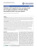
Fig. 1. Tregs play a significant role in virus persistence and the formation, progression, and metastasis of HCC. Teffs differentiate in response to
HBV and tumor antigens, and IFN-g-producing CD4ỵ Th1-cells and CD8ỵ T-cells are the principle immune cells responsible for inhibiting tumor
growth and development. Tregs are mainly induced from CD4ỵCD25 T-cells in the periphery, with cytokines such as TGF-b and IL-10
contributing to this process. iTregs and nTregs show similar suppression functions, inhibiting Teffs and reducing the anti-viral and antitumoral immune response. HBV: hepatitis B virus; APC: antigen-presenting cell; CD: cluster of differentiation; IL-10: interleukin-10; TGF-b:
transforming growth factor-b; iTreg: induced regulatory T-cell; nTreg: natural regulatory T cell; Th: T-helper; CHB: chronic hepatitis B; Tregs:
regulatory T-cells; HCC: hepatocellular carcinoma; Teffs: effector T-cells; IFN-g: interferon-g.
of chronic HBV infection to cirrhosis and HCC. In
addition, activated phenotypes and potent Tregs are
found in tumor sites. The suppressive environment
initiated by Tregs, therefore, is associated with the
chronicity of HBV infection, as well as HCC progression, metastasis, and prognosis (Fig. 1). Tregs should be
considered a target for HCC therapies. However, the
protocols for Treg management remain to be defined.
Conflicts of interest
The authors declare that they have no conflicts of
interest concerning this article.
References
1. Zhang H, Kong H, Zeng X, Guo L, Sun X, He S. Subsets of
regulatory T cells and their roles in allergy. J Transl Med.
2014;12:125.
2. Hiraoka N. Tumor-infiltrating lymphocytes and hepatocellular
carcinoma: molecular biology. Int J Clin Oncol.
2010;15:544e551.
3. Schmetterer KG, Neunkirchner A, Pickl WF. Naturally occurring regulatory T cells: markers, mechanisms, and manipulation. FASEB J. 2012;26:2253e2276.
4. Kakita N, Kanto T, Itose I, et al. Comparative analyses of
regulatory T cell subsets in patients with hepatocellular carcinoma: a crucial role of CD25- FOXP3- T cells. Int J Cancer.
2012;131:2573e2583.
W. Li et al. / Chronic Diseases and Translational Medicine 2 (2016) 67e80
5. Wang R, Kozhaya L, Mercer F, Khaitan A, Fujii H, Unutmaz D.
Expression of GARP selectively identifies activated human
FOXP3ỵ regulatory T cells. Proc Natl Acad Sci USA.
2009;106:13439e13444.
6. Kryczek I, Liu R, Wang G, et al. FOXP3 defines regulatory T
cells in human tumor and autoimmune disease. Cancer Res.
2009;69:3995e4000.
7. von Boehmer H. Mechanisms of suppression by suppressor T
cells. Nat Immunol. 2005;6:338e344.
8. Fontenot JD, Rudensky AY. A well adapted regulatory contrivance: regulatory T cell development and the forkhead family
transcription factor Foxp3. Nat Immunol. 2005;6:331e337.
9. Peng G, Li S, Wu W, Sun Z, Chen Y, Chen Z. Circulating CD4ỵ
CD25ỵ regulatory T cells correlate with chronic hepatitis B
infection. Immunology. 2008;123:57e65.
10. Billerbeck E, Bottler T, Thimme R. Regulatory T cells in viral
hepatitis. World J Gastroenterol. 2007;13:4858e4864.
11. Corthay A. How do regulatory T cells work? Scand J Immunol.
2009;70:326e336.
12. Camisaschi C, Casati C, Rini F, et al. LAG-3 expression
defines a subset of CD4ỵCD25highFoxp3ỵ regulatory T cells
that are expanded at tumor sites. J Immunol.
2010;184:6545e6551.
13. Liu W, Putnam AL, Xu-Yu Z, et al. CD127 expression inversely
correlates with FoxP3 and suppressive function of human
CD4ỵ Treg cells. J Exp Med. 2006;203:1701e1711.
14. Yan J, Zhang Y, Zhang JP, Liang J, Li L, Zheng L. Tim-3
expression defines regulatory T cells in human tumors. PLoS
One. 2013;8:e58006.
15. Knolle PA, Thimme R. Hepatic immune regulation and its
involvement in viral hepatitis infection. Gastroenterology.
2014;146:1193e1207.
16. Sakaguchi S, Setoguchi R, Yagi H, Nomura T. Naturally arising
Foxp3-expressing CD25ỵCD4ỵ regulatory T cells in selftolerance and autoimmune disease. Curr Top Microbiol
Immunol. 2006;305:51e66.
17. Hori S, Carvalho TL, Demengeot J. CD25ỵCD4ỵ regulatory T
cells suppress CD4ỵ T cell-mediated pulmonary hyperinflammation driven by Pneumocystis carinii in immunodeficient mice. Eur J Immunol. 2002;32:1282e1291.
18. Piao WH, Jee YH, Liu RL, et al. IL-21 modulates CD4ỵ
CD25ỵ regulatory T-cell homeostasis in experimental autoimmune encephalomyelitis. Scand J Immunol. 2008;67:37e46.
19. Stoop JN, Claassen MA, Woltman AM, et al. Intrahepatic
regulatory T cells are phenotypically distinct from their peripheral counterparts in chronic HBV patients. Clin Immunol.
2008;129:419e427.
20. Facciabene A, Motz GT, Coukos G. T-regulatory cells: key
players in tumor immune escape and angiogenesis. Cancer Res.
2012;72:2162e2171.
21. Li X, Wang Y, Chen Y. Cellular immune response in patients
with chronic hepatitis B virus infection. Microb Pathog.
2014;74:59e62.
22. Vignali DA, Collison LW, Workman CJ. How regulatory T cells
work. Nat Rev Immunol. 2008;8:523e532.
23. Pang YL, Zhang HG, Peng JR, et al. The immunosuppressive
tumor microenvironment in hepatocellular carcinoma. Cancer
Immunol Immunother. 2009;58:877e886.
24. Gondek DC, Lu LF, Quezada SA, Sakaguchi S, Noelle RJ.
Cutting edge: contact-mediated suppression by CD4ỵCD25ỵ
regulatory cells involves a granzyme B-dependent, perforinindependent mechanism. J Immunol. 2005;174:1783e1786.
77
25. Tanaka M, Katayama F, Kato H, et al. Hepatitis B and C virus
infection and hepatocellular carcinoma in China: a review of
epidemiology and control measures. J Epidemiol.
2011;21:401e416.
26. Stoop JN, Woltman AM, Biesta PJ, et al. Tumor necrosis factor
alpha inhibits the suppressive effect of regulatory T cells on the
hepatitis B virus-specific immune response. Hepatology.
2007;46:699e705.
27. Trehan Pati N, Geffers R, Sukriti, et al. Gene expression signatures of peripheral CD4ỵ T cells clearly discriminate between patients with acute and chronic hepatitis B infection.
Hepatology. 2009;49:781e790.
28. Xu D, Fu J, Jin L, et al. Circulating and liver resident
CD4ỵCD25ỵ regulatory T cells actively influence the antiviral
immune response and disease progression in patients with
hepatitis B. J Immunol. 2006;177:739e747.
29. Zhang Z, Zhang JY, Wang LF, Wang FS. Immunopathogenesis
and prognostic immune markers of chronic hepatitis B virus
infection. J Gastroenterol Hepatol. 2012;27:223e230.
30. Fu JL, Xu DP, Zhao P, et al. The characterization of regulatory
T cells in peripheral blood of HBV-infected patients. Natl Med
J China. 2006;86:1522e1525.
31. Oo YH, Weston CJ, Lalor PF, et al. Distinct roles for CCR4 and
CXCR3 in the recruitment and positioning of regulatory T cells
in the inflamed human liver. J Immunol. 2010;184:2886e2898.
32. Rehermann B. Chronic infections with hepatotropic viruses:
mechanisms of impairment of cellular immune responses.
Semin Liver Dis. 2007;27:152e160.
33. Sprengers D, van der Molen RG, Kusters JG, et al. Analysis of
intrahepatic HBV-specific cytotoxic T-cells during and after
acute HBV infection in humans. J Hepatol. 2006;45:182e189.
34. Belkaid Y. Regulatory T cells and infection: a dangerous necessity. Nat Rev Immunol. 2007;7:875e888.
35. Stross L, Guănther J, Gasteiger G, et al. Foxp3ỵ regulatory T
cells protect the liver from immune damage and compromise
virus control during acute experimental hepatitis B virus
infection in mice. Hepatology. 2012;56:873e883.
36. Bauer T, Sprinzl M, Protzer U. Immune control of hepatitis B
virus. Dig Dis. 2011;29:423e433.
37. Trehanpati N, Hissar S, Shrivastav S, Sarin SK. Immunological
mechanisms of hepatitis B virus persistence in newborns. Indian J Med Res. 2013;138:700e710.
38. Shrivastava S, TrehanPati N, Patra S, et al. Increased regulatory
T cells and impaired functions of circulating CD8 T lymphocytes is associated with viral persistence in Hepatitis B viruspositive newborns. J Viral Hepat. 2013;20:582e591.
39. Kim PS, Ahmed R. Features of responding T cells in cancer and
chronic infection. Curr Opin Immunol. 2010;22:223e230.
40. Fuse S, Molloy MJ, Usherwood EJ. Immune responses against
persistent viral infections: possible avenues for immunotherapeutic interventions. Crit Rev Immunol. 2008;28:159e183.
41. Ganem D, Prince AM. Hepatitis B virus infectionenatural
history and clinical consequences. N Engl J Med.
2004;350:1118e1129.
42. Li J, Qiu SJ, She WM, et al. Significance of the balance between regulatory T (Treg) and T helper 17 (Th17) cells during
hepatitis B virus related liver fibrosis. PLoS One.
2012;7:e39307.
43. Koay LB, Feng IC, Sheu MJ, et al. Hepatitis B virus (HBV)
core antigen-specific regulatory T cells confer sustained
remission to anti-HBV therapy in chronic hepatitis B with acute
exacerbation. Hum Immunol. 2011;72:687e698.
78
W. Li et al. / Chronic Diseases and Translational Medicine 2 (2016) 67e80
44. Xu HT, Xing TJ, Li H, Ye J. Association of T regulatory cells
with natural course and response to treatment with interferonalpha in patients with chronic hepatitis B infection. Chin Med
J (Engl). 2012;125:1465e1468.
45. Franzese O, Kennedy PT, Gehring AJ, et al. Modulation of the
CD8ỵ-T-cell response by CD4ỵ CD25ỵ regulatory T cells in
patients with hepatitis B virus infection. J Virol.
2005;79:3322e3328.
46. Zhang M, Zhou J, Zhao T, et al. Dissection of a circulating
and intrahepatic CD4ỵFoxp3ỵ T-cell subpopulation in chronic
hepatitis B virus (HBV) infection: a highly informative strategy
for distinguishing chronic HBV infection states. J Infect Dis.
2012;205:1111e1120.
47. Liang XS, Li CZ, Zhou Y, Yin W, Liu YY, Fan WH. Changes in
circulating Foxp3ỵ regulatory T cells and interleukin-17producing T helper cells during HBV-related acute-on-chronic
liver failure. World J Gastroenterol. 2014;20:8558e8571.
48. Dong X, Gong Y, Zeng H, et al. Imbalance between circulating
CD4ỵ regulatory T and conventional T lymphocytes in patients
with HBV-related acute-on-chronic liver failure. Liver Int.
2013;33:1517e1526.
49. Niu YH, Yin DL, Liu HL, et al. Restoring the Treg cell to Th17
cell ratio may alleviate HBV-related acute-on-chronic liver
failure. World J Gastroenterol. 2013;19:4146e4154.
50. Zhai S, Zhang L, Dang S, et al. The ratio of Th-17 to Treg cells
is associated with survival of patients with acute-on-chronic
hepatitis B liver failure. Viral Immunol. 2011;24:303e310.
51. Shen C, Yan WZ, Zhao CY, et al. Increased CD4ỵCD25ỵ
regulatory T cells correlate with poor short-term outcomes in
hepatitis B virus-related acute-on-chronic liver failure patients.
J Microbiol Immunol Infect. 2015;48:137e146.
52. Yang J, Yi P, Wei L, et al. Phenotypes and clinical significance
of circulating CD4ỵCD25ỵ regulatory T cells (Tregs) in patients with acute-on-chronic liver failure (ACLF). J Transl Med.
2012;10:193.
53. El-Badawy O, Sayed D, Badary MS, Abd-Alrahman ME, ElFeky MA, Thabit AG. Relations of regulatory T cells with
hepatitis markers in chronic hepatitis B virus infection. Hum
Immunol. 2012;73:335e341.
54. Yang GL, Xu LM, Yao HY, et al. Association between
CD4ỵCD25ỵFoxp3ỵ regulatory T cells and serum transforming growth factor beta 1 in patients with chronic hepatitis
B. Chin J Hepatol. 2009;17:831e834.
55. Wang F, Jing X, Li G, et al. Foxp3ỵ regulatory T cells are
associated with the natural history of chronic hepatitis B and
poor prognosis of hepatocellular carcinoma. Liver Int.
2012;32:644e655.
56. Tang Y, Jiang L, Zheng Y, Ni B, Wu Y. Expression of CD39 on
FoxP3ỵ T regulatory cells correlates with progression of HBV
infection. BMC Immunol. 2012;13:17.
57. Yang G, Liu A, Xie Q, et al. Association of
CD4ỵCD25ỵFoxp3ỵ regulatory T cells with chronic activity
and viral clearance in patients with hepatitis B. Int Immunol.
2007;19:133e140.
58. Miyaaki H, Zhou H, Ichikawa T, et al. Study of liver-targeted
regulatory T cells in hepatitis B and C virus in chronically
infected patients. Liver Int. 2009;29:702e707.
59. Zhang HH, Guo F, Fei R, et al. Inhibition of CD4ỵ CD25ỵ
regulatory T cells in chronic hepatitis B patients. Natl Med J
China. 2008;88:511e515.
60. Zhang HH, Fei R, Mei MH, et al. The frequency, phenotypes
and functions of CD4ỵ CD25ỵ regulatory T cells in
61.
62.
63.
64.
65.
66.
67.
68.
69.
70.
71.
72.
73.
74.
75.
76.
77.
hepatocellular carcinoma patients. Chin J Hepatol.
2007;15:266e272.
Stoop JN, van der Molen RG, Baan CC, et al. Regulatory T
cells contribute to the impaired immune response in patients
with chronic hepatitis B virus infection. Hepatology.
2005;41:771e778.
Fu JL, Xu DP, Shi M, et al. The phenotype and function of
CD4ỵ CD25ỵ regulatory T cells in hepatitis B patients. Chin J
Intern Med. 2006;45:642e645.
Kondo Y, Kobayashi K, Ueno Y, et al. Mechanism of T cell
hyporesponsiveness to HBcAg is associated with regulatory T
cells in chronic hepatitis B. World J Gastroenterol.
2006;12:4310e4317.
Unitt E, Rushbrook SM, Marshall A, et al. Compromised
lymphocytes infiltrate hepatocellular carcinoma: the role of Tregulatory cells. Hepatology. 2005;41:722e730.
Miroux C, Vausselin T, Delhem N. Regulatory T cells in HBV
and HCV liver diseases: implication of regulatory T lymphocytes in the control of immune response. Expert Opin Biol Ther.
2010;10:1563e1572.
Barboza L, Salmen S, Goncalves L, et al. Antigen-induced
regulatory T cells in HBV chronically infected patients.
Virology. 2007;368:41e49.
Tang TJ, Kwekkeboom J, Laman JD, et al. The role of intrahepatic immune effector cells in inflammatory liver injury and
viral control during chronic hepatitis B infection. J Viral Hepat.
2003;10:159e167.
Speletas M, Argentou N, Germanidis G, et al. Foxp3 expression
in liver correlates with the degree but not the cause of
inflammation. Mediat Inflamm. 2011;2011:827565.
Ferri S, Lalanne C, Lanzoni G, et al. Redistribution of regulatory T-cells across the evolving stages of chronic hepatitis C.
Dig Liver Dis. 2011;43:807e813.
Su ZJ, Yu XP, Guo RY, et al. Changes in the balance between
Treg and Th17 cells in patients with chronic hepatitis B. Diagn
Microbiol Infect Dis. 2013;76:437e444.
Niu Y, Liu H, Yin D, et al. The balance between intrahepatic
IL-17ỵ T cells and Foxp3ỵ regulatory T cells plays an
important role in HBV-related end-stage liver disease. BMC
Immunol. 2011;12:47.
Claassen MA, de Knegt RJ, Tilanus HW, Janssen HL,
Boonstra A. Abundant numbers of regulatory T cells localize to
the liver of chronic hepatitis C infected patients and limit the
extent of fibrosis. J Hepatol. 2010;52:315e321.
Castello G, Scala S, Palmieri G, Curley SA, Izzo F. HCVrelated hepatocellular carcinoma: from chronic inflammation to
cancer. Clin Immunol. 2010;134:237e250.
Yang P, Li QJ, Feng Y, et al. TGF-b-miR-34a-CCL22
signaling-induced Treg cell recruitment promotes venous metastases of HBV-positive hepatocellular carcinoma. Cancer
Cell. 2012;22:291e303.
Curiel TJ, Coukos G, Zou L, et al. Specific recruitment of
regulatory T cells in ovarian carcinoma fosters immune privilege
and
predicts
reduced
survival.
Nat
Med.
2004;10:942e949.
Facciabene A, Peng X, Hagemann IS, et al. Tumour hypoxia
promotes tolerance and angiogenesis via CCL28 and T(reg)
cells. Nature. 2011;475:226e230.
Chen KJ, Lin SZ, Zhou L, et al. Selective recruitment of regulatory T cell through CCR6-CCL20 in hepatocellular carcinoma fosters tumor progression and predicts poor prognosis.
PLoS One. 2011;6:e24671.
W. Li et al. / Chronic Diseases and Translational Medicine 2 (2016) 67e80
78. Riezu-Boj JI, Larrea E, Aldabe R, et al. Hepatitis C virus induces the expression of CCL17 and CCL22 chemokines that
attract regulatory T cells to the site of infection. J Hepatol.
2011;54:422e431.
79. Kalathil S, Lugade AA, Miller A, Iyer R, Thanavala Y. Higher
frequencies of GARPỵCTLA-4ỵFoxp3ỵ T regulatory cells and
myeloid-derived suppressor cells in hepatocellular carcinoma
patients are associated with impaired T-cell functionality.
Cancer Res. 2013;73:2435e2444.
80. Zamarron BF, Chen W. Dual roles of immune cells and their
factors in cancer development and progression. Int J Biol Sci.
2011;7:651e658.
81. Ye YB, Peng F, Li JY, et al. Significance of the expression of
lymphocytes and cytokines infiltrating in HCC. Chin J Cell Mol
Imm. 2011;27:1056e1060.
82. Shirabe K, Motomura T, Muto J, et al. Tumor-infiltrating
lymphocytes and hepatocellular carcinoma: pathology and
clinical management. Int J Clin Oncol. 2010;15:552e558.
83. Yang MC, Chang CP, Lei HY. Induction of liver fibrosis in a
murine hepatoma model by thioacetamide is associated with
enhanced tumor growth and suppressed antitumor immunity.
Lab Invest. 2010;90:1782e1793.
84. Zhou J, Ding T, Pan W, Zhu LY, Li L, Zheng L. Increased
intratumoral regulatory T cells are related to intratumoral
macrophages and poor prognosis in hepatocellular carcinoma
patients. Int J Cancer. 2009;125:1640e1648.
85. Ormandy LA, Hillemann T, Wedemeyer H, Manns MP,
Greten TF, Korangy F. Increased populations of regulatory T
cells in peripheral blood of patients with hepatocellular carcinoma. Cancer Res. 2005;65:2457e2464.
86. Fu J, Xu D, Liu Z, et al. Increased regulatory T cells correlate
with CD8 T-cell impairment and poor survival in hepatocellular
carcinoma patients. Gastroenterology. 2007;132:2328e2339.
87. Yang XH, Yamagiwa S, Ichida T, et al. Increase of CD4ỵ
CD25ỵ regulatory T-cells in the liver of patients with hepatocellular carcinoma. J Hepatol. 2006;45:254e262.
88. Kobayashi N, Hiraoka N, Yamagami W, et al. FOXP3ỵ regulatory T cells affect the development and progression of hepatocarcinogenesis. Clin Cancer Res. 2007;13:902e911.
89. Han YF, Zhao J, Ma LY, et al. Factors predicting occurrence
and prognosis of hepatitis-B-virus-related hepatocellular carcinoma. World J Gastroenterol. 2011;17:4258e4270.
90. Ki MR, Goo MJ, Park JK, et al. Helicobacter pylori accelerates
hepatic fibrosis by sensitizing transforming growth factor-b 1induced
inflammatory
signaling.
Lab
Invest.
2010;90:1507e1516.
91. Huang Y, Wang FM, Wang T, et al. Tumor-infiltrating FoxP3ỵ
Tregs and CD8ỵ T cells affect the prognosis of hepatocellular
carcinoma patients. Digestion. 2012;86:329e337.
92. Huang Y, Wang FM, Wang T, et al. Tumor-infiltrating FoxP3ỵ
Tregs are associated with CD34 expression and prognosis of
hepatocellular carcinoma. Chin J Hepatol. 2012;20:25e29.
93. Feng X, Li B, Ye H, Long D. Increased frequency of
CD4ỵCD25highFoxP3ỵ regulatory T cells in patients with hepatocellular carcinoma. Arch Immunol Ther Exp (Warsz).
2011;59:309e314.
94. Wang SY, Fu JL, Lv JY, Chen LM, Lv S, Wang FS. Increase in
peripheral and liver infiltrating regulatory T cells favors
development of primary hepatocellular carcinoma. Chin J Cell
Mol Imm. 2011;27:668e670.
95. Wu H, Chen P, Liao R, et al. Intratumoral regulatory T cells
with higher prevalence and more suppressive activity in
96.
97.
98.
99.
100.
101.
102.
103.
104.
105.
106.
107.
108.
109.
110.
111.
79
hepatocellular carcinoma patients. J Gastroenterol Hepatol.
2013;28:1555e1564.
Guo CL, Yang HC, Yang XH, et al. Associations between
infiltrating lymphocyte subsets and hepatocellular carcinoma.
Asian Pac J Cancer Prev. 2012;13:5909e5913.
Huang Y, Wang F, Wang Y, et al. Intrahepatic interleukin-17ỵ T
cells and FoxP3ỵ regulatory T cells cooperate to promote
development and affect the prognosis of hepatocellular carcinoma. J Gastroenterol Hepatol. 2014;29:851e859.
Thakur S, Singla A, Chawla Y, Rajwanshi A, Kalra N,
Arora SK. Expansion of peripheral and intratumoral regulatory
T-cells in hepatocellular carcinoma: a case-control study. Indian J Pathol Microbiol. 2011;54:448e453.
Flecken T, Schmidt N, Hild S, et al. Immunodominance and
functional alterations of tumor-associated antigen-specific
CD8ỵ T-cell responses in hepatocellular carcinoma. Hepatology. 2014;59:1415e1426.
Han Y, Yang Y, Chen Z, et al. Human hepatocellular
carcinoma-infiltrating CD4ỵCD69ỵFoxp3- regulatory T cell
suppresses T cell response via membrane-bound TGF-b1. J
Mol Med (Berl). 2014;92:539e550.
Wang Y, Deng B, Tang W, Liu T, Shen X. TGF-b1 secreted by
hepatocellular carcinoma induces the expression of the Foxp3
gene and suppresses antitumor immunity in the tumor microenvironment. Dig Dis Sci. 2013;58:1644e1652.
Cao M, Cabrera R, Xu Y, et al. Hepatocellular carcinoma cell
supernatants increase expansion and function of CD4ỵCD25ỵ
regulatory T cells. Lab Invest. 2007;87:582e590.
Peng QQ, Li SP, Xu L, Li JQ. Clinical significance of the
proportion of CD4ỵCD25ỵ regulatory T cells in peripheral
blood of hepatocellular carcinoma patients: a report of 117
cases. Ai Zheng. 2007;26:748e751.
Chen T, Song D, Min Z, et al. Perioperative dynamic alterations
in peripheral regulatory T and B cells in patients with hepatocellular carcinoma. J Transl Med. 2012;10:14.
Nan XP, Zhang Y, Yu HT, et al. Circulating CD4ỵCD25high
regulatory T cells and expression of PD-1 and BTLA on CD4ỵ
T cells in patients with chronic hepatitis B virus infection. Viral
Immunol. 2010;23:63e70.
Pedroza-Gonzalez A, Verhoef C, Ijzermans JN, et al. Activated
tumor-infiltrating CD4ỵ regulatory T cells restrain antitumor
immunity in patients with primary or metastatic liver cancer.
Hepatology. 2013;57:183e194.
Huang Y, Gan JH, Luo EP, Wang XH, Chen L, Yang L. Effect
and clinical significance of glucocorticoid on CD4ỵCD25ỵ
regulatory T cells in patients with hepatitis B virus-related preliver failure. Chin J Hepatol. 2014;22:577e579.
Gao YW, Chen YX, Wang ZM, et al. Increased expression of
cyclooxygenase-2 and increased infiltration of regulatory T
cells in tumors of patients with hepatocellular carcinoma.
Digestion. 2009;79:169e176.
Yang XH, Liu BR, Jiang HC. The presence and the significance
of CD4ỵCD25ỵ regulatory T cells in livers of patients with
hepatocellular carcinoma. Chin J Hepatol. 2007;15:279e282.
Zhang HH, Mei MH, Fei R, et al. Regulatory T cell depletion
enhances tumor specific CD8 T-cell responses, elicited by
tumor antigen NY-ESO-1b in hepatocellular carcinoma patients, in vitro. Int J Oncol. 2010;36:841e848.
Lin SZ, Chen KJ, Xu ZY, et al. Prediction of recurrence and
survival in hepatocellular carcinoma based on two Cox models
mainly determined by FoxP3ỵ regulatory T cells. Cancer Prev
Res (Phila). 2013;6:594e602.
80
W. Li et al. / Chronic Diseases and Translational Medicine 2 (2016) 67e80
112. Zhang HH, Mei MH, Fei R, et al. Regulatory T cells in chronic
hepatitis B patients affect the immunopathogenesis of hepatocellular carcinoma by suppressing the anti-tumour immune
responses. J Viral Hepat. 2010;17(suppl 1):34e43.
113. Gao Q, Qiu SJ, Fan J, et al. Intratumoral balance of regulatory
and cytotoxic T cells is associated with prognosis of hepatocellular carcinoma after resection. J Clin Oncol.
2007;25:2586e2593.
114. Shen SL, Liang LJ, Peng BG, He Q, Kuang M, Lai JM. Foxp3ỵ
regulatory T cells and the formation of portal vein tumour
thrombus in patients with hepatocellular carcinoma. Can J
Surg. 2011;54:89e94.
115. Hu L, Xue F, Li Y, Shao M, Sun Y, Wei G. A long-term followup and comprehensive observation of risk and prognosis factors
of recurrence and survival after resection of hepatocellular
carcinoma. Cell Biochem Biophys. 2014;69:421e431.
116. Lin GH, Wang J, Li SH, Wang J, Xu L, Li SP. Relationship and
clinical significance of TGF-beta1 expression with Treg cell
infiltration in hepatocellular carcinoma. Chin J Cancer.
2010;29:403e407.
117. Litzinger MT, Fernando R, Curiel TJ, Grosenbach DW,
Schlom J, Palena C. IL-2 immunotoxin denileukin diftitox reduces regulatory T cells and enhances vaccine-mediated T-cell
immunity. Blood. 2007;110:3192e3201.
118. Stoop JN, van der Molen RG, Kuipers EJ, Kusters JG,
Janssen HL. Inhibition of viral replication reduces regulatory T
cells and enhances the antiviral immune response in chronic
hepatitis B. Virology. 2007;361:141e148.
119. Li J, Shi J, Ren W, Wu W, Chen Z. Regulatory role of
CD4ỵCD25ỵFoxp3ỵ regulatory T cells on IL-17-secreting T
cells in chronic hepatitis B patients. Dig Dis Sci.
2014;59:1475e1483.
120. Aalaei-Andabili SH, Alavian SM. Regulatory T cells are the
most important determinant factor of hepatitis B infection
prognosis: a systematic review and meta-analysis. Vaccine.
2012;30:5595e5602.
121. Chen KJ, Zhou L, Xie HY, Ahmed TE, Feng XW, Zheng SS.
Intratumoral regulatory T cells alone or in combination with
cytotoxic T cells predict prognosis of hepatocellular carcinoma
after resection. Med Oncol. 2012;29:1817e1826.
122. Lee WC, Wu TJ, Chou HS, et al. The impact of CD4ỵ CD25ỵ
T cells in the tumor microenvironment of hepatocellular carcinoma. Surgery. 2012;151:213e222.
123. Mathai AM, Kapadia MJ, Alexander J, Kernochan LE,
Swanson PE, Yeh MM. Role of Foxp3-positive tumorinfiltrating lymphocytes in the histologic features and clinical
124.
125.
126.
127.
128.
129.
130.
131.
132.
133.
134.
135.
136.
outcomes of hepatocellular carcinoma. Am J Surg Pathol.
2012;36:980e986.
Sasaki A, Tanaka F, Mimori K, et al. Prognostic value of tumorinfiltrating FOXP3ỵ regulatory T cells in patients with hepatocellular carcinoma. Eur J Surg Oncol. 2008;34:173e179.
Li SP, Peng QQ, Ding T, et al. Clinical significance of regulatory T cells proportion in the peripheral blood and tumor
tissue in primary hepatocellular carcinoma. Chin J Oncol.
2008;30:523e527.
Yu X, Guo R, Ming D, et al. Ratios of regulatory T cells/Thelper 17 cells and transforming growth factor-b1/interleukin17 to be associated with the development of hepatitis B virusassociated liver cirrhosis. J Gastroenterol Hepatol.
2014;29:1065e1072.
Sutmuller RP, van Duivenvoorde LM, van Elsas A, et al.
Synergism of cytotoxic T lymphocyte-associated antigen 4
blockade and depletion of CD25ỵ regulatory T cells in antitumor therapy reveals alternative pathways for suppression of
autoreactive cytotoxic T lymphocyte responses. J Exp Med.
2001;194:823e832.
Bertoletti A, Gehring AJ. The immune response during hepatitis B virus infection. J Gen Virol. 2006;87:1439e1449.
Zhou S, Chen L, Qin J, et al. Depletion of CD4ỵ CD25ỵ regulatory T cells promotes CCL21-mediated antitumor immunity.
Plos One. 2013;8:e73952.
Chen L, Zhou S, Qin J, et al. Combination of SLC administration and Tregs depletion is an attractive strategy for targeting
hepatocellular carcinoma. Mol Cancer. 2013;12:153.
Cany J, Tran L, Gauttier V, et al. Immunotherapy of hepatocellular carcinoma: is there a place for regulatory T-lymphocyte
depletion? Immunotherapy. 2011;3:32e34.
Phan GQ, Weber JS, Sondak VK. CTLA-4 blockade with
monoclonal antibodies in patients with metastatic cancer: surgical issues. Ann Surg Oncol. 2008;15:3014e3021.
Oo YH, Sakaguchi S. Regulatory T-cell directed therapies in
liver diseases. J Hepatol. 2013;59:1127e1134.
Alatrakchi N, Koziel M. Regulatory T cells and viral liver
disease. J Viral Hepat. 2009;16:223e229.
Greten TF, Ormandy LA, Fikuart A, et al. Low-dose cyclophosphamide treatment impairs regulatory T cells and unmasks
AFP-specific CD4ỵ T-cell responses in patients with advanced
HCC. J Immunother. 2010;33:211e218.
Nagayama Y, Hase W, Motoyoshi Y, Saitoh O, Sogawa R,
Nakao K. Distinct responses of two hepatocellular carcinoma
cell lines of a similar origin to immunotherapies targeting
regulatory or effector T cells. Oncol Rep. 2007;17:1269e1273.
Edited by Pei-Fang Wei