nanostructure lipid carriers a modish contrivance to overcome the ultraviolet effects
Bạn đang xem bản rút gọn của tài liệu. Xem và tải ngay bản đầy đủ của tài liệu tại đây (1.34 MB, 12 trang )
Egyptian Journal of Basic and Applied Sciences xxx (2017) xxx–xxx
Contents lists available at ScienceDirect
Egyptian Journal of Basic and Applied Sciences
journal homepage: www.elsevier.com/locate/ejbas
Review Article
Nanostructure lipid carriers: A modish contrivance to overcome
the ultraviolet effects
Priyanka Jain, Prerna Rahi, Vikas Pandey, Saket Asati, Vandana Soni ⇑
Department of Pharmaceutical Sciences, Dr. Hari Singh Gour University, Sagar, Madhya Pradesh 470 003, India
a r t i c l e
i n f o
Article history:
Received 28 September 2016
Received in revised form 2 February 2017
Accepted 4 February 2017
Available online xxxx
Keywords:
UV blocker
UV radiation
Nanostructured lipid carriers (NLCs)
Sun Protection Factor (SPF)
a b s t r a c t
Protection of the skin from the ultraviolet radiation is the prime concern of society. An increase in the
adverse effects by ultraviolet (UV) radiation on the skin promoted cosmetic formulators to work in the
area of UV blockers and their effective means of delivery. Nanostructured lipid carriers (NLCs) is a modern
and successful lipid carrier system in the cosmetic world associated with various advantages i.e., stability,
effective drug loading capacity etc. NLCs also permits to load 70% of UV blockers which are sufficient to
obtain recommended Sun Protection Factor (SPF) which makes them suitable delivery systems for topical
application of the UV blockers.
Ó 2017 Production and hosting by Elsevier B.V. on behalf of Mansoura University. This is an open access
article under the CC BY-NC-ND license ( />
Contents
1.
2.
3.
4.
5.
Introduction . . . . . . . . . . . . . . . . . . . . . . . . . . . . . . . . . . . . . . . . . . . . . . . . . . . . . . . . . . . . . . . . . . . . . . . . . . . . . . . . . . . . . . . . . . . . . . . . . . . . . . . . . .
1.1.
Skin and radiation . . . . . . . . . . . . . . . . . . . . . . . . . . . . . . . . . . . . . . . . . . . . . . . . . . . . . . . . . . . . . . . . . . . . . . . . . . . . . . . . . . . . . . . . . . . . . . . .
Adverse effects of UV radiations . . . . . . . . . . . . . . . . . . . . . . . . . . . . . . . . . . . . . . . . . . . . . . . . . . . . . . . . . . . . . . . . . . . . . . . . . . . . . . . . . . . . . . . . . .
2.1.
Sunburn (erythema) and tanning . . . . . . . . . . . . . . . . . . . . . . . . . . . . . . . . . . . . . . . . . . . . . . . . . . . . . . . . . . . . . . . . . . . . . . . . . . . . . . . . . . . .
2.2.
Immune response . . . . . . . . . . . . . . . . . . . . . . . . . . . . . . . . . . . . . . . . . . . . . . . . . . . . . . . . . . . . . . . . . . . . . . . . . . . . . . . . . . . . . . . . . . . . . . . .
2.3.
Skin photoaging. . . . . . . . . . . . . . . . . . . . . . . . . . . . . . . . . . . . . . . . . . . . . . . . . . . . . . . . . . . . . . . . . . . . . . . . . . . . . . . . . . . . . . . . . . . . . . . . . .
2.4.
Skin cancer. . . . . . . . . . . . . . . . . . . . . . . . . . . . . . . . . . . . . . . . . . . . . . . . . . . . . . . . . . . . . . . . . . . . . . . . . . . . . . . . . . . . . . . . . . . . . . . . . . . . . .
2.5.
Eye diseases . . . . . . . . . . . . . . . . . . . . . . . . . . . . . . . . . . . . . . . . . . . . . . . . . . . . . . . . . . . . . . . . . . . . . . . . . . . . . . . . . . . . . . . . . . . . . . . . . . . . .
Sunscreen agents . . . . . . . . . . . . . . . . . . . . . . . . . . . . . . . . . . . . . . . . . . . . . . . . . . . . . . . . . . . . . . . . . . . . . . . . . . . . . . . . . . . . . . . . . . . . . . . . . . . . . .
3.1.
Chemical sunscreens (Organic). . . . . . . . . . . . . . . . . . . . . . . . . . . . . . . . . . . . . . . . . . . . . . . . . . . . . . . . . . . . . . . . . . . . . . . . . . . . . . . . . . . . . .
3.2.
Physical sunscreens (Inorganic) . . . . . . . . . . . . . . . . . . . . . . . . . . . . . . . . . . . . . . . . . . . . . . . . . . . . . . . . . . . . . . . . . . . . . . . . . . . . . . . . . . . . .
Novel drug delivery systems and formulations . . . . . . . . . . . . . . . . . . . . . . . . . . . . . . . . . . . . . . . . . . . . . . . . . . . . . . . . . . . . . . . . . . . . . . . . . . . . . .
4.1.
Liposomes . . . . . . . . . . . . . . . . . . . . . . . . . . . . . . . . . . . . . . . . . . . . . . . . . . . . . . . . . . . . . . . . . . . . . . . . . . . . . . . . . . . . . . . . . . . . . . . . . . . . . .
4.2.
Transfersomes . . . . . . . . . . . . . . . . . . . . . . . . . . . . . . . . . . . . . . . . . . . . . . . . . . . . . . . . . . . . . . . . . . . . . . . . . . . . . . . . . . . . . . . . . . . . . . . . . . .
4.3.
Niosomes . . . . . . . . . . . . . . . . . . . . . . . . . . . . . . . . . . . . . . . . . . . . . . . . . . . . . . . . . . . . . . . . . . . . . . . . . . . . . . . . . . . . . . . . . . . . . . . . . . . . . . .
4.4.
Ethosomes . . . . . . . . . . . . . . . . . . . . . . . . . . . . . . . . . . . . . . . . . . . . . . . . . . . . . . . . . . . . . . . . . . . . . . . . . . . . . . . . . . . . . . . . . . . . . . . . . . . . . .
4.5.
Solid lipid nanoparticles (SLNs) . . . . . . . . . . . . . . . . . . . . . . . . . . . . . . . . . . . . . . . . . . . . . . . . . . . . . . . . . . . . . . . . . . . . . . . . . . . . . . . . . . . . .
Nanostructured lipid carriers (NLCs). . . . . . . . . . . . . . . . . . . . . . . . . . . . . . . . . . . . . . . . . . . . . . . . . . . . . . . . . . . . . . . . . . . . . . . . . . . . . . . . . . . . . . .
5.1.
Imperfect NLCs . . . . . . . . . . . . . . . . . . . . . . . . . . . . . . . . . . . . . . . . . . . . . . . . . . . . . . . . . . . . . . . . . . . . . . . . . . . . . . . . . . . . . . . . . . . . . . . . . .
5.2.
Amorphous NLCs . . . . . . . . . . . . . . . . . . . . . . . . . . . . . . . . . . . . . . . . . . . . . . . . . . . . . . . . . . . . . . . . . . . . . . . . . . . . . . . . . . . . . . . . . . . . . . . . .
5.3.
Multiple NLCs . . . . . . . . . . . . . . . . . . . . . . . . . . . . . . . . . . . . . . . . . . . . . . . . . . . . . . . . . . . . . . . . . . . . . . . . . . . . . . . . . . . . . . . . . . . . . . . . . . .
5.4.
Advantages over other lipid carriers . . . . . . . . . . . . . . . . . . . . . . . . . . . . . . . . . . . . . . . . . . . . . . . . . . . . . . . . . . . . . . . . . . . . . . . . . . . . . . . . .
5.5.
Method of preparation of NLCs . . . . . . . . . . . . . . . . . . . . . . . . . . . . . . . . . . . . . . . . . . . . . . . . . . . . . . . . . . . . . . . . . . . . . . . . . . . . . . . . . . . . .
5.5.1.
Hot homogenization method . . . . . . . . . . . . . . . . . . . . . . . . . . . . . . . . . . . . . . . . . . . . . . . . . . . . . . . . . . . . . . . . . . . . . . . . . . . . . . . .
5.5.2.
Cold homogenization method . . . . . . . . . . . . . . . . . . . . . . . . . . . . . . . . . . . . . . . . . . . . . . . . . . . . . . . . . . . . . . . . . . . . . . . . . . . . . . .
5.5.3.
Solvent-emulsification evaporation method . . . . . . . . . . . . . . . . . . . . . . . . . . . . . . . . . . . . . . . . . . . . . . . . . . . . . . . . . . . . . . . . . . . .
00
00
00
00
00
00
00
00
00
00
00
00
00
00
00
00
00
00
00
00
00
00
00
00
00
00
⇑ Corresponding author.
E-mail addresses: (P. Jain), (P. Rahi), (V. Pandey), (S. Asati),
(V. Soni).
/>2314-808X/Ó 2017 Production and hosting by Elsevier B.V. on behalf of Mansoura University.
This is an open access article under the CC BY-NC-ND license ( />
Please cite this article in press as: Jain P et al. Nanostructure lipid carriers: A modish contrivance to overcome the ultraviolet effects. Egyp. Jour. Bas. App.
Sci. (2017), />
2
P. Jain et al. / Egyptian Journal of Basic and Applied Sciences xxx (2017) xxx–xxx
6.
7.
5.6.
Characterization of NLCs. . . . . . . . . . . . . . . . . . . . . . . . . . . . . . . . . . . . . . . . . . . . . . . . . . . . . . . . . . . . . . . . . . . . . . . . . . . . . . . . . . . . . . . . . . . 00
Patents on nanostructured lipid carriers. . . . . . . . . . . . . . . . . . . . . . . . . . . . . . . . . . . . . . . . . . . . . . . . . . . . . . . . . . . . . . . . . . . . . . . . . . . . . . . . . . . . 00
6.1.
Composite sun-screening agent nano-structure, lipid carrier and its preparation method (Application Number: CN 102697663 B). . . . . 00
6.2.
Formulation of anti-screening agent with nanostructured lipid carrier as its carrier system and its preparation method (Application Number:
CN 102688152 A) . . . . . . . . . . . . . . . . . . . . . . . . . . . . . . . . . . . . . . . . . . . . . . . . . . . . . . . . . . . . . . . . . . . . . . . . . . . . . . . . . . . . . . . . . . . . . . . . . . . 00
6.3.
Anionic lipids and lipid nano-structures and methods of producing and using same (Application Number: US20110059157 A1) . . . . . . 00
6.4.
Nanostructured lipid carriers containing riluzole and pharmaceutical formulations containing said particles (Application Number:
US20100247619 A1, WO2008000448 A3). . . . . . . . . . . . . . . . . . . . . . . . . . . . . . . . . . . . . . . . . . . . . . . . . . . . . . . . . . . . . . . . . . . . . . . . . . . . . . . . 00
6.5.
Sunscreen formulation containing triethanolamine neutralized 2-hydroxy-4-methoxy-benzophenone-5-sulfonic acid (Application Number:
US3670074 A) . . . . . . . . . . . . . . . . . . . . . . . . . . . . . . . . . . . . . . . . . . . . . . . . . . . . . . . . . . . . . . . . . . . . . . . . . . . . . . . . . . . . . . . . . . . . . . . . . . . . . . 00
6.6.
Disappearing color sunscreen compositions (Application Number: US6007797 A) . . . . . . . . . . . . . . . . . . . . . . . . . . . . . . . . . . . . . . . . . . . . 00
6.7.
Amorphous silicon film as a uv filter (Application Number: US3743847 A) . . . . . . . . . . . . . . . . . . . . . . . . . . . . . . . . . . . . . . . . . . . . . . . . . . 00
6.8.
Use of Benzophenone Uv Filters for Preventing Tanning (Application Number: US20070219275 A1) . . . . . . . . . . . . . . . . . . . . . . . . . . . . . 00
Conclusion and future perspective . . . . . . . . . . . . . . . . . . . . . . . . . . . . . . . . . . . . . . . . . . . . . . . . . . . . . . . . . . . . . . . . . . . . . . . . . . . . . . . . . . . . . . . . 00
References . . . . . . . . . . . . . . . . . . . . . . . . . . . . . . . . . . . . . . . . . . . . . . . . . . . . . . . . . . . . . . . . . . . . . . . . . . . . . . . . . . . . . . . . . . . . . . . . . . . . . . . . . . . 00
1. Introduction
UV rays are the component of sunlight, which exerts both
positive and negative effects on living beings. There are three types
of UV radiations, which include UV-A (400–320 nm), UV-B
(320–290 nm) and UV-C (100–290 nm) radiations (Fig. A1). About
95% of UV radiations enters into the earth are about UV-A radiations and form the part of solar radiation, which penetrates deeper
on skin tissues or cells as compared to UV-B radiations [1]. UV-A is
responsible for skin aging, wrinkles, tanning and can lead to the
development of skin cancer. On the other hand UV-B radiation
causes sunburn, weakening of the skin inner tissues, affects human
eye lens and immune system [2]. It is also reported that when the
human body is exposed to the UV-B rays, they are absorbed by the
human cells and results DNA (deoxyribonucleic acid) impairments
which will ultimately lead to death of cells. An excessive exposure
of UV-B radiation, leads to suppression of the immune system
which in turn make the body more vulnerable to herpes simplex
virus, acne, and skin lesion, etc [3]. UV-C is completely absorbed
by the ozone layer [4].
1.1. Skin and radiation
The structure of human skin consists of three main layers (1)
Epidermis (2) Dermis (3) Subcutaneous (Fig. A2). Epidermis consists of five layers, namely stratum basale/germinativum, stratum
spinosum, stratum granulosum, stratum lucidum and stratum corneum [5]. The stratum corneum is the uppermost layer of human
skin made up of flattened dead cells and hold about 25% of total
epidermis. In the stratum corneum due to continuous proliferation
of keratinocytes, corneocytes are formed which are covered by
Fig. A2. Effect of UV radiation on human skin.
cornified protein [6]. Corneocytes tightly bound together to form
a barrier of the skin. Proliferating keratinocytes releases lipid in
this layer which make up the lipid barrier of the skin [7]. In stratum
granulosum layer ‘‘Cornification” takes place which is a unique
process of differentiation and programmed death of the cell in keratinocytes. Next layer, i.e. stratum spinosum consists of immune
cells (Langerhens cells). Langerhans cells are responsible for the
protection against the infections. These cells present about 3–6%
in the epidermis excluding the stratum corneum and over
expressed in stratum spinosum. They play an important role in
immunity in several diseases and involved in maintaining the
immune homeostasis in skin by activating skin resident regulatory
T Cells. [8,9]. The deepest layer, stratum basale/germinativum is
the most germinative part of the epidermis, which shows the
highest mitotic activity. This layer consists of various cells, such
Fig. A1. Schematic representation of various layers of human skin and penetration of UV radiation to the various layers of human skin.
Please cite this article in press as: Jain P et al. Nanostructure lipid carriers: A modish contrivance to overcome the ultraviolet effects. Egyp. Jour. Bas. App.
Sci. (2017), />
P. Jain et al. / Egyptian Journal of Basic and Applied Sciences xxx (2017) xxx–xxx
as pigment producing cells known as melanocytes and merkel cells
(touch receptor) [10]. Next to the epidermis is the dermis layer of
the skin, which provides the mechanical stability to the skin.
Dermis is enriched with blood vessels and lymphatic vessels along
with hair follicles, sweat glands and sebaceous glands. Cells such as
mast cell, lymphocytes and macrophages are also observed in the
dermis. Beneath the dermis, subcutaneous layer is found, which
is attached to the bones and muscles. As similar to the dermis, this
layer is also enriched with blood vessels and nerves, but as they are
bigger as compared to the dermis and consists of adipose tissue,
which provides mechanical protection. Fibroblasts, (responsible
for the production of extracellular matrix and collage) are also present in this layer and is responsible for maintaining the structural
integrity within the connective tissue by secreting extracellular
matrix precursors required for the formation of the connective tissue and various fibres [11].
2. Adverse effects of UV radiations
If the spectrum of radiation is observed closely in concern to the
adverse effects, UV-C is completely washed out by a protective
shield of the ozone layer and UV-A and UV-B creates major problems to human. Some of the adverse effects of these radiations
are discussed below.
2.1. Sunburn (erythema) and tanning
Sunburn is mostly observed in light skin individuals. The
increased blood flow at dermis due to UV radiations is responsible
for sunburning. Further exposure also leads blister and edema [12].
Sunburn is usually seen after 24 h of exposure, caused predominantly by UV-B and short-wavelength UV-A. Sunburn is related
to molecular and cellular changes of inflammatory cells in the dermis i.e. activation of mediators of inflammation such as chemokines, cytokines, prostaglandins, histamine, and nitric oxide. The
severity of sunburn depends on the action of the spectrum. UV-B
is much more energetic than UV-A; therefore very small exposure
to UV-B radiation is responsible for the majority of the erythemal
response.
Tanning is one of the common effects that are observed mainly
due to the elevated response of the melanocytes, which resides in
the basal cell layer [13]. Melanin is a complex polymer of tyrosine
derivatives, which is produced by melanocytes and packaged in
melanosomes. Melanosomes consist of two pigments that are
responsible for skin colour, that is eumelanin (brown/black pigment) and pheomelanin (yellow/red pigment). Skin colour is
decided by the amount of these two pigments within melanosomes
[14]. Tanning, darkening of skin occurs due to the UV rays exposure
of skin for a few hours or days, involves three phases which are (a)
immediate pigment darkening (b) persistent pigment darkening
and (c) delayed tanning. Immediate pigment darkening is observed
within few minutes of UV exposure. Persistent pigment darkening
is observed after 1–2 h of exposure to UV rays and may last for
3–5 days and delayed tanning are seen after 2–3 days of exposure.
Among all the three, delayed tanning effect lasts for several weeks
to months because of the synthesis of new melanin occurs during
this phase. Although, tanning depends on the time of UV exposure
and the individual’s skin type [15].
2.2. Immune response
UV radiation also affects the immune system. Langerhens cells
(LC) present in the skin are an element of the immune system
which on interaction with UV radiation leads to specific responses
i.e. delayed type hypersensitivity, contact hypersensitivity etc.
3
Kripke et al, 1992 reported that DNA damage is the main cause
of delayed and contact type hypersensitivity [16].
2.3. Skin photoaging
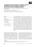
Human skin consists of collagen fiber and various other proteins
which contribute to the formation of extracellular matrix. Complex
network of collagen prevents deformation and elastic fiber provides elasticity to the skin. On the other side UV radiation induces
oxidation and the consequential reactive oxygen species (ROS)
affect the expression of several key transcription factors especially
activator protein 1 (AP-1) and Transforming Growth Factor-b
(TGF-b). AP-1 and TGF-b triggers the synthesis of matrix metalloproteases (MMP) which degrades dermal collagen and affects other
skin molecules. It also affects the elasticity of the skin which, leads
to photoaging and it is identified by wrinkling of skin, persistent
hyper pigmentation, roughness, and irregular pigmentation. When
radiation UV-A and UV-B are compared in case of skin photoaging,
UV-A photons are more energetic and most responsible for skin
photoaging, sun burning, and tanning etc [17]. This whole process
is summarized with the help of Fig. A2.
2.4. Skin cancer
Skin cancer is the result of mutations induced by UV radiations.
Genes involved in the development of skin cancer are (1) p53,
which is involved in tumor suppression, induction of DNA repair
as well as apoptosis (2) patched gene, involved in regulation of cell
proliferation and differentiation (3) ras (retrovirus-associated DNA
sequences), involved in protooncogenes in cell membranes. The
number of investigations has detected p53 gene mutation in Squamous Cell Carcinoma (SCC), Basal Cell Carcinoma (BCC) and Actinic
ketososis. UV exposure involved in the development of BCC is
detected by patched (Ptc) mutation. Ptc gene works by repressing
the activity of gene which are involved in cell growth and differentiation. This is also possible by the opposition of hedgehog (hh)
gene activity. Hedgehog gene is one which encodes for signaling
protein that induces cell growth and differentiation [18,19].
2.5. Eye diseases
UV rays cause deleterious effects on the eye which may include
cataract, pterygium, and photokeratitis etc. Cataract which is a
cloudiness of the lens inside the eye is a major cause of blindness
worldwide. Pterygium is a development of tissue on the white of
the eye that may extend onto the clear cornea so responsible for
blocking vision. Photokeratitis is caused by excessive UV-B exposure of the cornea results in temporary loss of vision [1].
All these are the detrimental effects of UV exposure. The use of
protective clothing, avoiding sun exposure, and the application of
sunscreen is the most common practice to protect the exposure
from excessive sun rays. Out of these, the application of sunscreens
remains the most popular protection used by the public. There are
various sunscreen agents that effectively work to protect the skin
from the harmful effect of UV radiation are discussed in the following section.
3. Sunscreen agents
Skin which is largely affected by the solar radiations has its own
mechanism to combat its harmful effect of UV by the perforation of
the stratum corneum and increased melanin secretion. These two
mechanisms are not sufficient to prevent or subside solar radiation
effects as a result the use of sunscreen is now become the most
popular method which is used by a large population of the society
Please cite this article in press as: Jain P et al. Nanostructure lipid carriers: A modish contrivance to overcome the ultraviolet effects. Egyp. Jour. Bas. App.
Sci. (2017), />
4
P. Jain et al. / Egyptian Journal of Basic and Applied Sciences xxx (2017) xxx–xxx
Fig. A3. Diagrammatic representation of the chemical sunscreen mechanism of action.
Fig. A4. Diagrammatic representation of the physical sunscreen mechanism of action.
[20,21]. In the last few years sunscreen products are introduced in
the cosmetic world to protect the skin from the harmful effects of
UV rays. Sunscreen products consist of both organic as well as inorganic UV blockers out of which organic sunscreen compounds
remains on the upper epidermis and absorbs solar radiations,
whereas inorganic compounds reflects and scatter the radiations.
Various broad spectrum sunscreen agents are incorporated in different formulations such as gels, lotions, creams, ointments and
hydrogels by various scientists [22]. To show its maximum efficiency an ideal sunscreen agent should have certain properties
included (a) Neutral (b) Non- toxic (c) Compatible with various
adjutants (d) Effective at 200–400 nm wavelength (e) Photostable
(f) Non-irritant (g) No systemic effect. Nowadays UV exposure
and its harmful effects are inescapable. Therefore, these sunscreen
agents have proven excellent in preventing the damaging effects of
UV radiations.
Sunscreen agents or UV blockers are broadly divided into two
groups (1) Chemical sunscreens (2) Physical sunscreens.
3.1. Chemical sunscreens (Organic)
Chemical sunscreens are the agents which absorb UV radiation
and convert them into a harmless energy which does not have any
damaging effects on the skin as shown in (Fig. A3). Their action is
restricted to the superficial layer of the skin rather than the systemic
action. Molecules grouped in this category are always found superior to that of the physical ones as they are easily applied and effectively interact with the UV radiations. But due to penetration into
the superficial part of skin, may leads some of the adverse effects
upon regular use. Chemical (organic) sunscreens works by absorbing high-energy UV rays to protect from these harmful rays. They
are called as chemical sunscreen because chemical changes will
be there in sunscreens molecules to prevent UV radiation reaching
the skin [23]. Examples include para-aminobenzoic acid (PABA)
and PABA esters, salicylates, cinnamates, benzophenones etc.
3.2. Physical sunscreens (Inorganic)
These are the sunscreen which forms layer over the skin and
reflects the incident solar radiations as shown in Fig. A4. Physical
(inorganic) sunscreens work by scattering the microparticles in
the upper layers of skin, which may able to divert the optical pathway of photons (of UV radiation). Physical phenomena, such as
scattering and reflection of radiation are involved in the protection
of the skin from the UV radiation. Examples include titanium
dioxide, zinc oxide, iron oxide, kaolin etc., which are inert and
non-irritant substances. The most common inorganic UV filters
include titanium dioxide (TiO2) and zinc oxide (ZnO) [23–25].
Please cite this article in press as: Jain P et al. Nanostructure lipid carriers: A modish contrivance to overcome the ultraviolet effects. Egyp. Jour. Bas. App.
Sci. (2017), />
P. Jain et al. / Egyptian Journal of Basic and Applied Sciences xxx (2017) xxx–xxx
5
Table A1
FDA approved sunscreen agents.
U.V Blockers
Range
covered
(nm)
Description
Para amino benzoic acid (PABA)
290–340
Sulisobenzone(Benzophenone-4)
290–340
3-Benzylidene camphor
290–320
Bemotrizinol(BEMT)
290–340
Butyl Methoxydibenzoylmethane
(Avobenzone)
340–400
Camphor benzalkonium methosulphate
290–340
Diethylamino hydroxybenzoyl hexyl
benzoate
Diethylhexyl Butamido Triazone
340–400
Drometriazole trisiloxane (silatriazole;
Mexoryl XL)
Cinoxate
290–340
Effective UV-B filters when used in a 5% concentration in 50–60% alcohol base. It penetrates deep into the
dermis, acidic in nature. Current use in sunscreen formulations is limited due to its in vitro carcinogenicity
and allergic reactions (contact and photoallergic) [27]
It is a broad spectrum sunscreen agent approved by the FDA in a concentration of 5%. Highly stable and
strong oxidizing agent. Sulisobenzone is water insoluble in its acid form [27]
3-Benzylidene camphor is used as a sunscreen agent at levels up to 2%; highly stable. The dermal
absorption of the 3-Benzylidene camphor is very low. It is a potential endocrine disruptor and also shows
multiple hormonal activities. Soluble in absolute alcohol and isopropanol. Insoluble in water [27]
Bemotrizinol absorbs ultraviolet radiations in both UV-A and UV-B range. It is oil soluble chemical. Highly
photostable. Its presence in formulation also protects less photostable UV blockers, such as avobenzone
[27]
Highly effective for UV-A radiations. It is used in combination with other sunscreen agents to cover a broad
spectrum of UV radiation. Concentrations up to 3% is used as an effective sunscreen agent. Insoluble in
water, soluble in ethanol [27]
Camphor benzalkonium methosulphate is used as UV-filter at a maximum concentration of 6.0%. Mild
irritating potential to the eye when used undiluted or as a 20% aqueous solution. It has a borderline margin
of safety [27]
An effective UV-A filter; oil-soluble yellow liquid. Widely used as sun protective agent at concentrations up
to 10%. It has good compatibility with other UV filters and other ingredients. High Photostability [27]
In European country, it is used as a UV filter in cosmetics and personal care products at a maximum
concentration of 10%. Although FDA approved up to 3 percent only. At 3% concentration, it is efficient and is
not irritant as well as non sensitizer, photosensitizer or photoirritant [27]
Photostable broad spectrum UV filter. An allergic reaction is rarely seen [27]
Dioxybenzone
290–340
Ecamsule (Mexoryl SX)
340–400
Homomenthyl salicylate (Homosalate)
290–320
Meradimate (Menthyl Anthranilate)
Octocrylene
340–400
290–320
Ethylhexyl methoxycinnamate and
octinoxate
IMC (Amiloxate)
4–4-Methylbenzylidene camphor
(enzacamene)
Methylene-bis-benzotriazolyl
tetramethylbutylphenol (Tinasorb M)
290–320
340–400
290–320
290–320
290–320
290–340
Ethylhexyl Triazone
290–320
Ethylhexyl salicylate(octylsaliclate;
octisalate)
Oxybenzone
290–320
Padimate O
290–320
Titanium Dioxide
290–340
Zinc oxide
290–340
290–340
A light yellowish liquid, insoluble in water, but soluble in glycerol and various vegetable oil. Covers the
spectrum of UV-B [23]
Broad spectrum UV Blocker from the family of benzophenone used in various other cosmetic formulations,
but it is reported as sensitizing agent [23]
Photostable. Has a good safety profile as compared to other UV-A blockers. It is not absorbed by the skin
and effectively covers the entire spectrum of UV-A [23]
Salycilate derivative, oil soluble. Has a good safety profile, but used in combination with other UV blocker
for effective sun blocking activity. Covers, UV-B spectrum. Up to 10% w/w used as a sun screen agent [23]
Rarely used anthranilates and it covers only a specific spectrum of UV-A [23]
Effective for UV-B range. Photostable, moisturizing effect is observed due to its ethyhexol portion of the
molecule. Used in combination with other UV blockers [23]
Used for the protection against UV-B radiation. Water insoluble cinnamate. Used in combination due to its
instability [23]
Insoluble in water, soluble in ethanol and other organic solvents, effective UV-B sunscreen agent, safe [23]
Ability to protect the skin against UV-B radiation. It also showed estrogenic effect [23]
Absorb both UV-A and UB-A radiation It is a new class of UV filters that combine the properties of both UV
conventional filters (organic and inorganic) – it scatters, reflects and absorbs UV light. It is colorless organic
microfine particles and photostable. Very less systemic absorption [23]
Absorb UV light at maximum 5% concentration. Oil-soluble UV-B filter. Insoluble in water, which makes it
suitable for water-resistant products. It has excellent photostability[23]
Octyl salicylate is an oil soluble sunscreen agent, efficient for UV-B radiation. Salicylates are weak UV-B
absorbers. Used in combination with other UV filters. Has a good safety profile [23]
Broad spectrum UV filters from the family of benzophenone. Most popular UV blocker. Photostable.
Carcinogenic activity is also observed hence its regular use is still argued [28]
PABA derivative, compatible with various cosmetic ingredients. Used in combination with other UV blocker
to attain greater and effective sun blocking agent [28]
A physical sunscreen agent. Photostable. Less reactive in nature. Used in micronized and nanosized for their
maximum efficacy [28]
Used with titanium dioxide in majority of marketed formulation. zinc oxide is the only sunscreen
ingredient that appears on more than one FDA monograph. Microfine zinc oxide effective in the UV-A
protection [28]
The molecules of these physical sunscreens are smaller in size,
which helps in the reflection and scattering of the radiation [26].
Skin can be protected from UV-A and UV-B by both inorganic
and organic UV filters. Some of the sunscreen agents which are
approved by Food and Drug Administration (FDA) are enlisted in
Table 1.
4. Novel drug delivery systems and formulations
Nanotechnology is one of the flourishing technologies in the
pharmaceutical industries in which drug and their delivery systems are designed and structured by controlling their size in nano
range. The delivery system bearing sunscreen/UV filters must be
suitable enough to deliver sun protectants to the predetermined
site in the sufficient amount [29]. An ideal drug delivery system
must have following qualities[30].
a) Maximum drug loading capacity
b) Enhance drug stability
c) Provide targeted and sustained action.
The nano ranged UV filters provide better action with long lasting effects. The utilization of the nano sized material was increased
due to the fact that nano ranged substance have different properties than larger sized particle, and have altered physiochemical
Please cite this article in press as: Jain P et al. Nanostructure lipid carriers: A modish contrivance to overcome the ultraviolet effects. Egyp. Jour. Bas. App.
Sci. (2017), />
6
P. Jain et al. / Egyptian Journal of Basic and Applied Sciences xxx (2017) xxx–xxx
properties. The carrier systems may be modified according to the
use and the cosmetic drugs/agents to be delivered [31].
Various drug carrier systems used for sunscreen agents and
basically for topical drug delivery systems includes (1) Liposomes
(2) Transferosomes (3) Niosomes (4) Ethosomes (5)Solid Lipid
Nanoparticles, NLCs etc [32–33].
4.1. Liposomes
Liposomes are the spherical vesicles with an aqueous core in
the center, surrounded by lipid layer. Lipid layer is formed by phospholipids and cholesterol [34]. Structurally liposomes are equipped
with both lipid and aqueous phases which make them to carry
both lipophilic and hydrophilic drugs. Liposomes can be classified
on the basis of their size which includes (a) Small unilamellar vesicles (SUVs) (b) Large unilamellar vesicles (LUVs) (c) Multilamellar
vesicles (MLVs) (d) Oligolamellar (OLVs) [35]. The role of phospholipids is to produce bilayer whereas, cholesterol is used to provide
stability to the bilayer. There are various methods adopted to prepare liposomes are lipid film hydration, solvent injection, emulsification and reverse phase evaporation method. When the size of the
liposomes are required to optimized then some other techniques
such as sonication, extrusion and high pressure homogenization
are commonly used. The structural benefits of liposomes are their
similarity to biological membranes of the body which helps them
to easily penetrate and deliver the content. The drugs, depending
upon their characteristic and affinity either get into the aqueous
phase or bilipid layer. But the disadvantage which associated with
liposomes is the low entrapment efficiency of hydrophilic cosmetic
agents and unstability due to phospholipids [36].
Liposomes are effective drug delivery systems for antibiotics,
proteins as well as for sunscreen agents and other cosmetic agents.
More than 10% of the cosmetic market consists of liposomes as a
drug delivery system. Lipid used for the liposomes are specifically
stratum corneum compatible, which helps in effectively depositing
the drug topically. Liposomes also provide a moisturizing effect
due to the presence of skin friendly phospholipids. Formulations
with liposomes has good adherence to the skin surface and therefore they are not easily washed away. In one study the liposome
bearing Sodium ascorbyl phosphate was prepared which showed
enhanced sodium ascorbyl phosphate penetration through the epidermal membrane as compared to sodium ascorbyl phosphate as
in simple water solution [37]. Liposomes are biodegradable and
provide sustained delivery of UV blockers. Toxicity effects of
encapsulated agents are also minimized to a greater extent [38].
Kitagawa et al prepared the cationic liposomes bearing retinoic
acid by using double-chained cationic surfactant(dimethyldipalmi
tylammonium) and phosphatidylcholine. These cationic liposomes
enhance the delivery of retinoic acid about two-fold, which
indicates the potential use of the cationic liposomes for the intradermal delivery of lipophilic drugs like retinoic acid [39].
4.2. Transfersomes
Various vesicular systems have already developed as an effective drug delivery system in the cosmetic industry and transferosomes is one of the such vesicular system and also known as
‘‘elastic vesicular” system. The difference between liposome and
transfersomes is the use of edge activator which is basically the
surfactant. Surfactants are used in the preparation to deform the
lipid layer of vesicles. This deformation induced by the added surfactant helps in the better penetration into the skin [40]. Non
occlusive nature enhances the effective function of transfersomes
and perfect deposition of the drug [41]. Due to the deformation
observed in the lipid layer provide transfersomes a structural
benefit to form a depot, which help in slow release of drug as well
as extrude themselves from the pores of intracellular lipid of stratum corneum. Transfersomes are the best vesicular systems for
topical administration of various drugs which are needed to be
localized. An improved skin deposition and photostability was
observed when a–tocopherol was administered topically in the
form of transferosome [42]. Another reported drugs like triamicinolone acetonide [43], oestradiol [44] and cyclosporin A [45] which
are successfully encapsulated in transfersomes for topical delivery.
4.3. Niosomes
Niosomes are those vesicular systems which are similar to liposome, but made from nonionic surfactant and cholesterol for the
formation of bilayer. They are superior to other vesicular system
in terms of stability and ultimately its shelf life [46]. The advantage
of using a nonionic surfactant in the formation of bilayer is to
increase permeability and bioavailability of the drug entrapment
[47]. Method of preparation are same as that of liposomes, which
includes sonication, extrusion etc. Vesicular formulation are advantageous in cosmetic application as various ingredients such as
antioxidant, fatty acids, vitamins and UV blockers are successfully
encapsulated either in the bilayer or aqueous core. The content of
the carrier resides on the skin surface, i.e. the upper layer of the
stratum corneum and provide effective localized action. Drugs such
as enoxacin [48], b-galactosidase [49], interferon a, cyclosporine
[50], estradiol [51], have also been delivered transdermally through
niosomes.
4.4. Ethosomes
As similar to liposomes and niosomes, ethosomes are also composed of phospholipids, but vesicles are prepared with the help of
ethanol and water. The size of vesicular systems depends on the
concentration of phospholipids and ethanol [52]. The striking feature of these carrier systems is the use of ethanol, which allows
the entrapment of the different nature of drugs such as hydrophilic,
lipophilic and amphiphilic molecule [53]. The cholesterol is the fluidity buffer used in liposomes as well as in ethosomes. Ethanol
helps in deformation or disruption of the upper layer of skin and
provides the entry of ethosomes to enhance drug delivery of trihexylphenidyl hydrochloride [54] testosterone [55], acyclovir [56]
ammonium glycyrhizinate [57] and bacitracin [58]. The release of
drug in the deep layers of the skin and transdermal absorption is
the result of fusion of ethosomes with skin lipids and drug release
is observed at various points along the penetration pathway [59].
Improved therapeutic effectiveness and permeation of antibiotics
and antibacterial from these vesicular systems are also reported
[60,61].
4.5. Solid lipid nanoparticles (SLNs)
SLNs are the nano sized lipophilic matrix in which drug is
effectively encapsulated. Lipids utilized in the preparation of SLNs
Fig. A5(a). Imperfect NLCs.
Please cite this article in press as: Jain P et al. Nanostructure lipid carriers: A modish contrivance to overcome the ultraviolet effects. Egyp. Jour. Bas. App.
Sci. (2017), />
P. Jain et al. / Egyptian Journal of Basic and Applied Sciences xxx (2017) xxx–xxx
Fig. A5(b). Amorphous NLCs. (c) Multiple NLCs.
7
NLCs are one such kind of nano-carriers that has conquered a
better place than other carrier systems mainly in topical preparations. The structural aspect of NLCs due to which higher loading
capacity is observed can be justified by the fact that a larger
fraction of drugs are soluble in liquid lipids and when solid and liquid lipids are blended together the liquid lipids accommodated in
the core space surrounded by solid lipids, in this way drugs are
accurately encapsulated [71].
Identified structure of NLCs obtained by matching various
compositions as well as parameters are: (1) Imperfect NLCs (2)
Amorphous NLCs (3) Multiple NLCs.
5.1. Imperfect NLCs
These NLCs are produced by the mixing of solid lipids and
chemically vary different liquid lipids. To increase the drug loading
capacity, glycerides composed of different fatty acids are used.
Because of the distance in the fatty acid chain leads to the formation of imperfections in the crystal (Fig. A5(a)). The imperfections
are the result of the incompatibility between lipids and intentionally utilized for the achievement of higher loading capacity, hence
thus makes important to choose incompatible lipids [72].
Fig. A5(c). Multiple NLCs.
5.2. Amorphous NLCs
include fatty acids, waxes, glycerides, triglycerides etc. Method of
preparation of SLNs includes hot homogenization, high pressure
homogenization, high shear homogenization, ultra sonication, melt
dispersion, microemulsion dilution, microemulsion cooling, coacervation, solvent injection, solvent evaporation, supercritical fluid
extraction of emulsion and spray drying etc. SLNs are termed as
the first generation, lipid based nanoparticles and have wide applicability in pharmaceutical medicine due to chemical stability,
physical stability and biocompatibility with large number of drugs.
For providing stability to the system, emulsifiers such as poloxamer 188, polysorbate 80, fatty acid ester etc are used [62]. Cold
homogenization is better suited for heat sensitive compounds or
substances which can be easily partitioned from the melted lipid
[63]. It is well reported that the therapeutic active substance such
as clobetasol propionate [64], antiandrogen etc [65] was delivered
successfully with SLNs. The formulation of SLNs was found to be
localized in the outer layer skin with minimum systemic circulation. Retinol, tocopherol and coenzyme Q10 compounds are protected from degradation when successfully incorporated into
SLNs [66]. SLNs have several other advantages such as modified
release of the active compound, lipophilic and hydrophilic drugs
incorporated easily and increased in skin hydration. However,
drawbacks associated with SLNs are uncontrolled drug expulsion
from the carrier and limited drug loading capacity. To overcome
these limitations, a second generation of lipid nanoparticles, NLCs,
has been developed [67,68].
The crystallization process leads to the expulsion of drugs and
therefore NLCs which are solid are preferred over crystalline one
with the use of special lipids (hydroxyoctacosanylhydroxy-stea
rate, isopropylmyristate) by which particle acquires solid state
rather than crystalline (Fig. A5(b)) [73].
5.3. Multiple NLCs
Multiple NLCs are prepared by mixing solid lipids with large
amount of oil (liquid lipids), small nanocompartments within
nanoparticles are created by a phase separation process during
particles production. The solid matrix of the lipid nanoparticles
contains tiny liquid nanocompartments of oil. In these oil compartments the drug has high solubility (Fig. A5(c)). The oil compartments formed are surrounded by solid lipids and hence
controlled drug release was observed [74].
5.4. Advantages over other lipid carriers
Comparison between NLCs and SLNs reflects that SLNs require
pure solid lipids, which leads to the formation of the perfect carrier
structure, whereas in the case of NLCs imperfect/distorted structure was observed, which allows more of the drugs to be fitted in
5. Nanostructured lipid carriers (NLCs)
For the delivery of drug and cosmetics, further improvement in
lipid based carrier systems, paved way to a new generation of SLNs,
which was termed as nanostructured lipid carriers (NLCs) [69].
NLCs composed of both solid and liquid lipids. Liquid Lipids (oil)
incorporation causes structural imperfections of solid lipids due
to which a perfect crystalline structure is deviated to form a crystal
lattice with many spaces. The spaces are assumed to be imperfection, but these are the actual spaces where drug homes itself.
Hence the liquid lipid being used, determines the state of nanocarriers as well as its loading capacity [70]. Release pattern of the
active constituents is also based on the blend of solid and liquid
lipids.
Fig. A6(a). Flow chart of the hot homogenization method.
Please cite this article in press as: Jain P et al. Nanostructure lipid carriers: A modish contrivance to overcome the ultraviolet effects. Egyp. Jour. Bas. App.
Sci. (2017), />
8
P. Jain et al. / Egyptian Journal of Basic and Applied Sciences xxx (2017) xxx–xxx
5.5.1. Hot homogenization method
High pressure homogenization is the conventional method for
the fabrication of NLCs. The advantages associated with this
method includes, short production time, limited use of various
other chemicals and easy scale up. In this method active pharmaceutical ingredient is dissolved in a mixture of melted lipids, the
resulted mixture is quickly dispersed in aqueous emulsifier with
high speed stirring. Temperature is maintained constant during
the whole process. The prepared emulsion is subjected to high
pressure homogenization with high ultrasonic intensity which
converts the emulsion to nano range emulsion. Cooling is done
either in cold water or by a heat exchanger and precipitate of
nanoparticles is collected (Fig. A6(a)). Disadvantage associated
with this method is the degradation of heat sensitive ingredients
due to the temperature [81].
Fig. A6(b). Flow chart of cold homogenization method.
the imperfect site and an overall increase in the drug loading
capacity was seen. Liposomes and various emulsions are also studied in terms of stability of the active constituents in comparison to
NLCs and it was observed that liposomes have limited protection
against the chemical degradation. Active drugs in case of liposomal
formulations can be placed either in the aqueous core or phospholipids bilayer and if the drug partitions in the phase which is
incompatible for drug, degradation may occur. Some experiments
were performed to demonstrate stability of lipid nanoparticles
[75]. Presently, NLCs are used as novel drug delivery system owing
to its several advantages which includes solubility enhancement of
poorly soluble drugs, reduces skin irritation, better physical stability, ease of manufacturing and scale-up, high entrapment efficiency of both the lipophilic s and hydrophilic drugs, controlled
particle size, occlusive in nature and provide extended release of
the drug [76,77,26].
When topical formulations are concerned, adhesiveness is
required for film formation. Adhesiveness is the property of fine
material which is directly related to occlusion. Adhesiveness
increases with decreasing particle size. NLCs have enhanced adhesive property; they adhere to the skin surface which ultimately
leads to the formation of a film over the skin and provide occlusion
effect. The occlusion can be increased by reducing the particle size
or at a given particle size by increasing the number of particles i.e.
increasing lipid concentration. Therefore, nanoparticles provide
‘‘controlled occlusion effect”. Skin hydration is another important
factor because it promotes penetration of the drug in the skin. NLCs
also maintain sufficient skin hydration by the formation of occlusive layer over the skin. Other important parameters such as the
size of the carriers as well as drugs which are an important tool
to avoid systemic effects. If the physical stability of the system is
concerned, SLNs and NLCs have proven themselves far better than
any other system due to the presence of solid matrix [78].
NLCs are suitable carriers for the sunscreen agents because
these agents are the active material place itself in the solid matrix
causes delayed and prolonged drug release [79]. Also the lipids utilized for NLCs act as UV filters which provides synergistic effect
and because of this synergistic effect, the required quantity of sunscreen agent will decrease for sunscreen action.
5.5. Method of preparation of NLCs
Production of NLCs are closely related to SLNs. The most common methods used for their preparation are hot homogenization
method, cold homogenization method and solvent emulsification
evaporation method [80].
5.5.2. Cold homogenization method
As the name suggests that the temperature used in the whole
process is lower than that used in a hot homogenization process
which ultimately rule out disadvantage that may be produced
due to heat. The mixture of the lipids with the drug is rapidly
cooled by the utilization of liquid nitrogen. The lipid matrixes
obtained are milled and then the particles are dispersed in the
emulsifier solution and subsequently homogenized to produce fine
particle (Fig. A6(b)). Various advantages of this process over the
hot homogenization process are: 1. Thermal degradation is minimized. 2. Improved drug entrapment efficiency 3. Uniform distribution of drug within the lipid [82]. In comparison to the hot
homogenization method, larger particle sizes and a broader size
distribution are observed in cold homogenized method. Although
cold homogenization minimizes the thermal exposure of the sample, but it cannot be completely avoidable as melting of the lipid/
drug mixture is required in the initial step.
5.5.3. Solvent-emulsification evaporation method
In this method, lipids and drugs are mixed with certain solvent
and resulted mixture is quickly dispersed in the emulsifier solution
and the solvent is evaporated by the reduction in pressure which
leaves behind required nanoparticles[83].
Some of common lipids used for the preparation of NLC’s are
discussed with their structure and properties in Table 2.
5.6. Characterization of NLCs
Characterization is an important aspect to understand nanomaterials and their possible applications. Nanostructures have a physical size, which is the important characteristic for their applications.
Structure and properties of any nanoparticles formulations depend
on the environment exposed, which leads to structural transformation, agglomeration, etc. These changes in the nanostructure have
to be identified and studied for their better implications. Table 3
describes the characterization parameters required for NLCs.
6. Patents on nanostructured lipid carriers
6.1. Composite sun-screening agent nano-structure, lipid carrier and
its preparation method (Application Number: CN 102697663 B)
The invention discloses a novel sunscreen composite nanostructured lipid carriers, the carrier loaded with ethylhexyl triazone and
diethylamino hydroxylbenzoyl hexyl benzoate. Nanostructured
lipid carrier system was prepared according to weight percentage
of ethylhexyl triazone 1–10% and diethylamino hydroxyl benzoyl
hexyl benzoate 2–20% and 3–15% of emulsifier. The rest is deionized water; the composition material is a mixture of solid lipid
Please cite this article in press as: Jain P et al. Nanostructure lipid carriers: A modish contrivance to overcome the ultraviolet effects. Egyp. Jour. Bas. App.
Sci. (2017), />
P. Jain et al. / Egyptian Journal of Basic and Applied Sciences xxx (2017) xxx–xxx
9
Table A2
Lipids Used in the NLCs preparation.
Structure
A. Liquid lipid
Medium Chain
Triglyceride/Caprylic
Triglyceride
Corn Oil
Description
Fatty acid with 6–12 carbon atom and glycerol as backbone. Stable against oxidation,
used as solvent, emulsifier and vehicle. These are the lipids with different molecular
weight and are easily digestible [84]
Natural oil and an antioxidant, protects the drug from oxidation due to high unsaturation.
Highly viscous [85]
a-Tocopherol
Yellowish viscous liquid, soluble in acetone, ethanol and chloroform. They are unstable in
UV light and used as antioxidant and vitamin E supplement [86]
Squalene‘
Translucent liquid. Not used in high concentration. It is a good moisturizer and less
susceptible to oxidation. It is an important part of steroid synthesis [87]
Oleic Acid
Yellowish oil, water insoluble. It has low viscosity. On exposure to air it oxidizes. It has
been reported to effectively penetrate into the skin and also through hair follicle [88]
B. Solid lipid
Cetyl Palmitate
Stearic Acid
Tristearin
Ester of palmitic acid. Naturally occurs in the wax found in the skull of sperm whale.
Water insoluble, pharmaceutically used for skin conditioning and emollient action. Also
used due to its property of excellent film former [89]
Saturated fatty acid with 18 carbon backbone. Soluble in acetone and slightly soluble in
ethanol. Ability to penetrate skin and even mucous membrane make good candidate for
NLC preparations [90]
White, odorless powder, insoluble, emollient in nature, solvent and skin conditioning
agent. Controls viscosity of the formulation [91]
Propylene Glycol
Monostearate
Soluble in water, colorless liquid. Used as humectants and solvent in various
formulations. It is a common ingredient in personal care product [92]
Glyceryl Monostearate
It is glycerol ester of stearic acid. Used as emulsifier as well as thickening agent. It is nontoxic and non-irritant [93]
and liquid lipid material. Lipid material is selected from at least one
of the following compounds: acetylation monoglycerides,
glyceryl stearate, grape seed oil, glycerol L. The prepared ethylhexyl
triazone and diethylamino hydroxyl benzoyl hexyl benzoate loaded
NLC can be effectively used in cosmetics with excellent properties
such as stability, simple method of preparation and reproducible
results.
6.2. Formulation of anti-screening agent with nanostructured lipid
carrier as its carrier system and its preparation method (Application
Number: CN 102688152 A)
The invention discloses about the composition of anti-screening
agent bearing nanostructured lipid carrier. The formulation of
nanostructured lipid carrier comprises the following components
in percentage by weight: 3–40% of anti-screening agent, 2–15% of
emulsifier, 2–20% of lipid material and the water. UV-A
anti-screening agent is avobenzone and consists of at least one of
the following compounds: octocrilene and iso-octyl p-methoxycinnamate. The composition of lipid material is the mixture of solid
lipid material and liquid lipid material. The lipid material consists
of at least one of the following compounds: glyceryl triacetate,
diethyl sebacate, caprylic/capric triglyceride, acetylate monoglyceride, diisopropyl sebacate, glyceryl monostearate, and carnauba
wax. The preparation method is simple and good repeatability.
The nanostructured lipid carrier is used for preparing antiscreening cosmetics.
6.3. Anionic lipids and lipid nano-structures and methods of producing
and using same (Application Number: US20110059157 A1)
This invention explained the development of anionic lipid and
liposome/lipid nanostructures as well as study the effect of various
anionic lipids on hemoglobin encapsulation.
Please cite this article in press as: Jain P et al. Nanostructure lipid carriers: A modish contrivance to overcome the ultraviolet effects. Egyp. Jour. Bas. App.
Sci. (2017), />
10
P. Jain et al. / Egyptian Journal of Basic and Applied Sciences xxx (2017) xxx–xxx
Table A3
Characterization parameters for NLCs.
S. NO
Parameter
Instrument
Importance
1.
Particle size and
charges
Particle size analysis is important for quality assurance and in consideration of stability
aspect [94]
2.
Particle Morphology
3.
Encapsulation
efficiency
4
Thermal Analysis
5.
Interaction between
drug and exipient
Drug release pattern
Laser Diffractometry
Photo Correlation spectroscopy (PCS)
Zetasizer
Scanning Electron Microscopy (SEM),
Transmission Electron
Microscopy (TEM) Atomic Force
Microscopy (AFM)
Centrifuge
Ultra centrifuge
HPLC(High Performance Liquid
Chromatography)
Differential Scanning
Calorimetry
X-ray Diffraction
Fourier transform infrared (FTIR)
spectroscopy
In-vitro drug release study
6.
7.
Drug availability in
body
In vivo study
These high magnification microscopy provides information about surface as well as a
three dimensional structure of the nanoparticle. Controlling the morphology of the
nanostructure directly affects the properties of the material such as drug loading
efficiency, drug release potential etc [95]
Measurement of the active ingredient encapsulated is necessary to validate the delivery
system for being appropriate in carrying the drug to the target site that too in sufficient
quantity [96].
Thermal stress leads crystal changes which indirectly affect the particle size and drug
loading efficiency, this method also provides information regarding the maximum
temperature in which the delivery system is stable and retain to be solid in nature [97]
Characteristic peaks of drug give the information of any possible interaction between the
drug and excipients in NLCs formulation [98]
Provide the drug release profile from different formulations and help in determining the
release of drug from system and its availability [99]
Gives the information about the better bioavailibilty of poorly water soluble drugs and
proper availability of drugs in different tissues [100]
6.4. Nanostructured lipid carriers containing riluzole and pharmaceutical formulations containing said particles (Application Number:
US20100247619 A1, WO2008000448 A3)
This invention relates to nanoparticles consisting of riluzole
trapped in lipids, and their use to prepare medicinal products for
the treatment of Amyotrophic Lateral Sclerosis and Multiple
Sclerosis.
6.5. Sunscreen formulation containing triethanolamine neutralized
2-hydroxy-4-methoxy-benzophenone-5-sulfonic acid (Application
Number: US3670074 A)
This invention describes an active sunscreen ingredient which
is having the composition of 2-hydroxy-4-methoxy-benzophe
none-5-sulfonic acid, neutralized with triethanolamine, and
formulated with various compatible vehicles. They describe the
production of effective sunscreens for human use.
6.6. Disappearing color sunscreen compositions (Application Number:
US6007797 A)
This invention describes the colored sunscreen emulsion which
includes an oil-soluble phase, at least one sunscreen active agent,
water, and an emulsifier. The oil-soluble phase comprises about
0.0005–0.5% by weight of the complete emulsion of at least one
oil-soluble dye. The dye imparts a color other than white to the
sunscreen emulsion.
6.7. Amorphous silicon film as a uv filter (Application Number:
US3743847 A)
This invention describe the morphous silicon film as a uv filter
and use of a thin amorphous silicon film as a narrow-band rejection filter protect from to UV light.
6.8. Use of Benzophenone Uv Filters for Preventing Tanning
(Application Number: US20070219275 A1)
The invention describes the use of Benzophenone as a UV filters.
7. Conclusion and future perspective
NLCs seem to be suitable delivery systems intended for topical
administration of drugs and cosmetic agents. NLCs are considered
as a second and smarter generation of nanoparticles, which has
enhanced and improved properties for drug loading and stability
of drug incorporation throughout the storage period. NLCs are considered useful for the administration of lipophilic agents. Thus the
NLCs have very promising future for the delivery of drugs and cosmetic agents. In future, the pharmaceutical and cosmetic companies
will prefer to formulate NLCs because of their various advantages
but pre-clinical and clinical studies are needed to be performed to
establish formulations in the market on the basis of low risk/high
benefit ratio as compared to high risk/low benefit ratio.
References
[1] Gallagher RP, Lee TK. Adverse effect of ultraviolet radiation: a brief review.
Prog Biophys Mol Biol 2006;92:199.
[2] Matsumura Y, Ananthaswamy HN. Toxic effect of ultraviolet radiation on the
skin. Toxicol Appl Pharmacol 2001;195:298–308.
[3] Hönigsmann H, Szeimies RM, Knobler R, Fitzpatrick TB, Pathak MA, Wolff K.
Photochemotherapy and photodynamic therapy. Fitzpatrick’s dermatology in
general medicine. fifth ed. New York: McGraw-Hill; 1999. p. 2880–900.
[4] Pathak SK, Mason NJ. Our shrinking ozone layer. Resonance 2002;7
(12):71–80.
[5] Gauglitz GC, Schauber J. Skin architecture and function. In: Dermal
replacement in general, burn, and plastic surgery. Springer-Verlag; 2013. p.
1–11.
[6] Radner FP, Fischer J. The important role of epidermal triacylglycerol
metabolism for maintenance of the skin permeability barrier function.
Biochim Biophys Acta 2014;1841(3):409–15.
[7] Meckfessel MH, Brandt S. The structure, function, and importance of
ceramides in skin and their use as therapeutic agents in skin-care products.
J Am Acad Dermatol 2014;71(1):177–84.
[8] Stoitzner P. The Langerhans cell controversy: are they immunostimulatory or
immunoregulatory cells of the skin immune system? Immunol Cell Biol
2010;88(4):348.
[9] Seneschal J, Clark RA, Gehad A, Baecher-Allan CM, Kupper TS. Human
epidermal Langerhans cells maintain immune homeostasis in skin by
activating skin resident regulatory T cells. Immunity 2012;36(5):873–84.
[10] Lavker RM, Sun TT. Epidermal stem cells. J Invest Dermatol 1983;81(s
1):121–7.
[11] McGrath JA, Eady RA, Pope FM. Anatomy and organization of human skin. In:
Burns T, Breathnach S, Cox N, Griffiths C, editors. Rook’s textbook of
dermatology. p. 25–8.
[12] Parrish JA, Jaenicke KF, Anderson R. Erythema and melanogenesis action
spectra of normal human skin. Photochem Photobiol 1982;36(2):187–91.
[13] Pathak MA, Jimbow K, Parrish JA, Kaidbey KH, Kligman AL, Fitzpatrick TB.
Effect of UV-A, UV-B and psoralen on in vivo human melanin pigmentation.
Pigment cell 1976;3:291–8.
Please cite this article in press as: Jain P et al. Nanostructure lipid carriers: A modish contrivance to overcome the ultraviolet effects. Egyp. Jour. Bas. App.
Sci. (2017), />
P. Jain et al. / Egyptian Journal of Basic and Applied Sciences xxx (2017) xxx–xxx
[14] Cichorek M, Wachulska M, Stasiewicz A, Tyminska A. Skin melanocytes:
biology and development. Postepy Dermatol Alergol 2013;30(1):30–41.
[15] Brenner M, Hearing VJ. The protective role of melanin against UV damage in
human skin. Photochem Photobiol 2008;84(3):539–49.
[16] Kripke ML. Immunological unresponsiveness induced by ultraviolet radiation.
Immunol Rev 1984:87–102.
[17] Kochevar JE. Molecular and cellular effect of UV radiation relevant to chronic
photodamage. In: Bilchrest BA, editor. Photodamage. M.A.: Blackwell; 1995.
p. 51–8.
[18] Ziegler A, Jonason AS, Leffellt DJ, Simon JA, Sharma HW, Kimmelman J, et al.
Sunburn and p53 in the onset of skin cancer. Nature 1994;372(6508):
773–6.
[19] Narayanan DL, Saladi RN, Fox JL. Ultraviolet radiation and skin cancer. Int J
Dermatol 2010;49:978–86.
[20] Pathak MA. Sunscreens: topical and systemic approaches for protection of
human skin against harmful effects of solar radiation. J Am Acad Dermatol
1982;7(3):285.
[21] Jablonski NG, Chaplin G. The evolution of human skin coloration. J Hum Evol
2000;39(1):57–106.
[22] Morabito K, Shapley NC, Steeley KG, Tripathi A. Review of sunscreen and the
emergence of non-conventional absorbers and their applications in
ultraviolet protection. Int J Cosmet Sci 2011;33(5):385–90.
[23] Latha MS, Martis J, Shobha V, Shinde RS, Bangera S, Krishnankutty B, et al.
Sunscreening agents: a review. J Clin Aesthet Dermatol 2013;6(1):16.
[24] Lademann J, Schanzer S, Jacobi U, Schaefer H, Pflu F, Driller H, et al. Synergy
effects between organic and inorganic UV filters in sunscreens. J Biomed Opt
2005;10(1):014008.
[25] Saewan N, Jimtaisong A. Natural products as photoprotection. J Cosmet
Dermatol 2015;14(1):47–63.
[26] Jain SK, Jain NK. Multiparticulate carriers for sun-screening agents. Int J
Cosmet Sci 2010;32(2):89–98.
[27] Barel AO, Paye M, Maibach HI, editors. Handbook of cosmetic science and
technology. CRC Press; 2014.
[28] Tuchinda C, Lim HW, Osterwalder U, Rougier A. Novel emerging sunscreen
technologies. Dermatol Clin 2006;24(1):105–17.
[29] Nash JF, Tanner PR, Matts PJ. Ultraviolet A radiation: testing and labeling for
sunscreen products. Dermatol Clin 2006;24(1):63–74.
[30] Gabard B, Elsner P, Surber C, Treffel P, editors. Dermatopharmacology of
topical preparations: a product development-oriented approach. Springer
Science & Business Media; 2011.
[31] Raj S, Jose S, Sumod US, Sabitha M. Nanotechnology in cosmetics:
opportunities and challenges. J Pharm Bioall Sci 2012;4(3):186.
[32] Wu X, Guy RH. Applications of nanoparticles in topical drug delivery and in
cosmetics. J Drug Deliv Sci Technol 2009;19(6):371–84.
[33] Kaur IP, Kapila M, Agrawal R. Role of novel delivery systems in developing
topical antioxidants as therapeutics to combat photoageing. Ageing Res Rev
2007;6(4):271–88.
[34] Touitou E, Junginger HE, Weiner ND, Nagai T, Mezei M. Liposomes as
carriers for topical and transdermal delivery. J Pharm Sci 1994;83
(9):1189–203.
[35] Fang J. Nano-or submicron-sized liposomes as carriers for drug delivery.
Chang Gung Med J 2006;29(4):358.
[36] Brandl M. Liposomes as drug carriers: a technological approach. Biotechnol
Annu Rev 2001;7:59–85.
[37] Focˇo A, Gašperlin M, Kristl J. Investigation of liposomes as carriers of sodium
ascorbyl phosphate for cutaneous photoprotection. Int J Pharm 2005;291
(1):21–9.
[38] Lasic DD. Application of liposome. In: Lipowsky R, Sackmann E, editors.
Handbook of Biological Physics: Structure and Dynamics of
Membrane. Amsterdam: Elsevier Science BV; 1995. p. 491–591.
[39] Kitagawa S, Kasamaki M. Enhanced delivery of retinoic acid to skin by
cationic liposomes. Chem Pharm Bull 2006;54(2):242–4.
[40] Van den Bergh BA, Vroom J, Gerritsen H, Junginger HE, Bouwstra JA.
Interactions of elastic and rigid vesicles with human skin in vitro: electron
microscopy and two-photon excitation microscopy. BBA Biomembr
1999;1461(1):155–73.
[41] Cevc G. Material transport across permeability barriers by means of lipid
vesicles. Handbook of biological physics 1995;1:465–90.
[42] Chapman SJ, Walsh A. Desmosomes, corneosomes and desquamation. An
ultrastructural study of adult pig epidermis. Arch Dermatol Res 1990;282
(5):304–10.
[43] Cevc G, Blume G. Biological activity and characteristics of triamcinoloneacetonide formulated with the self-regulating drug carriers, TransfersomesÒ.
BBA Biomembr 2003;1614(2):156–64.
[44] El Maghraby GM, Williams AC, Barry BW. Oestradiol skin delivery from
ultradeformable liposomes: refinement of surfactant concentration. Int J
Pharm 2000;196(1):63–74.
[45] Guo J, Ping Q, Sun G, Jiao C. Lecithin vesicular carriers for transdermal
delivery of cyclosporin A. Int J Pharm 2000;194(2):201–7.
[46] Uchegbu IF, Vyas SP. Non-ionic surfactant based vesicles (niosomes) in drug
delivery. Int J Pharm 1998;172(1):33–70.
[47] Jayaraman SC, Ramachandran C, Weiner N. Topical delivery of erythromycin
from various formulations: an in vivo hairless mouse study. J Pharm Sci
1996;85(10):1082–4.
[48] Fang JY, Hong CT, Chiu WT, Wang YY. Effect of liposomes and niosomes on
skin permeation of enoxacin. Int J Pharm 2001;219(1):61–72.
11
[49] Raghavachari N, Fahl WE. Targeted gene delivery to skin cells in vivo: a
comparative study of liposomes and polymers as delivery vehicles. J Pharm
Sci 2002;91(3):615–22.
[50] Niemiec SM, Ramachandran C, Weiner N. Influence of nonionic liposomal
composition on topical delivery of peptide drugs into pilosebaceous units: an
in vivo study using the hamster ear model. Pharm Res 1995;12(8):1184–8.
[51] Fang JY, Yu SY, Wu PC, Huang YB, Tsai YH. In vitro skin permeation of
estradiol from various proniosome formulations. Int J Pharm 2001;215
(1):91–9.
[52] Touitou E, Dayan N, Bergelson L, Godin B, Eliaz M. Ethosomes—novel vesicular
carriers for enhanced delivery: characterization and skin penetration
properties. J Control Release 2000;65(3):403–18.
[53] Touitou E, inventor; Yissum Research Development Company of the Hebrew
University of Jerusalem, assignee. Composition for applying active substances
to or through the skin. United States patent US 5,716,638 1998.
[54] Dayan N, Touitou E. Carriers for skin delivery of trihexyphenidyl HCl:
ethosomes vs. liposomes. Biomaterials 2000;21(18):1879–85.
[55] Touitou E, Dayan N, Bergelson L, Godin B, Eliaz M. Ethosomes—novel vesicular
carriers for enhanced delivery: characterization and skin penetration
properties. J Control Release 2000;65(3):403–18.
[56] Ainbinder D, Touitou E. Testosterone ethosomes for enhanced transdermal
delivery. Drug delivery 2005;12(5):297–303.
[57] Horwitz E, Pisanty S, Czerninski R, Helser M, Eliav E, Touitou E. A clinical
evaluation of a novel liposomal carrier for acyclovir in the topical treatment
of recurrent herpes labialis. Oral Surg Oral Med Oral Pathol Oral Radiol Endod
1999;87(6):700–5.
[58] Paolino D, Lucania G, Mardente D, Alhaique F, Fresta M. Ethosomes for skin
delivery of ammonium glycyrrhizinate: in vitro percutaneous permeation
through human skin and in vivo anti-inflammatory activity on human
volunteers. J Control Release 2005;106(1):99–110.
[59] Godin B, Touitou E. Mechanism of bacitracin permeation enhancement
through the skin and cellular membranes from an ethosomal carrier. J Control
Release 2004;94(2):365–79.
[60] Godin B, Touitou E, Rubinstein E, Athamna A, Athamna M. A new approach for
treatment of deep skin infections by an ethosomal antibiotic preparation: an
in vivo study. J Antimicrob Chemother 2005;55(6):989–94.
[61] Godin B, Touitou E. Erythromycin ethosomal systems: physicochemical
characterization and enhanced antibacterial activity. Curr Drug Deliv
2005;2(3):269–75.
[62] Schäfer-Korting M, Mehnert W, Korting HC. Lipid nanoparticles for improved
topical application of drugs for skin diseases. Adv Drug Deliv Rev 2007;59
(6):427–43.
[63] Dingler A, Blum RP, Niehus H, Muller RH, Gohla S. Solid lipid nanoparticles
(SLNTM/LipopearlsTM) a pharmaceutical and cosmetic carrier for the
application of vitamin E in dermal products. J Microencapsul 1999;16
(6):751–67.
[64] Müller RH, Radtke M, Wissing SA. Solid lipid nanoparticles (SLN) and
nanostructured lipid carriers (NLC) in cosmetic and dermatological
preparations. Adv Drug Deliv Rev 2002;54. S131-55.
[65] Santos Maia C, Mehnert W, Schaller M, Korting HC, Gysler A, Haberland A,
Schäfer-Korting M. Drug targeting by solid lipid nanoparticles for dermal use.
J Drug Target 2002;10(6):489–95.
[66] Kalariya M, Padhi BK, Chougule M, Misra A. Clobetasol propionate solid lipid
nanoparticles cream for effective treatment of eczema: formulation and
clinical implications. Indian J Exp Biol 2005;43(3):233–40.
[67] Wissing SA, Müller RH. Cosmetic applications for solid lipid nanoparticles
(SLN). Int J Pharm 2003;254(1):65–8.
[68] Wissing SA, Müller RH. Solid lipid nanoparticles as carrier for sunscreens:
in vitro release and in vivo skin penetration. J Control Release 2002;81
(3):225–33.
[69] Tamjidi F, Shahedi M, Varshosaz J, Nasirpour A. Nanostructured lipid carriers
(NLC): A potential delivery system for bioactive food molecules. Innov Food
Sci Emerg Technol 2013;19:29–43.
[70] Müller RH, Petersen RD, Hommoss A, Pardeike J. Nanostructured lipid carriers
(NLC) in cosmetic dermal products. Adv Drug Deliv Rev 2007;59(6):522–30.
[71] Müller RH, Radtke M, Wissing SA. Nanostructured lipid matrices for improved
microencapsulation of drugs. Int J Pharm 2002;242(1):121–8.
[72] Das S, Chaudhury A. Recent advances in lipid nanoparticle formulations with
solid matrix for oral drug delivery. Aaps Pharmscitech 2011;12(1):62–76.
[73] Muller RH, Jenning V. Zusammensetzung, Herstellung and Verwendungeiner
Suspension verfestigter, amorpher Oltropfchen. Deutsche Patentanmeldung
1999;12:199–383.
[74] Jenning V, Gohla SH. Encapsulation of retinoids in solid lipid nanoparticles
(SLN). J Microencapsul 2001;18(2):149–58.
[75] Jenning, Feste V. Lipid-Nanopartikel (SLN) als Trägersystem für die dermale
applilkation von Retinol [Ph.D. thesis]. Free University Berlin; 1999.
[76] Sanad RA, AbdelMalak NS, Badawi AA. Formulation of a novel oxybenzoneloaded nanostructured lipid carriers (NLCs). Aaps Pharmscitech 2010;11
(4):1684–94.
[77] Kaur S, Nautyal U, Singh R, Singh S, Devi A. Nanostructure Lipid Carrier (NLC):
the new generation of lipid nanoparticles. Asian Pac J Health Sci 2015;2
(2):76–93.
[78] Xia Q, Saupe A, Müller RH, Souto EB. Nanostructured lipid carriers as novel
carrier for sunscreen formulations. Int J Cosmet Sci 2007;29(6):473–82.
[79] Müller-Goymann CC. Physicochemical characterization of colloidal drug
delivery systems such as reverse micelles, vesicles, liquid crystals and
Please cite this article in press as: Jain P et al. Nanostructure lipid carriers: A modish contrivance to overcome the ultraviolet effects. Egyp. Jour. Bas. App.
Sci. (2017), />
12
[80]
[81]
[82]
[83]
[84]
[85]
[86]
[87]
[88]
[89]
[90]
P. Jain et al. / Egyptian Journal of Basic and Applied Sciences xxx (2017) xxx–xxx
nanoparticles for topical administration. Eur J Pharm Biopharm 2004;58
(2):343–56.
Pardeike J, Hommoss A, Müller RH. Lipid nanoparticles (SLN, NLC) in cosmetic
and pharmaceutical dermal products. Int J Pharm 2009;366(1):170–84.
Severino P, Santana MH, Souto EB. Optimizing SLN and NLC by 2 2 full
factorial design: Effect of homogenization technique. Mater Sci Eng C 2012;32
(6):1375–9.
Weiss J, Gaysinsky S, Davidson M, McClements J. Nanostructured
encapsulation systems food antimicrobials. Global Issues Food Sci Technol
2009:1.
Wissing SA, Kayser O, Müller RH. Solid lipid nanoparticles for parenteral drug
delivery. Adv Drug Deliv Rev 2004;56(9):1257–72.
Porter CJ, Trevaskis NL, Charman WN. Lipids and lipid-based formulations:
optimizing the oral delivery of lipophilic drugs. Nat Rev Drug Discov 2007;6
(3):231–48.
Gramdorf S, Hermann S, Hentschel A, Schrader K, Müller RH, KumpugdeeVollrath M, Kraume M. Crystallized miniemulsions: Influence of operating
parameters during high-pressure homogenization on size and shape of
particles. Colloids Surf A 2008;331(1):108–13.
Souto EB, Müller RH. SLN and NLC for topical delivery of ketoconazole. J
Microencapsul 2005;22(5):501–10.
Araújo J, Nikolic S, Egea MA, Souto EB, Garcia ML. Nanostructured lipid
carriers for triamcinolone acetonide delivery to the posterior segment of the
eye. Colloids Surf B Biointerfaces 2011;88(1):150–7.
Lopez-Huertas E. Health effects of oleic acid and long chain omega-3 fatty
acids (EPA and DHA) enriched milks. A review of intervention studies.
Pharmacol Res 2010;61(3):200–7.
Nawar WW. Lipids. In: Fennema OR, editor. Food chemistry. New
York: Marcel Dekker; 1996. p. 225–319.
Severino P, Pinho SC, Souto EB, Santana MH. Polymorphism, crystallinity and
hydrophilic–lipophilic balance of stearic acid and stearic acid–capric/caprylic
[91]
[92]
[93]
[94]
[95]
[96]
[97]
[98]
[99]
[100]
triglyceride matrices for production of stable nanoparticles. Colloids Surf B
2011;86(1):125–30.
Esposito E, Mariani P, Ravani L, Contado C, Volta M, Bido S, Drechsler M,
Mazzoni S, Menegatti E, Morari M, Cortesi R. Nanoparticulate lipid
dispersions for bromocriptine delivery: characterization and in vivo study.
Eur J Pharm Biopharm 2012;80(2):306–14.
Fathi M, Varshosaz J, Mohebbi M, Shahidi F. Hesperetin-loaded solid lipid
nanoparticles and nanostructure lipid carriers for food fortification:
preparation, characterization, and modeling. Food Bioprocess Technol
2013;6(6):1464–75.
Hentschel A, Gramdorf S, Müller RH, Kurz T. Β-Carotene-loaded
nanostructured lipid carriers. J Food Sci 2008;73(2):N1–6.
Williams PA. Food emulsions: principles, practice, and techniques. Int J Food
Sci Technol 2001;36(2):223–4.
Klang V, Matsko NB, Valenta C, Hofer F. Electron microscopy of
nanoemulsions: an essential tool for characterization and stability
assessment. Micron 2012;43(2):85–103.
Kumbhar DD, Pokharkar VB. Engineering of a nanostructured lipid carrier for
the poorly water-soluble drug, bicalutamide: physicochemical investigations.
Colloids Surf A 2013;416:32–42.
Mäder K, Mehnert W. 1—solid lipid nanoparticles—concepts, procedures, and
physicochemical aspects. In: Lipospheres in drug targets and delivery:
approaches, methods, and applications. CRC Press; 2004. p. 1–22.
Shah NV, Seth AK, Balaraman R, Aundhia CJ, Maheshwari RA, Parmar GR.
Nanostructured lipid carriers for oral bioavailability enhancement of
raloxifene: Design and in vivo study. J Adv Res 2016;7(3):423–34.
Wang W, Chen L, Huang X, Shao A. Preparation and characterization of
minoxidil loaded nanostructured lipid carriers. AAPS PharmSciTech
2016;27:1–8.
Poonia N, Kharb R, Lather V, Pandita D. Nanostructured lipid carriers:
versatile oral delivery vehicle. Future Sci OA 2016;2(3):FSO135.
Please cite this article in press as: Jain P et al. Nanostructure lipid carriers: A modish contrivance to overcome the ultraviolet effects. Egyp. Jour. Bas. App.
Sci. (2017), />