koumine enhances spinal cord 3 hydroxysteroid oxidoreductase expression and activity in a rat model of neuropathic pain
Bạn đang xem bản rút gọn của tài liệu. Xem và tải ngay bản đầy đủ của tài liệu tại đây (3.12 MB, 13 trang )
Qiu et al. Mol Pain (2015) 11:46
DOI 10.1186/s12990-015-0050-1
Open Access
RESEARCH
Koumine enhances spinal cord
3α‑hydroxysteroid oxidoreductase expression
and activity in a rat model of neuropathic pain
Hong‑Qiang Qiu1†, Ying Xu1,2†, Gui‑Lin Jin1, Jian Yang1,2, Ming Liu1, Su‑Ping Li1 and Chang‑Xi Yu1*
Abstract
Background: Koumine is an alkaloid monomer found abundantly in Gelsemium plants. It has been shown to reverse
thermal hyperalgesia and mechanical allodynia induced by sciatic nerve chronic constriction injury (CCI) in rats in a
dose-dependent manner. Interestingly, this effect is mediated by elevated allopregnanolone levels in the spinal cord
(SC). Since 3α-hydroxysteroid oxidoreductase (3α-HSOR), the key synthetase of allopregnanolone, is responsible for
allopregnanolone upregulation in the SC, the objective of the present study was to investigate the role of its expres‑
sion in the SC in koumine-induced analgesia using a rat model of neuropathic pain following peripheral nerve injury.
Results: Time-course investigations of immunohistochemistry and real-time polymerase chain reaction revealed
that the immunoreactivity and mRNA expression of 3α-HSOR markedly increased in a time-dependent manner in
the SC of koumine-treated CCI rats. Furthermore, 3α-HSOR activity in the SC of koumine-treated CCI rats increased by
15.8% compared to the activity in untreated CCI rats. Intrathecal injection of medroxyprogesterone acetate, a selective
3α-HSOR inhibitor, reversed the analgesic effect of koumine on CCI-induced mechanical pain perception. Our results
confirm that koumine alleviates neuropathic pain in rats with CCI by enhancing 3α-HSOR mRNA expression and
bioactivity in the SC.
Conclusion: This study demonstrates that 3α-HSOR is an important molecular target of koumine for alleviating neu‑
ropathic pain. Koumine may prove a promising compound for the development of novel analgesic agents effective
against intractable neuropathic pain.
Keywords: Koumine, Neuropathic pain, Neurosteroids, Allopregnanolone, 3α-Hydroxysteroid dehydrogenase
Background
Neuropathic pain is pain resulting from an injury or
disease of the somatosensory system [1]. A wide variety of insults to the peripheral and central nervous systems, including cerebrovascular accident, chemotherapy,
nutritional deficiencies, surgery, systemic diseases, and
trauma, can result in neuropathic pain. Neuropathic pain
can cause abnormal pain sensations, including allodynia,
hyperalgesia, dysesthesia, and spontaneous pain, which
are difficult to treat. Current pharmacologic therapy
*Correspondence:
†
Hong-Qiang Qiu and Ying Xu contributed equally to this work
1
Department of Pharmacology, College of Pharmacy, Fujian Medical
University, 350108 Fuzhou, Fujian, People’s Republic of China
Full list of author information is available at the end of the article
for neuropathic pain consists mainly of nonsteroidal
anti-inflammatory drugs (NSAIDs), opioid analgesics,
anticonvulsants, antidepressants, and topical remedies.
Unfortunately, the treatments available for neuropathic
pain are far from satisfactory: nearly two-thirds of
patients experiencing neuropathic pain receive insufficient relief [2]. Therefore, novel analgesics may contribute to the development of effective treatment strategies
against neuropathic pain.
Gelsemium is a genus of the family Loganiaceae; it
comprises 3 species: (1) Gelsemium elegans Benth.
(Fig. 1), native to Asia; (2) Gelsemium sempervirens
Ait.; and (3) Gelsemium rankinii Small., native to North
America [3, 4]. An increasing body of evidence indicates that alkaloidal extracts from G. elegans Benth. elicit
© 2015 Qiu et al. This article is distributed under the terms of the Creative Commons Attribution 4.0 International License (http://
creativecommons.org/licenses/by/4.0/), which permits unrestricted use, distribution, and reproduction in any medium, provided
you give appropriate credit to the original author(s) and the source, provide a link to the Creative Commons license, and indicate
if changes were made. The Creative Commons Public Domain Dedication waiver ( />zero/1.0/) applies to the data made available in this article, unless otherwise stated.
Qiu et al. Mol Pain (2015) 11:46
Page 2 of 13
actions. Since allopregnanolone biosynthesis is dependent on the activity of 3α-hydroxysteroid oxidoreductase (3α-HSOR), we performed molecular time-course
experiments to analyze 3α-HSOR’s cellular distribution,
gene expression, and bioactivity in the lumbar SC following koumine treatment of CCI-induced pain symptoms.
The aim of this study was to investigate the relationship
between the analgesic effect of koumine on neuropathic
pain and 3α-HSOR in SC after peripheral nerve injury in
rats to clarify koumine’s analgesic mechanism of action.
Results
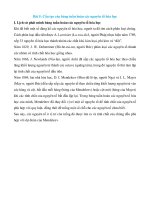
Fig. 1 Chemical structure of koumine. The chemical structure of
koumine. Molecular formula, C20H22N2O; molecular weight, 306.40;
CAS registry number, 1358-76-5.
numerous biological effects, including analgesic, antidepressant, anxiolytic, and antitumor effects [5–9]. G. elegans Benth. has long been used in Chinese folk medicine
to alleviate pain, inflammation, and cancer [9]. Consistently, alkaloids of G. elegans Benth. are thought to have
analgesic properties and exhibit pharmaceutical potential
[10, 11]. The most abundant alkaloid in G. elegans Benth.
is koumine (molecular formula, C20H22N2O; molecular weight, 306.30; CAS registry number, 1358-76-5)
(Fig. 1). According to our previous behavioral observations in animals, koumine reverses chronic constriction
injury (CCI) to the sciatic nerve and thermal hyperalgesia
induced by lumbar 5 (L5) spinal nerve ligation (SNL) in a
dose-dependent manner. Furthermore, mechanical allodynia in rats is reduced by koumine in a dose-dependent
manner [12]. Koumine differs substantially from the currently available analgesics, since it belongs to a class of
chemicals known as indole alkaloids. Moreover, it lacks
the adverse effects associated with most analgesic agents
[6, 11]. Therefore, we hypothesized that the analgesic
profile and underlying mechanism by which koumine
induces analgesia are unique.
Allopregnanolone,
also
known
as
3α,
5α-tetrahydroprogesterone (3α, 5α-THP), is one of the
most important neuroactive steroids. Upregulation of
allopregnanolone was shown to induce significant analgesia, implying that allopregnanolone in the spinal cord
(SC) may be an important key modulator of neuropathic
pain. Interestingly, our previous work has demonstrated
that increased allopregnanolone levels in the SC mediated the analgesic effect of koumine on neuropathic
pain [12]. Although allopregnanolone has been found
to be upregulated in the SC of rats with CCI following
koumine treatment, little is known about the cellular and
molecular mechanisms underlying its antinociceptive
The effect of koumine on CCI‑induced neuropathic pain
in rats
We have previously demonstrated that koumine has no
effects in sham CCI rats [12]. In the current study, twoway repeated measures ANOVA of the thermal withdrawal
latency (TWL) and mechanical withdrawal threshold
(MWT) measurement values of the hind paw ipsilateral
to the CCI demonstrated a significant treatment effect
between subjects (F5,210 = 1,463.57, P < 0.001 for TWL and
F5,210 = 167.03, P < 0.001 for MWT) and treatment time
(F6,210 = 1,816.41, P < 0.001 for TWL and F6,210 = 451.51,
P < 0.001 for MWT). Furthermore, a significant interaction
was found between treatment and time (F30,210 = 171.84,
P < 0.001 for TWL and F30,210 = 36.35, P < 0.001 for MWT).
Analysis with post hoc Dunnett’s T3 tests indicated that
CCI significantly decreased the thermal withdrawal latency
to thermal stimulation (P < 0.001, vs. the sham group) and
the mechanical withdrawal threshold to mechanical stimulation (P < 0.001, vs. the sham group). These findings demonstrated that the development of thermal hyperalgesia
and mechanical allodynia peaked on postoperative day 8
and 10, respectively. Furthermore, both conditions persisted for the entire observation period.
In these experiments, gabapentin [40 mg kg−1 body
weight (bw)] significantly attenuated thermal hyperalgesia
(P < 0.001) and mechanical allodynia (P < 0.05) compared
to the effect of the vehicle. Twice-daily subcutaneous (s.c.)
administration of koumine (7 mg kg−1 bw) between postoperative day 4 and 10 also significantly reversed thermal hyperalgesia (P < 0.001) and mechanical allodynia
(P < 0.01) compared to the effect of the vehicle. Interestingly, koumine exhibited more potent suppression of
thermal hyperalgesia (P < 0.01) and mechanical allodynia
(P < 0.01) than was observed for gabapentin. Koumine
exerted dose-dependent (two-way repeated measures ANOVA, F2,105 = 290.07, P < 0.001 for TWL and
F2,105 = 424.15, P < 0.001 for MWT) and time-dependent
(two-way repeated measures ANOVA, F6,105 = 1,666.7,
P < 0.001 for TWL and F2,105 = 22.34, P < 0.001 for MWT)
analgesic effects. As shown in Fig. 2, koumine administration (7 mg kg−1 bw, s.c.) induced analgesia within 2 days.
Qiu et al. Mol Pain (2015) 11:46
Fig. 2 The effects of a repeated subcutaneous administra‑
tion of koumine in sciatic nerve chronic constriction injury rats.
Koumine (0.28, 1.4, or 7 mg kg−1 body weight (bw)), gabapentin
(40 mg kg−1 bw), or vehicle was administered twice daily for 7 con‑
secutive days, starting on postoperative day 4. The time course of the
effect of koumine on the thermal withdrawal latency (TWL, a) and
mechanical withdrawal threshold (MWT, b) revealed that repeated
subcutaneous (s.c.) injections of koumine dose-dependently
reversed hyperalgesia and allodynia induced by sciatic nerve chronic
constriction injury (CCI) neuropathy. The data are presented as the
mean ± SEM (n = 6 per group) and were analyzed using two-way
repeated measures ANOVA. The significant differences between
the groups were determined by Bonferroni post hoc test at each
time point. ###P < 0.001 vs. the sham group; *P < 0.05, **P < 0.01,
***P < 0.001 vs. the vehicle control group.
The maximum analgesic effect was reached on day 7 of
treatment, and was maintained for 2 days after koumine
withdrawal. Together, these findings suggest that koumine
may reverse neuropathic pain.
The effect of koumine on 3α‑HSOR immunoreactivity
in the dorsal horn of L5–L6 SC of rats with CCI neuropathy
We previously determined that elevated levels of
allopregnanolone, a neurosteroid present in the SC,
Page 3 of 13
mediated the analgesic effect of koumine [12]. To further
investigate the exact mechanism of allopregnanolone
upregulation, the spinal distribution of 3α-HSOR, the
key synthetase of allopregnanolone, was assessed by
fluorescence immunohistochemistry in koumine-treated
CCI rats. The immunoreactivity of 3α-HSOR in the
dorsal horn of L5–L6 SC ipsilateral to CCI is shown in
Fig. 3a. A two-way ANOVA performed on the 3α-HSOR
fluorescence density data revealed a significant treatment effect between subjects (F4,100 = 61.21, P < 0.001)
and that 3α-HSOR fluorescence density significantly
changed with treatment time (F3,100 = 11.35, P < 0.001).
Furthermore, there was a significant interaction between
treatment and time (F12,100 = 3.53, P < 0.001). Bonferroni
post hoc test revealed that CCI treatment significantly
increased 3α-HSOR immunofluorescence staining density (P < 0.001 vs. the sham group) from postoperative
day 7 to the end of the observation period. In contrast
to the findings in the CCI group, the treatment- and
surgery-naïve (“naïve”) and sham groups showed no
changes in 3α-HSOR immunofluorescence density during the entire observation period. Twice-daily administration of koumine (7 mg kg−1 bw s.c.) between day 4
and 10 further increased 3α-HSOR immunofluorescence
staining density, reaching a maximum after 7 consecutive days of treatment (P < 0.001 vs. the vehicle group).
As shown in Fig. 3b, no differences in dorsal horn
3α-HSOR immunofluorescence density were observed
between the CCI and koumine-treated group on postoperative day 14 (4 days after koumine withdrawal). Using
immunofluorescence double labeling we found that
3α-HSOR was widely distributed in the dorsal horn of
the SC, and was co-expressed mainly with neurons and
microglia (Fig. 4).
The effect of koumine on 3α‑HSOR mRNA expression in the
dorsal horn of rat L5–L6 SC after CCI‑induced neuropathic
pain
Since 3α-HSOR immunostaining in the dorsal horn
of the SC of CCI rats was increased after koumine
administration, we determined lumbar 3α-HSOR
mRNA expression by reverse transcription polymerase chain reaction (RT-PCR) in the same kouminetreated CCI rats. Spinal RNA was extracted and its
integrality and concentration were determined. A
two-way ANOVA performed on 3α-HSOR mRNA
expression in the SC ipsilateral to CCI also demonstrated a significant treatment effect between subjects (F4,100 = 75.89, P < 0.001) and treatment time
(F3,100 = 18.34, P < 0.001), and a significant interaction
between treatment and time (F12,100 = 4.72, P < 0.001)
(Fig. 5). In agreement with the results of the immunochemistry analysis, Bonferroni post hoc test showed
Qiu et al. Mol Pain (2015) 11:46
Page 4 of 13
Fig. 3 3α-Hydroxysteroid oxidoreductase immunohistochemical staining in the spinal cord of koumine-treated sciatic nerve chronic constriction
injury rats. a 3α-Hydroxysteroid oxidoreductase (3α-HSOR) immunohistochemical staining in the ipsilateral dorsal horn of the lumbar spinal cord
(SC, L5–L6). Scale bar 100 μm. b Quantification of the 3α-HSOR expression in the SC after chronic constriction injury (CCI) by fluorescence density
analysis. A time-dependent increase in 3α-HSOR fluorescence density was observed within the ipsilateral SC dorsal horn after CCI. The data are
presented as the means ± SEM from 5 to 7 rats per group and were analyzed using two-way ANOVA followed by Bonferroni post hoc test at each
time point. ##P < 0.01 vs. the sham group; *P < 0.05 vs. the CCI group.
Qiu et al. Mol Pain (2015) 11:46
Page 5 of 13
Fig. 4 Immunostaining of the nerve cellular distribution of 3α-hydroxysteroid oxidoreductase in the dorsal horn of rat spinal cord. Left (nerve cells):
Photomicrograph of the dorsal horn section labeled with anti-neuronal nuclei (NeuN), anti-ionized calcium binding adaptor molecule 1 (Iba1), and
anti-glial fibrillary acidic protein (GFAP) antibody (green). Center (3α-HSOR): The same section was labeled with anti-3α-hydroxysteroid oxidoreduc‑
tase (3α-HSOR) antibody (red). Right (merged): Photomicrograph of the same section labeled with anti-3α-HSOR antibody and either anti-NeuN,
anti-Iba1, or anti-GFAP antibody. Scale bar 5 μm.
that CCI significantly enhanced 3α-HSOR mRNA
expression (P < 0.001 vs. sham group) from postoperative day 7 to the end of the observation period. In
contrast, the naïve and sham groups showed no difference in 3α-HSOR mRNA expression during the
observation period (P > 0.05, vs. the CCI group).
Twice-daily administration of koumine (7 mg kg−1 bw,
s.c.) between postoperative day 4 and day 10 further
increased 3α-HSOR mRNA expression and reached
a maximum after 7 consecutive days of treatment
(P < 0.05 vs. the CCI group). However, as shown in
Fig. 5, compared to the 3α-HSOR mRNA expression
in the CCI group, koumine-treated rats demonstrated
a noticeable 3α-HSOR mRNA expression upregulation on postoperative day 14, i.e., 4 days after koumine
withdrawal (P < 0.05).
The effect of koumine on 3α‑HSOR catalytic activity in rat
SC after CCI‑induced neuropathic pain
The effect of koumine on spinal 3α-HSOR activity in CCI
rats was determined by enzyme kinetics analysis. A significant treatment effect on the 3α-HSOR catalytic activity was observed between the rats (one-way ANOVA,
F4,25 = 19.19, P < 0.001). As shown in Fig. 6, 3α-HSOR
activity was significantly increased by 17.4% in CCI rats
(P < 0.05 vs. the sham group). After 7 consecutive days of
koumine administration (7 mg kg−1 bw, s.c.), 3α-HSOR
activity in the SC of CCI rats was further enhanced by
15.8% (P < 0.05 vs. the CCI group). This finding implies
that the increased 3α-HSOR mRNA expression and
immunostaining in the SC of koumine-treated CCI rats
may enhance 3α-HSOR bioactivity and upregulate allopregnanolone in the SC.
Qiu et al. Mol Pain (2015) 11:46
Page 6 of 13
The effect of medroxyprogesterone acetate on the
analgesic effect of koumine in mechanical allodynia tests
in CCI rats
Fig. 5 The effect of koumine on 3α-hydroxysteroid oxidoreductase
mRNA expression in the spinal cord of sciatic nerve chronic constric‑
tion injury rats. The 3α-hydroxysteroid oxidoreductase (3α-HSOR)
mRNA level is expressed as the ratio of glyceraldehyde-3-phosphate
dehydrogenase (GAPDH) mRNA in the spinal cord (SC) of naïve, shamoperated, sciatic nerve chronic constriction injury (CCI), or kouminetreated rats. The data are presented as the mean ± SEM (n = 6 per
group) and were analyzed by two-way ANOVA followed by Bonferroni
post hoc test at each time point. ##P < 0.01 vs. the sham group;
*P < 0.05 vs. the CCI group.
Fig. 6 The effect of koumine on 3α-hydroxysteroid oxidoreductase
catalytic activity in the spinal cord of sciatic nerve chronic constric‑
tion injury rats. The 3α-hydroxysteroid oxidoreductase (3α-HSOR)
catalytic activity in the spinal cord (SC) lumbar region L5–L6 was
assessed spectrophotometrically by measuring the oxidation rate of
nicotinamide adenine dinucleotide phosphate (NADPH) at 340 nm
and 37°C. The data are presented as the mean ± SEM (n = 6 per
group) and were analyzed by one-way ANOVA followed by Bonferroni
post hoc test at each time point. *P < 0.05 vs. the CCI group, #P < 0.05
vs. the sham group.
We used the 3α-HSOR inhibitor medroxyprogesterone
acetate (MPA, administered by intrathecal injection) to
confirm the hypothesis that the enhanced expression and
activity of 3α-HSOR, which increase allopregnanolone
levels in the SC of koumine-treated CCI rats, mediates analgesia. As shown in Fig. 7, the MWT in the hind
paw ipsilateral to the CCI revealed a significant intergroup treatment effect (F5,30 = 9.496, P < 0.001, one-way
ANOVA). Furthermore, s.c.-injected koumine significantly relieved mechanical allodynia compared to the
effect of the vehicle (dimethyl sulfoxide + normal saline,
DMSO + NS) group (P < 0.05). Conversely, intrathecal
injection of MPA dose-dependently reversed the analgesic effect of koumine (F2,15 = 14.511, P < 0.001). MPA (0.5
and 1.25 mg kg−1 bw) significantly reduced the MWT
(P < 0.001 vs. the DMSO + koumine group). However,
the highest dose of MPA tested (1.25 mg kg−1 bw) had
no effect on the MWT of untreated CCI rats (P > 0.05
vs. the DMSO + NS group). These data suggest that the
analgesic effect of koumine may be linked to increased
3α-HSOR activity in the SC.
Discussion
Gelsemium elegans Benth. has diverse biological effects;
however, its clinical use is hampered by its toxicity. The
crude alkaloid Gelsemium extract exhibits a high level
of toxicity with an LD50 of 15 and 4 mg kg−1 bw following intragastric and intraperitoneal (i.p.) administration, respectively, in rats [13]. Several research groups
are currently trying to derive alkaloid monomers with
high potency and low toxicity from G. elegans Benth.
In a previous study, we were able to obtain several different alkaloid monomers using the pH-zone–refining
counter-current chromatography technique [14], which
enabled us to perform pharmacodynamic screening. Our
preliminary findings suggested that the most toxic alkaloid derived from G. elegans Benth., gelsenicine, exerted
analgesic activity during inflammatory and neuropathic
pain [5]. Recently, Zhang et al. reported that gelsemine,
another G. elegans Benth.-derived main alkaloid, displayed potent and specific antinociceptive properties in
chronic pain [15]. Furthermore, we previously demonstrated that koumine exhibited potent analgesic effects
with a relatively low toxicity compared to that of other G.
elegans Benth. alkaloid extracts [12, 16]. Koumine’s acute
toxicity was previously investigated in both mice and
rats. The LD50 of s.c.-administered koumine to mice was
Qiu et al. Mol Pain (2015) 11:46
Fig. 7 The analgesic effect of koumine was reversed in sciatic
nerve chronic constriction injury rats by intrathecal treatment with
medroxyprogesterone acetate. Medroxyprogesterone acetate (MPA)
or dimethyl sulfoxide (DMSO, vehicle) was administered via an
intrathecal catheter 10 days after sciatic nerve chronic constriction
injury (CCI) surgery. After 30 min, koumine (7 mg kg−1 bw) or normal
saline (NS) was administered by subcutaneous (s.c.) injection. The
mechanical withdrawal threshold (MWT) of the hind paws was meas‑
ured 1 h after completion of the drug or vehicle administration. The
data are presented as the mean ± SEM (n = 6 per group) and were
analyzed by one-way ANOVA followed by Bonferroni post hoc test at
each time point. *P < 0.05 vs. the DMSO + NS group; #P < 0.05 vs. the
DMSO + koumine group.
99.0 mg kg−1 bw [12]. Acute lethality generally occurred
within 10 min of s.c. injection in mice, with labored
respiration and brief coordinated clonic convulsions
occurring immediately before death [12]. Interestingly,
koumine’s toxicity is much lower than that of the crude
G. elegans extract. The LD50 of intragastrically administered koumine was 300 mg kg−1 bw in rats (unpublished
data), a level much higher than that estimated for the
crude alkaloidal extract (15 mg kg−1 bw) using the same
administration route [13]. Moreover, in a previous study,
the spontaneous travel velocity was not affected after
administration of koumine in mice as determined by
the spontaneous motor activity test [17], and no adverse
effects were observed in the rats treated with a range of
koumine doses (0.28–7 mg kg−1 bw) in the current study.
The relatively wide therapeutic index of koumine suggests that it may be a promising agent in clinical applications. In this study, the TWL and MWT behavioral
data indicated that koumine may ameliorate sciatic nerve
CCI-induced chronic pain. Repeated administration of
koumine reversed thermal hyperalgesia and mechanical
allodynia in a dose-dependent manner in our CCI model.
Additionally, our previous work has provided evidence of
koumine’s analgesic effect in a number of animal models
of pain, including SNL and diabetic-induced neuropathic
Page 7 of 13
pain [12, 18], indicating a role for koumine in the treatment of peripheral neuropathic pain.
Repeated s.c. administration of koumine was not associated with adverse effects commonly associated with
opioids, such as physical and psychological dependence [19]. Interestingly, LaBuda and Little [20] revealed
that agents such as gabapentin and morphine completely or partially reversed tactile allodynia in L5 SNL,
another animal model of painful peripheral neuropathy. Conversely, indomethacin had no effect. Koumine
also exhibited positive analgesic effects in the L5 SNL
model [12]. Taken together, these findings suggest that
koumine’s analgesic mechanism of action differs from
that of NSAIDs and opioids. Interestingly, we found that
koumine exhibited its analgesic effect by upregulating
allopregnanolone, one of the most potent neurosteroids
in the SC [12].
The pathophysiology underlying neuropathic pain is
complicated, with many aspects remaining unclear. A
striking finding in the past three decades was the discovery that neurons are capable of synthesizing neurosteroids independently of the classical mechanism
involving endocrine glands [21, 22]. The involvement of
endogenous neurosteroids is well established in the area
of chronic pain modulation [21–25]. Inflammatory and
neuropathic pain states are associated with the upregulated synthesis of endogenous neurosteroids in the SC,
such as allopregnanolone and pregnenolone [21, 23, 26].
The administration of exogenous allopregnanolone has
been shown to prevent and suppress oxaliplatin-evoked
painful neuropathy [25]. Allopregnanolone is an allosteric modulator that controls several important neurophysiological mechanisms through its actions on the
γ-aminobutyric acid A (GABAA) channel [27]. Indeed,
neurosteroids modulate pain perception by potentiating the effects at GABAA channels and inhibiting T-type
calcium channels located in peripheral sensory neurons [23, 28–30]. Furthermore, neurosteroid synthesis
has been reported in the SC, partly explaining the vital
role of SC in the control of pain processing. The SC contains key enzymes related to steroid synthesis, including cytochrome P450 side chain cleavage enzymes,
3α-reductase, and 3α-HSOR [22, 23, 31]. Therefore, the
key cellular and molecular components involved in neurosteroid biosynthesis in the SC may provide potential
targets that may be useful for the development of novel
analgesics for the treatment of persevering neuropathic
pain. Consequently, an increasing number of scientists
are focusing their research toward the discovery of novel
neurosteroid-directed analgesics [29, 30, 32–34].
3α-HSOR belongs to the aldo–keto reductase superfamily. It is crucial for the synthesis of neuroactive
3α-reduced steroids, such as allopregnanolone and
Qiu et al. Mol Pain (2015) 11:46
tetrahydrodeoxycorticosterone, in a reversible manner. The dorsal horn of the SC, a pivotal structure that
controls pain and nociception, exhibits intense immunostaining of 3α-HSOR [22, 35]. Furthermore, neurons
and glial cells synthesize various neurosteroids. Our
double immunolabeling experiments showed 3α-HSOR
immunostaining in SC neurons, microglia, and astrocytes. This prompted us to investigate the relationship
between koumine and 3α-HSOR and koumine’s effects
on allopregnanolone upregulation in the SC. The present
study draws on data generated by different experimental techniques that clearly show that koumine increases
the cellular immunoreactivity, gene expression, and bioactivity of 3α-HSOR in SC during chronic pain. Importantly, our time-course experiments revealed that the
koumine-induced upregulation of 3α-HSOR cellular
immunoreactivity and gene expression correlated with
the development of neuropathic pain symptoms.
Consistent with these findings, our in vivo enzymatic activity assays revealed that the catalytic activity of 3α-HSOR in the SC was increased in CCI rats
after 7 days of koumine treatment. Furthermore, we
observed that koumine produced significant analgesia and dose-dependently reversed the mechanical pain
thresholds induced by the intrathecal injection of the
3α-HSOR pharmacological inhibitor MPA at a dose of
0.5 or 1.25 mg kg−1 bw. It is worth noting that the highest dose of MPA used (1.25 mg kg−1 bw) had no effect
on the MWT values measured in naïve CCI rats. Thus,
inhibiting 3α-HSOR activity in the sensory nerve circuit
of the SC could reverse the analgesic effect of koumine
in CCI rats. Our findings demonstrate the direct role of
3α-HSOR in mediating pain modulation and koumine’s
influence thereon. Therefore, 3α-HSOR may be a fundamental molecular target for koumine in the modulation of pain sensation. However, although the 3α-HSOR
fluorescence density and mRNA expression were still
elevated on postoperative day 14, the analgesic effect
had disappeared by then (Figs. 3, 5). One possibility
is that koumine may regulate the catalytic activity of
3α-HSOR. Allopregnanolone biosynthesis in the SC may
thus be insufficient in the absence of koumine because of
low 3α-HSOR catalytic activity. However, this hypothesis needs to be confirmed by the determination of allopregnanolone levels in the SC on postoperative day 14.
Another explanation may be that a molecular target
other than 3α-HSOR, and whose function is also altered
by koumine, participates in the observed analgesic effect
of koumine.
The endogenous biosynthesis of neurosteroids is also
upregulated in the SC during inflammatory and neuropathic pain states [21, 36, 37]. We demonstrated a similar increase in allopregnanolone and pregnenolone levels
Page 8 of 13
in CCI rats [12]. The observed increase in allopregnanolone levels in CCI rats is consistent with the findings
of Kawano et al. in SNL neuropathy [37]. Similarly, we
found increased immunohistochemical staining, mRNA
expression, and bioactivity of 3α-HSOR in the SC during
sciatic nerve CCI-induced chronic pain. On the basis of
the results of our and other studies, we consider that a
state of chronic pain in animals is predominantly determined by two sets of factors. The first includes the pronociceptive mechanisms mediated by neurotransmitters
supporting the development and maintenance of pain
symptoms, such as substance P, bradykinin, prostaglandin, and histamine. The second group of factors opposes
the pro-nociceptive processes, and its effects are mediated by endorphins, neurosteroids, and neuroprotective factors that possess adaptive or antinociceptive
properties. The latter help the animal to cope with or
accelerate the recovery from the pathological pain state.
Consequently, the selective upregulation of the 3α-HSOR
immunoreactivity, mRNA expression, and bioactivity
and the 3α-HSOR-induced increase in spinal synthesis of
endogenous allopregnanolone may represent an intrinsic
adaptive response to neuropathic pain and elicit beneficial effects against diverse pathological pain symptoms.
However, these natural or adaptive mechanisms are not
sufficient to achieve the suppression of pain sensation
in rats with CCI [21]. As observed in rats with CCI, an
insufficient increase in 3α-HSOR and allopregnanolone
levels fails to reverse the development of neuropathic
pain. Only a sufficient increase in allopregnanolone
would offer adequate protection against the development
of neuropathic pain [12, 37]. Allopregnanolone that is
endogenously formed in the central nervous system significantly alters nociception through paracrine and autocrine mechanisms [36, 38]. Therefore, koumine and other
alkaloid extracts of G. elegans Benth. that are capable of
stimulating allopregnanolone formation in neural networks may provide a novel approach for the development
of analgesic therapies [39].
Our fluorescence immunohistochemistry experiments also revealed that 3α-HSOR was widely distributed in the SC dorsal horn and co-localized with neural
and non-neural cells, including neurons and microglia
(Fig. 4, merged photomicrograph). In recent years, there
has been a growing consensus that glial cells located in
the spinal dorsal horn are activated following peripheral nerve injury [40–42]. Microglial activation induces
the release of proinflammatory cytokines, such as
interleukin-β, tumor necrosis factor-α, and interleukin-6,
which play important roles in the development of neuropathic pain. The protective property of allopregnanolone
in pain perception in relation to glial cells has been well
documented [25, 34, 43]. Allopregnanolone treatment
Qiu et al. Mol Pain (2015) 11:46
not only significantly reduced astrocyte proliferation
and microglial activation, but also enhanced myelination
in mice [44]. Neurosteroids, such as allopregnanolone,
could reduce inflammatory cytokine levels, which were,
for example, elevated following traumatic brain injury
[45]. Our findings demonstrated that koumine administration increased 3α-HSOR activity, which could contribute to increased allopregnanolone levels in the SC of CCI
rats. Consequently, we hypothesized that elevated allopregnanolone levels may exert analgesic effects through
allosteric modulation of GABAA and by suppressing the
release of microglia activation-induced inflammatory
cytokines. Further studies are warranted to determine
whether koumine influences the activation of glial cells
during the neuropathic pain state.
Conclusion
This study demonstrated that koumine could relieve neuropathic pain in rats by enhancing the mRNA expression
and bioactivity of 3α-HSOR in the SC. Our study also
suggested that by targeting 3α-HSOR, koumine altered
3α-HSOR-regulated allopregnanolone levels in the SC of
rats. Therefore, koumine may be promising in the search
for novel analgesic agents that are protective against
painful neuropathy.
Methods
Chemicals and reagents
Koumine (99% purity) was isolated from G. elegans
Benth. by pH-zone–refining counter-current chromatography as described previously [14]. Gabapentin (purity:
99%; Shanghai Sunheat Chemicals Co., Ltd, Shanghai,
China) was used as positive control. Rabbit polyclonal
antibody against 3α-HSOR was purchased from Biosynthesis Biotechnology (Beijing, China) and mouse
anti-neuronal nuclei monoclonal antibody (anti-NeuN)
was purchased from GeneTex (Irvine, CA, USA). Goat
anti-ionized calcium binding adaptor molecule 1 antibody (anti-Iba1; catalog no. ab5076) and mouse anti-glial
fibrillary acidic protein antibody (anti-GFAP; catalog no.
ab4648) were purchased from Abcam (Cambridge, UK).
Fluorescein (FITC)-conjugated donkey anti-mouse, tetramethylrhodamine (TRITC)-conjugated goat anti-rabbit,
and Alexa Fluor® 488-conjugated donkey anti-mouse
were purchased from Jackson ImmunoResearch (West
Grove, PA, USA). Normal rabbit and donkey serum were
purchased from Biosynthesis Biotechnology (Beijing,
China). PCR reagent kits, PrimeScript® RT Reagent Kit,
and SYBR® Premix Ex Taq™ II Real-Time PCR Reagent
Kit, were purchased from Takara Biotechnology Co.,
Ltd. (Dalian, China). Nicotinamide adenine dinucleotide
phosphate-oxidase (NADPH; Roche, Pleasanton, CA,
USA), 5α-dihydroprogesterone (5α-DHP; Sigma-Aldrich,
Page 9 of 13
St. Louis, MO, USA), medroxyprogesterone acetate
(MPA; Selleck, Houston, TX, USA), and chloral hydrate
(Sigma-Aldrich, St. Louis, MO, USA) were of pharmaceutical grade. All other reagents used were of analytical
grade.
Koumine was prepared daily prior to use in sterile physiological saline (0.9% w/v sodium chloride),
and was administered by s.c. injection at a dose of
4 mL kg−1 rat bw.
Animals
Male adult Sprague–Dawley rats (180–200 g) were
obtained from the Shanghai Laboratory Animal Center
at the Chinese Academy of Sciences (Shanghai, China).
All rat experiments were performed in accordance with
the National Institutes of Health Guide for Care and Use
of Laboratory Animals (Publication No. 85-23, revised
1985) and were conducted under the authority of the
Committee of Ethics of the Fujian Medical University
(Fujian, China). All procedures complied with the guidelines for animal care and use established at the Fujian
Medical University. The rats were housed in a temperature-controlled room (25 ± 2°C) under a 12-h light/dark
cycle (lights on: 08:00 AM), with access to standard laboratory food and water ad libitum, except during behavioral observations. The rats were acclimatized for at least
1 week before undergoing any experiments. Each rat was
assigned to one specific behavioral experiment, and the
experiments were performed between 09:00 and 17:00.
In vivo intrathecal catheter implantation and drug
administration
Intrathecal implantation of polyethylene (PE) tubing
(Intramedic PE-10, Clay Adams, Parsippany, NJ, USA)
into the subarachnoid space of the lumbar enlargement
was performed in rats as described previously [46]. This
method permits the direct administration of a drug of
interest. After 1 day of recovery post surgery, the rats
that were considered neurologically healthy received 2%
lidocaine (20 μL) through the intrathecal catheter to confirm post-surgical placement of the PE tubing within the
subarachnoid space. Those rats that displayed complete
paralysis of both hind limbs and the tail following the
administration of lidocaine were used for the subsequent
experiments.
After recovery from intrathecal catheter placement, peripheral neuropathy was induced by CCI. The
3α-HSOR selective inhibitor MPA was used to evaluate
the analgesic actions of koumine. The rats undergoing
mechanical allodynia were assigned to groups receiving either DMSO (vehicle for MPA) with NS, MPA
(1.25 mg kg−1 bw) with NS, DMSO with koumine, or
MPA (0.25, 0.5, or 1.25 mg kg−1 bw) with koumine. On
Qiu et al. Mol Pain (2015) 11:46
postoperative (CCI) day 10, DMSO or MPA was administered via the intrathecal catheter. After 30 min, koumine
or NS was administered by s.c. injection. The MWT of
the hind paws was measured 1 h after completion of the
drug administration.
Visual confirmation of the placement of the PE tubing
in the intrathecal space at the lumbar enlargement was
performed by exposing the lumbar SC at the end of each
experiment. The data obtained from rats with an incorrect PE tubing position were excluded from the study.
Rat CCI model
The rats were prepared for the induction of CCI according to the method described by Bennett et al. [47].
The rats were anesthetized by i.p. administration of
400 mg kg−1 bw chloral hydrate. Subsequently, the right
common sciatic nerve isolated at the level of the midthigh was loosely ligated using four chromic gut (5-0) ties
at 1-mm intervals. The same procedure was performed
without ligation in the rats assigned to the sham group.
The rats were monitored for 3 days following surgery
and were only used in the subsequent studies if the baseline thermal hyperalgesia and mechanical allodynia test
scores (described in “Measurement of thermal hyperalgesia and mechanical allodynia in rats” below) surpassed
the acceptance threshold. Baseline threshold scores were
calculated as the CCI ipsilateral paw baseline score/contralateral paw baseline score. Rats displaying baseline
scores between 0.8 and 1.2 were accepted into the study.
Rats with thermal predose latency scores >0.8, mechanical predose threshold scores >0.75, and/or demonstrating
motor deficits after surgery were excluded from the subsequent experiments.
Measurement of thermal hyperalgesia and mechanical
allodynia in rats
Thermal hyperalgesia was measured with a commercial thermal paw stimulator (PL-200, Chengdu Technology & Market Co., Ltd., Sichuan, China) as described by
Hargreaves et al. [48]. The rats were placed in individual
plastic cubicles mounted on a glass surface in a temperature-controlled room (25 ± 2°C). The plantar surface of
each hind paw was subsequently exposed to a thermal
stimulus, i.e., radiant heat emitted from a focused projection bulb, for a maximum exposure time of 16 s to minimize potential tissue damage. The second hind paw of
each rat was tested after a 10-min interval. The paw TWL
was calculated as the mean of the 2 hind paw withdrawal
times.
CCI rats were assigned to groups receiving the vehicle, koumine (0.28, 1.4, or 7.0 mg kg−1 bw), or gabapentin (40 mg kg−1 bw) twice daily by s.c. injection for
7 consecutive days starting on postoperative day 4.
Page 10 of 13
Sham-operated rats underwent identical treatments.
TWL was measured before surgery (baseline), before
drug treatment (pre-dosing), and 30 min after drug
administration (post-dosing) on the morning of postoperative day 6, 8, 10, 12, and 14.
Mechanical allodynia was determined with a commercial electronic von Frey apparatus (Model 2390; IITC Life
Science Inc., Woodland Hills, CA, USA) as described
by Mitrirattanakul et al. [49], with minor modifications.
Briefly, the rats were placed in a Plexiglas box on a steel
mesh floor. The center of the hind paw was stimulated
using the von Frey filament applied up to a maximum
strength of 55 g or until the point of paw withdrawal. The
threshold at which withdrawal occurred was automatically registered. The procedure was performed twice for
each hind paw at 10-min intervals. The MWT was calculated as the mean of the 2 thresholds.
The MWT was measured 30 min after the TWL measurement in sham-operated rats and in rats receiving the
vehicle, koumine (0.28, 1.4, or 7.0 mg kg−1 bw), or gabapentin (40 mg kg−1 bw) twice daily by s.c. injection for 7
consecutive days starting on postoperative day 4.
Immunofluorescence
The rats were assigned to naïve, sham, CCI, CCI with
7 mg kg−1 bw koumine, and CCI with 0.28 mg kg−1 bw
koumine groups. Koumine and the vehicle were administered by s.c. injection at a volume of 0.25 mL/100 g bw
twice daily for 7 consecutive days starting from postoperative day 4. The rats were anesthetized by i.p. injection
of 400 mg kg−1 bw chloral hydrate 1 h after drug administration on the morning of postoperative day 5, 7, 10,
or 14. The L5–L6 of the SC were excised for analysis by
fluorescence immunohistochemistry as described previously, with minor modifications [21]. Briefly, 100 mL of
0.1 M phosphate buffer (PB, pH 7.4) was perfused transcardially followed by perfusion with 450 mL of 4% formaldehyde prepared in PB (fixative solution). The SC
located between L5 and L6 was rapidly dissected and
fixed in fixative solution for 24 h. The SC tissue was
immersed in 15% sucrose-containing PB for 12 h and was
subsequently transferred into 30% sucrose-containing
PB for 24 h. The SC tissue was then placed in TissueTek® OCT embedding medium (Sakura, Torrance, CA,
USA), and was immediately frozen at −22°C. Coronal
sections (16 μm thick) were cut on a Microm HM 525E
cryostat (Francheville, France) and were subsequently
mounted on glass slides coated with gelatin and chromium potassium sulfate.
The SC sections were preincubated for 1 h with the
following sera in preparation for subsequent immunohistochemical experiments: (1) for mono-labeling
with anti-3α-HSOR, anti-NeuN, or anti-GFAP, and
Qiu et al. Mol Pain (2015) 11:46
double-labeling with anti-3α-HSOR and anti-NeuN or
anti-GFAP, the SC sections were preincubated with 10%
nonimmune goat serum prepared in PB containing 0.3%
Triton X-100 (PBT); (2) for mono-labeling with anti-Iba1,
and double-labeling with anti-3α-HSOR and anti-Iba1,
the SC sections were preincubated with 10% nonimmune
donkey serum prepared in PBT.
The immunohistochemical mono-labeling experiments
were conducted by incubating the SC sections for 24 h
at 4°C with a single antibody (anti-3α-HSOR, 1:500 dilution; anti-NeuN, 1:1,000 dilution; anti-Iba1, 1:500 dilution; anti-GFAP, 1:400 dilution) prepared in PBT. In the
immunohistochemical double-labeling experiments, the
sections were incubated with anti-3α-HSOR in combination with anti-NeuN, anti-Iba1, or anti-GFAP prepared
in PBT at the same dilution ratios used for the mono
labeling experiments. After washing for 4 times in phosphate-buffered saline (PBS, 5 min per rinse), the sections
were transferred into a solution containing either a single
secondary antibody (mono-labeling) or multiple antibodies (double-labeling), and were incubated for 1 h at room
temperature. The mono-labeling solutions contained
either TRITC-conjugated goat anti-rabbit, FITC-conjugated goat anti-mouse, or FITC-conjugated donkey antigoat secondary antibody prepared in PBT at a dilution
ratio of 1:300. The double-labeling solutions contained
a mixture of the appropriate two secondary antibodies.
After rinsing 3 times in PBS (5 min per rinse), the sections were mounted with anti-fade mounting medium
(Beyotime, Haimen China), and imaged under a fluorescence DMR microscope equipped with a digital camera
(IX71-A12FL/PH, Olympus, Tokyo, Japan) connected to
a Pentium 4 PC. The images were adjusted using PhotoShop (Version 7; Adobe Systems, Inc., San Jose, CA,
USA) and the fluorescence density was analyzed using
the Image-Pro Plus software (Version 6.0; Media Cybernetics, Rockville, MD, USA).
Reverse transcription and real‑time PCR
The SC located between L5 and L6 was excised from rats
anesthetized by i.p. injection of 400 mg kg−1 bw chloral
hydrate at 5, 7, 10, or 14 days after sciatic nerve ligature
or the sham procedure. Total RNA was extracted from
tissue samples using TRIzol reagent (Invitrogen, Carlsbad, CA, USA) according to the manufacturer’s instructions. RNA quality was determined by electrophoresis
using ethidium bromide-stained agarose gels, and was
further confirmed by an optical density (OD) absorption
ratio (OD 260 nm/OD 280 nm) >1.7. A fixed quantity of
total RNA (1 μg) was subjected to reverse transcription
(RT) PCR performed at 37°C for 15 min. The reaction
for first-strand cDNA contained the following: 5 ì PrimeScriptđ Buffer (4àL), 50àM Oligo dT Primer (1àL),
Page 11 of 13
100àM random 6 mers (1àL), PrimeScriptđ RT Enzyme
Mix I (1 µL), and total RNA (1 µg) added to RNase Free
dH2O in a final volume of 20 μL. Real-time PCR (rt-PCR)
experiments were performed using a LightCycler system (Roche Diagnostics GmbH, Mannheim, Germany).
The primer sequences were as follows: 3α-HSOR sense:
5′-TTCATTCCTGTACTGGG-3′ and 3α-HSOR antisense: 5′-AGTAGCCTTGATAACTTCAT-3′ [50], glyceraldehyde-3-phosphate dehydrogenase (GAPDH) sense:
5′-ACCACAGTCCATGCCATCAC-3′
(nucleotides
3,069–3,088) and GAPDH antisense: 5′-TCCACCACCCTGTTGCTGTA-3′ (nucleotides 3,624–3,605). All
primers were chemically synthesized by Sangon Biotech
(Shanghai, China). The housekeeping gene, GAPDH, was
used as an internal control. The rt-PCR experiments were
performed in a total volume of 20 μL containing cDNA
(2 μL), 10 μM specific primers (1.6 μL), and SYBR Premix Ex Taq (10 μL) containing Taq polymerase, deoxyribonucleotide triphosphate, MgCl2, SYBR Green I dye,
and dH2O (6.4 µL). A sample without the cDNA template
(water) was used as a negative control and was run in
every assay. Each PCR reaction was performed in triplicate according to the following amplification protocol:
a 30-s denaturing step at 95°C, 5-s amplification cycle at
95°C, 5-s amplification cycle at 55°C, and 12-s amplification cycle at 62°C in a total of 45 cycles. The specificity
of the rt-RT-PCR products and the absence of non-specific products were confirmed by examining the melting
curve. The concentration of 3α-HSOR mRNA in each
sample was determined after normalizing the rt-RT-PCR
3α-HSOR product to that of GAPDH.
Enzymatic activity assay of 3α‑HSOR
To investigate the enzymatic activity of 3α-HSOR in the
L5 to L6 region of the SC following koumine administration, CCI- or sham-operated rats were assigned to vehicle, sham-operated, or koumine (0.28 or 7.0 mg kg−1
bw) treatment groups. A naïve group was used as additional control. Vehicle or koumine was administered by
s.c. injection twice daily for 7 consecutive days starting on postoperative day 4. On postoperative day 10,
the rats were euthanized by decapitation, and the SC
L5 to L6 were rapidly excised and stored at −80°C.
Enzyme activity assays for 3α-HSOR were performed
as described previously [51, 52], with minor modifications. Briefly, 3α-HSOR activity was determined by
spectrophotometric measurement (UV-2450, Shimadzu,
Kyoto, Japan) of NADPH oxidation at 340 nm and 37°C
using a 1.0-cm path length cuvette. The excised SC sections were homogenized in 2 mL of ice-cold 10 mM PB
(pH 6.5) containing 0.154 M KCl, 1 mM dithiothreitol, 0.5 mM ethylenediaminetetraacetic acid (EDTA),
and 1 μM phenylmethanesulfonyl fluoride (PMSF). The
Qiu et al. Mol Pain (2015) 11:46
homogenate was centrifuged at 105,000×g for 60 min at
4°C in an Eppendorf 5430R ultracentrifuge (Hamburg,
Germany). The supernatant (cytosolic) fraction was
stored at −80°C until required for the enzymatic activity assay and quantitative analysis. Reductase activity was
measured in 100 mM PB (pH 6.5) containing 0.1 mM
NADPH, 0.08 mM 5α-DHP (substrate), and enzyme
solution (80 μL of the cytosolic fraction) in a total volume of 0.7 mL. The reaction was initiated by addition
of the cofactor to the assay mixture, and a blank sample
without substrate was included in the measurements.
The protein concentrations were determined using the
Enhanced BCA Protein Assay Kit (Beyotime Biotech,
Haimen, China).
Statistical analysis
Continuous data were expressed as the mean ± SEM
unless otherwise indicated. The TWL and MWT
responses were analyzed using two-way repeated measures ANOVA (treatment and time). The significant
differences between the groups were determined by
Bonferroni post hoc test. The data from the 3α-HSOR
catalytic activity and MPA inhibition tests were analyzed using one-way ANOVA with Bonferroni post hoc
test. Immunohistochemistry and rtPCR data were analyzed using two-way ANOVA (treatment and time), followed by either Dunnett’s T3 or Bonferroni post hoc
test. Differences were considered statistically significant
if P < 0.05. Statistical analyses were performed with the
Statistical Package for the Social Sciences software (SPSS
version 13.0; SPSS Inc., Chicago, IL, USA).
Abbreviations
CCI: chronic constriction injury; DMSO: dimethyl sulfoxide; 3α-HSOR:
3α-hydroxysteroid dehydrogenase; EDTA: ethylenediaminetetraacetic acid;
GAPDH: glyceraldehyde-3-phosphate dehydrogenase; GFAP: glial fibrillary
acid protein; Iba1: ionized calcium binding adaptor molecule 1; L5-L6: lumbar
region 5 and 6; MWT: mechanical withdrawal threshold; MPA: medroxyproges‑
terone acetate; NADPH: nicotinamide adenine dinucleotide phosphate; NeuN:
neuronal nuclei; NS: normal saline; NSAIDs: nonsteroidal anti-inflammatory
drugs; OD: optical density; PB: phosphate buffer; PBS: phosphate-buffered
saline; PBT: PB containing 0.3% Triton X-100; PMSF: phenylmethanesulfo‑
nyl fluoride; rt-PCR: real-time polymerase chain reaction; RT-PCR: reverse
transcriptase PCR; SC: spinal cord; SNL: spinal nerve ligation; 3α, 5α-THP:
3α, 5α-tetrahydroprogesterone; PE: polyethylene; TWL: thermal withdrawal
latency.
Authors’ contributions
HQQ and YX performed the experiments, analyzed the data, prepared the
figures, and drafted the manuscript. HQQ and YX contributed equally to this
work. GLJ, JY, and SPL contributed to the behavioral, immunohistochemistry,
and rt-RT-PCR experiments. ML analyzed the data and prepared the figures.
CXY conceived and designed the study, and revised and verified the manu‑
script. All authors read and approved the final manuscript.
Author details
1
Department of Pharmacology, College of Pharmacy, Fujian Medical Univer‑
sity, 350108 Fuzhou, Fujian, People’s Republic of China. 2 Fujian Key Laboratory
Page 12 of 13
of Natural Medicine Pharmacology, College of Pharmacy, Fujian Medical
University, Fuzhou, Fujian, People’s Republic of China.
Acknowledgements
This research was supported by the National Natural Science Foundation of
China (grant no. 30973520, 81273493, and 81200868) and the Key Program of
Scientific Research of Fujian Medical University (grant no. 09ZD009).
Compliance with ethical guidelines
Competing interests
The authors declare that they have no competing interests. Sponsorships or
competing interests that may be relevant to the content of this article are
disclosed at the end of the article.
Received: 11 March 2015 Accepted: 28 July 2015
References
1. Horowitz SH. Response to commentary: a new definition of neuropathic
pain. Pain. 2012;153:935–6.
2. DeFrates S, Cook AM. Pharmacologic treatment of neuropathic pain fol‑
lowing spinal cord injury. Orthopedics. 2011;34:203–7.
3. Dutt V, Thakur S, Dhar VJ, Sharma A. The genus Gelsemium: an update.
Pharmacogn Rev. 2010;4:185–94.
4. Ornduff R. The systematics and breeding system of Gelsemium (Logani‑
aceae). J Arnold Arbor. 1970;51:1–17.
5. Liu M, Shen J, Liu H, Xu Y, Su YP, Yang J, et al. Gelsenicine from Gelsemium
elegans attenuates neuropathic and inflammatory pain in mice. Biol
Pharm Bull. 2011;34:1877–80.
6. Dutt V, Dhar VJ, Sharma A. Antianxiety activity of Gelsemium sempervirens.
Pharm Biol. 2010;48:1091–6.
7. Lu JM, Qi QZ, Liu GL, Shen ZY, Tu KC. Effect of Gelsemium elegans Benth.
injection on proliferation of tumor cells. Chin J Cancer. 1990;9:472–7.
8. Magnani P, Conforti A, Zanolin E, Marzotto M, Bellavite P. Dose-effect
study of Gelsemium sempervirens in high dilutions on anxiety-related
responses in mice. Psychopharmacology. 2010;210:533–45.
9. Jin GL, Su YP, Liu M, Xu Y, Yang J, Liao KJ, et al. Medicinal plants of the
genus Gelsemium (Gelsemiaceae, Gentianales—a review of their phyto‑
chemistry, pharmacology, toxicology and traditional use. J Ethnopharma‑
col. 2014;152:33–52.
10. Chen ZL. Extraction of Gelsemium alkaloids and the preliminary clinical
research. J Navy Med 1984;2(3):52–3.
11. Tan JQ, Qiu CZ, Zheng LZ. Analgesic effect and no physical dependence of
Gelsemium elegans Benth. Pharmacol Clin Chin Mater Med 1988;4(1):24–8.
12. Xu Y, Qiu HQ, Liu H, Liu M, Huang ZY, Yang J, et al. Effects of koumine, an
alkaloid of Gelsemium elegans Benth., on inflammatory and neuropathic
pain models and possible mechanism with allopregnanolone. Pharmacol
Biochem Behav. 2012;101:504–14.
13. Rujjanawate C, Kanjanapothi D, Panthong A. Pharmacological effect and
toxicity of alkaloids from Gelsemium elegans Benth. J Ethnopharmacol.
2003;89:91–5.
14. Su YP, Shen J, Xu Y, Zheng M, Yu CX. Preparative separation of alkaloids
from Gelsemium elegans Benth. using pH-zone-refining counter-current
chromatography. J Chromatogr A. 2011;1218:3695–8.
15. Zhang JY, Gong N, Huang JL, Guo LC, Wang YX. Gelsemine, a principal
alkaloid from Gelsemium sempervirens Ait., exhibits potent and specific
antinociception in chronic pain by acting at spinal alpha3 glycine recep‑
tors. Pain. 2013;154:2452–62.
16. Liu H, Xu Y, Shi DM, Yu CX. Pharmacognostical study on the Gelsemium
elegans Benth. from Fuzhou. Strait Pharm J. 2008;20(62–64):143.
17. Liu M, Huang HH, Yang J, Su YP, Lin HW, Lin LQ, et al. The active alkaloids
of Gelsemium elegans Benth. are potent anxiolytics. Psychopharmacology.
2013;225:839–51.
18. Ling Q, Liu M, Wu MX, Xu Y, Yang J, Huang HH, et al. Anti-allodynic and
neuroprotective effects of koumine, a Benth alkaloid, in a rat model of
diabetic neuropathy. Biol Pharm Bull. 2014;37:858–64.
Qiu et al. Mol Pain (2015) 11:46
19. Xu Y, Qiu HQ, Shen J, Zhuang WX, Zhuang JQ, Zheng GY, et al. No
morphine-like drug dependence potential of koumine. J Fujian Med
Univ. 2013;47:210–3.
20. LaBuda CJ, Little PJ. Pharmacological evaluation of the selective spinal
nerve ligation model of neuropathic pain in the rat. J Neurosci Methods.
2005;144:175–81.
21. Patte-Mensah C, Meyer L, Schaeffer V, Mensah-Nyagan AG. Selective
regulation of 3 alpha-hydroxysteroid oxido-reductase expression in dor‑
sal root ganglion neurons: a possible mechanism to cope with peripheral
nerve injury-induced chronic pain. Pain. 2010;150:522–34.
22. Patte-Mensah C, Kibaly C, Boudard D, Schaeffer V, Begle A, Saredi S, et al.
Neurogenic pain and steroid synthesis in the spinal cord. J Mol Neurosci.
2006;28:17–31.
23. Mensah-Nyagan AG, Kibaly C, Schaeffer V, Venard C, Meyer L, Patte-Men‑
sah C. Endogenous steroid production in the spinal cord and potential
involvement in neuropathic pain modulation. J Steroid Biochem Mol Biol.
2008;109:286–93.
24. Meyer L, Patte-Mensah C, Taleb O, Mensah-Nyagan AG. Cellular and
functional evidence for a protective action of neurosteroids against
vincristine chemotherapy-induced painful neuropathy. Cell Mol Life Sci.
2010;67:3017–34.
25. Meyer L, Patte-Mensah C, Taleb O, Mensah-Nyagan AG. Allopregnanolone
prevents and suppresses oxaliplatin-evoked painful neuropathy: multiparametric assessment and direct evidence. Pain. 2011;152:170–81.
26. Poisbeau P, Patte-Mensah C, Keller AF, Barrot M, Breton JD, Luis-Delgado
OE, et al. Inflammatory pain upregulates spinal inhibition via endogenous
neurosteroid production. J Neurosci. 2005;25:11768–76.
27. Jez JM, Penning TM. The aldo-keto reductase (AKR) superfamily: an
update. Chem Biol Interact. 2001;130–132:499–525.
28. Mensah-Nyagan AG, Do-Rego JL, Beaujean D, Luu-The V, Pelletier G,
Vaudry H. Neurosteroids: expression of steroidogenic enzymes and regu‑
lation of steroid biosynthesis in the central nervous system. Pharmacol
Rev. 1999;51:63–81.
29. Jevtovic-Todorovic V, Covey DF, Todorovic SM. Are neuroactive steroids
promising therapeutic agents in the management of acute and chronic
pain? Psychoneuroendocrinology. 2009;34(Suppl 1):S178–85.
30. Meyer L, Patte-Mensah C, Taleb O, Mensah-Nyagan AG. Allopregnanolone
prevents and suppresses oxaliplatin-evoked painful neuropathy: multiparametric assessment and direct evidence. Pain. 2011;152:170–81.
31. Zhang F, Vadakkan KI, Kim SS, Wu LJ, Shang Y, Zhuo M. Selective activation
of microglia in spinal cord but not higher cortical regions following nerve
injury in adult mouse. Mol Pain. 2008;4:15.
32. Kawano T, Soga T, Chi H, Eguchi S, Yamazaki F, Yokoyama M. The involve‑
ment of the neurosteroid allopregnanolone in the antihyperalgesic
effect of paroxetine in a rat model of neuropathic pain. Neuroreport.
2011;22:984–8.
33. Nadeson R, Goodchild CS. Antinociceptive properties of neurosteroids II.
Experiments with Saffan and its components alphaxalone and alphado‑
lone to reveal separation of anaesthetic and antinociceptive effects and
the involvement of spinal cord GABA(A) receptors. Pain. 2000;88:31–9.
34. Sasso O, Russo R, Vitiello S, Raso GM, D’Agostino G, Iacono A, et al. Impli‑
cation of allopregnanolone in the antinociceptive effect of N-palmitoyle‑
thanolamide in acute or persistent pain. Pain. 2012;153:33–41.
35. Patte-Mensah C, Penning TM, Mensah-Nyagan AG. Anatomical and cel‑
lular localization of neuroactive 5 alpha/3 alpha-reduced steroid-synthe‑
sizing enzymes in the spinal cord. J Comp Neurol. 2004;477:286–99.
36. Labombarda F, Pianos A, Liere P, Eychenne B, Gonzalez S, Cambourg
A, et al. Injury elicited increase in spinal cord neurosteroid content
analyzed by gas chromatography mass spectrometry. Endocrinology.
2006;147:1847–59.
Page 13 of 13
37. Kawano T, Soga T, Chi H, Eguchi S, Yamazaki F, Kumagai N, et al. Role of
the neurosteroid allopregnanolone in the hyperalgesic behavior induced
by painful nerve injury in rats. J Anesth. 2011;25:942–5.
38. Korneyev A, Costa E. Allopregnanolone (THP) mediates anesthetic effects
of progesterone in rat brain. Horm Behav. 1996;30:37–43.
39. Venard C, Boujedaini N, Mensah-Nyagan AG, Patte-Mensah C. Com‑
parative analysis of gelsemine and Gelsemium sempervirens activity on
neurosteroid allopregnanolone formation in the spinal cord and limbic
system. Evid Based Complement Alternat Med. 2011;2011:407617.
40. Ikeda H, Kiritoshi T, Murase K. Contribution of microglia and astrocytes
to the central sensitization, inflammatory and neuropathic pain in the
juvenile rat. Mol Pain. 2012;8:43.
41. Tsuda M, Inoue K, Salter MW. Neuropathic pain and spinal microglia: a big
problem from molecules in “small” glia. Trends Neurosci. 2005;28:101–7.
42. Aldskogius H, Kozlova EN. Microglia and neuropathic pain. CNS Neurol
Disord Drug Targets. 2013;12:768–72.
43. Afrazi S, Esmaeili-Mahani S, Sheibani V, Abbasnejad M. Neurosteroid allo‑
pregnanolone attenuates high glucose-induced apoptosis and prevents
experimental diabetic neuropathic pain: in vitro and in vivo studies. J
Steroid Biochem Mol Biol. 2014;139:98–103.
44. Liao G, Cheung S, Galeano J, Ji AX, Qin Q, Bi X. Allopregnanolone treat‑
ment delays cholesterol accumulation and reduces autophagic/lysoso‑
mal dysfunction and inflammation in Npc1−/− mouse brain. Brain Res.
2009;1270:140–51.
45. He J, Evans CO, Hoffman SW, Oyesiku NM, Stein DG. Progesterone and
allopregnanolone reduce inflammatory cytokines after traumatic brain
injury. Exp Neurol. 2004;189:404–12.
46. Storkson RV, Kjorsvik A, Tjolsen A, Hole K. Lumbar catheterization
of the spinal subarachnoid space in the rat. J Neurosci Methods.
1996;65:167–72.
47. Bennett GJ, Xie YK. A peripheral mononeuropathy in rat that produces
disorders of pain sensation like those seen in man. Pain. 1988;33:87–107.
48. Hargreaves K, Dubner R, Brown F, Flores C, Joris J. A new and sensitive
method for measuring thermal nociception in cutaneous hyperalgesia.
Pain. 1988;32:77–88.
49. Mitrirattanakul S, Ramakul N, Guerrero AV, Matsuka Y, Ono T, et al. Sitespecific increases in peripheral cannabinoid receptors and their endog‑
enous ligands in a model of neuropathic pain. Pain. 2006;126:102–14.
50. Lin HK, Hung CF, Moore M, Penning TM. Genomic structure of rat 3alphahydroxysteroid/dihydrodiol dehydrogenase (3alpha-HSD/DD, AKR1C9). J
Steroid Biochem Mol Biol. 1999;71:29–39.
51. Takahashi M, Iwata N, Hara S, Mukai T, Takayama M, Endo T. Cyclic change
in 3 alpha-hydroxysteroid dehydrogenase in rat ovary during the estrous
cycle. Biol Reprod. 1995;53:1265–70.
52. Escudero C, Casas S, Giuliani F, Bazzocchini V, Garcia S, Yunes R, et al. Allo‑
pregnanolone prevents memory impairment: effect on mRNA expression
and enzymatic activity of hippocampal 3-alpha hydroxysteroid oxidereductase. Brain Res Bull. 2011;87:280–5.
Submit your next manuscript to BioMed Central
and take full advantage of:
• Convenient online submission
• Thorough peer review
• No space constraints or color figure charges
• Immediate publication on acceptance
• Inclusion in PubMed, CAS, Scopus and Google Scholar
• Research which is freely available for redistribution
Submit your manuscript at
www.biomedcentral.com/submit