human farnesyl pyrophosphate synthase is allosterically inhibited by its own product
Bạn đang xem bản rút gọn của tài liệu. Xem và tải ngay bản đầy đủ của tài liệu tại đây (1.35 MB, 8 trang )
ARTICLE
Received 13 Sep 2016 | Accepted 2 Dec 2016 | Published 18 Jan 2017
DOI: 10.1038/ncomms14132
OPEN
Human farnesyl pyrophosphate synthase is
allosterically inhibited by its own product
Jaeok Park1, Michal Zielinski1, Alexandr Magder1, Youla S. Tsantrizos1,2 & Albert M. Berghuis1
Farnesyl pyrophosphate synthase (FPPS) is an enzyme of the mevalonate pathway and a wellestablished therapeutic target. Recent research has focused around a newly identified
druggable pocket near the enzyme’s active site. Pharmacological exploitation of this pocket is
deemed promising; however, its natural biological function, if any, is yet unknown. Here we
report that the product of FPPS, farnesyl pyrophosphate (FPP), can bind to this pocket and
lock the enzyme in an inactive state. The Kd for this binding is 5–6 mM, within a catalytically
relevant range. These results indicate that FPPS activity is sensitive to the product concentration. Kinetic analysis shows that the enzyme is inhibited through FPP accumulation.
Having a specific physiological effector, FPPS is a bona fide allosteric enzyme. This allostery
offers an exquisite mechanism for controlling prenyl pyrophosphate levels in vivo and thus
contributes an additional layer of regulation to the mevalonate pathway.
1 Department of Biochemistry, McGill University, 3649 Promenade Sir William Osler, Montreal, Quebec, Canada H3G 0B1. 2 Department of Chemistry, McGill
University, 801 Sherbrooke Street West, Montreal, Quebec, Canada H3A 0B8. Correspondence and requests for materials should be addressed to A.M.B.
(email: ).
NATURE COMMUNICATIONS | 8:14132 | DOI: 10.1038/ncomms14132 | www.nature.com/naturecommunications
1
ARTICLE
I
NATURE COMMUNICATIONS | DOI: 10.1038/ncomms14132
n mammalian cells, synthesis of many lipids originates
from the mevalonate pathway. At the first branching point
in this pathway lies farnesyl pyrophosphate synthase (FPPS).
FPPS catalyses the sequential condensation of dimethylallyl
pyrophosphate (DMAPP) with isopentenyl pyrophosphate (IPP)
and the resulting geranyl pyrophosphate (GPP) with another
unit of IPP, eventually producing the 15-carbon isoprenoid
farnesyl pyrophosphate (FPP; Fig. 1a). FPP serves as a starting
substrate for a number of biosynthetic processes. Cholesterol,
dolichol and ubiquinone are just a few examples of the numerous
downstream products (Fig. 1b). Alternatively, FPP undergoes
an additional condensation reaction to produce geranylgeranyl
pyrophosphate (GGPP; Fig. 1b). Attachment of a prenyl anchor
using FPP or GGPP (viz., prenylation) is essential for proper
localization of many proteins. Prenylated proteins constitute
up to 2% of the mammalian proteome and are best represented
by the small GTPases such as Ras and Rho1.
The molecular mechanism of FPPS action has been extensively
studied2–4. An allylic substrate (DMAPP or GPP) binds to
the enzyme first, with its pyrophosphate group coordinated
between two Asp-rich motifs by three Mg2 þ ions. The binding
of an allylic substrate induces an open-to-closed conformational
change in the enzyme, which reshapes its active site cleft
and thereby fully forms the IPP-binding site. IPP binding is not
metal dependent, occurring mainly through direct interactions
between its pyrophosphate head and surrounding protein
residues. This binding induces yet another conformational
change in the enzyme, which orders the four amino-acid
C-terminal tail and seals the active site cavity completely.
During catalysis, the prenyl portion of the allylic substrate
dissociates as a carbocation and condenses with IPP at its
homoallylic double bond. Subsequent proton abstraction by
the pyrophosphate leaving group introduces a new carbon double
bond in the condensed intermediate, completing the reaction.
The proton transfer also facilitates release of the pyrophosphate
from the enzyme, which then reverts back to its open
state. Translocation of the product (if GPP) to the allylic
substrate site or binding of a new DMAPP molecule following
its release (if FPP) readies the enzyme for IPP reloading and
a subsequent round of catalysis.
As a result of its vast implication for cellular activities, human
FPPS has major pharmacological relevance. Inhibition of
the enzyme has been well established as the mechanism of action
of nitrogen-containing bisphosphonates (N-BPs), blockbuster
drugs that are widely used against bone resorption disorders5.
In addition, there has been growing interest in the anticancer
effects of FPPS inhibition. Inhibition of the enzyme deprives
cells of FPP and bottlenecks protein prenylation. Without
prenylation, oncogenic small GTPases are unable to function
and lose their transforming activity6. FPPS inhibition also results
in accumulation of IPP, which indirectly kills cancer cells by
activating gd T cells7. At present, N-BPs comprise the only class
of clinically approved inhibitors of FPPS. As chemically stable
substrate analogues, all current N-BP drugs are competitive,
active site inhibitors.
Recently, Jahnke et al.8 identified non-BP FPPS inhibitors
that bind to a previously undescribed pocket adjacent to the
active site. Despite expanding research efforts for its therapeutic
exploitation, the intrinsic function of the newly found druggable
pocket has remained elusive. An allosteric regulatory role
was proposed, and the biological effector pursued, based on
its preference for lipophilic ligands with a negatively charged
substituent. However, neither cholesterol and bile acids
(downstream metabolites) nor nucleotides and their analogues
inhibited the enzyme8. More recently, our own efforts identified
a different series of non-BP inhibitors targeting the same
2
a
O
O
P
O
O–
O
O
O–
P
IPP
DMAPP
O
P
O
O–
O
P
O–
O–
O–
PPi
O
O
P
O
O–
O
O–
P
O
IPP
GPP
O
P
O–
O
O
P
O–
O–
O–
PPi
O
O
P
O
O–
O
P
O–
FPP
O–
HMG CoA
b
Statins
H
CoA reductase
HMG
Mevalonate
Mevalonate kinase
Mevalonate-5-phosphate
Mevalonate-5-phosp
IPP isomerase
IPP
DMAPP
APP
FPPS
F
Cholesterol
erol
steroidss
i
Ubiquinone
N-BPs
FPP
GGPP
GGPPS
Dolichol
heme
Figure 1 | FPP synthesis and mevalonate pathway. (a) Catalytic steps
of FPPS reaction. (b) Overview of mevalonate pathway and downstream
metabolites. Enzymes are shown in Italics. Dotted arrows represent
multi-enzyme steps. Sites of intervention by current clinical drugs are
indicated. Abbreviations: GGPPS, geranylgeranyl pyrophosphate synthase;
HMG CoA, hydroxylmethylglutaryl coenzyme A.
pocket9,10. Here we discovered that certain BPs—all of
them with bulky lipophilic side chains—could bind to this
pocket. This finding raised an interesting possibility: if the
identified pocket has a physiological function, the natural
allosteric inhibitor might be a prenyl pyrophosphate.
In the present work, we determine a crystal structure of
FPPS in complex with FPP. Intriguingly, the product is bound
not to the active site, but to the speculated allosteric pocket
of the enzyme. Complementary solution studies indicate that
this binding occurs in a catalytically relevant concentration
range. Indeed, reaction progress kinetic analyses demonstrate
product inhibition by FPP. These results strongly suggest
that FPP is the physiological allosteric effector of FPPS. The
allostery thus provides the enzyme with a negative feedback
mechanism, the implication of which extends to the entire
mevalonate pathway.
Results
Crystal structure of FPPS in complex with FPP. Human FPPS
was crystalized in the presence of FPP, and its X-ray structure
was determined at 1.9 Å resolution (Rwork/Rfree ¼ 0.172/0.211;
Table 1). Binding of the product at the previously speculated
allosteric pocket was unambiguous based on the electron
density and anomalous signals (Fig. 2a). The a-phosphate of
FPP occupies the entrance of this pocket, engaged in a H-bond
with Asn59 and a quadrupole–charge interaction with
Phe239 (Fig. 2b). The b-phosphate sits at the edge of the
IPP-binding site, forming salt bridges with Lys57 and Arg60
NATURE COMMUNICATIONS | 8:14132 | DOI: 10.1038/ncomms14132 | www.nature.com/naturecommunications
ARTICLE
NATURE COMMUNICATIONS | DOI: 10.1038/ncomms14132
Table 1 | Data collection and structure refinement statistics.
Data set 1
(synchrotron)
Data set 2
(home source)
Data collection
Space group
P41212
P41212
Cell dimensions
a, b, c (Å)
110.89, 110.89, 77.48
110.70, 110.70, 77.40
a, b, g (°)
90.0, 90.0, 90.0
90.0, 90.0, 90.0
Resolution (Å)
49.59–1.90 (1.95–1.90) 45.02–2.60 (2.67–2.60)
0.039 (1.111)
0.034 (0.192)*
Rmerge
I/sI
31.5 (2.4)
59.5 (8.5)
Redundancy
9.7 (9.7)
12.7 (6.0)
Completeness (%)
99.8 (99.0)
99.1 (96.8)
Refinement
Resolution (Å)
No. reflections
Rwork/Rfree
No. atoms
Protein
Ligand/ion
Water
B-factors (Å2)
Protein
Ligand/ion
Water
R.m.s.d.’s
Bond lengths (Å)
Bond angles (°)
49.59–1.90
36,427
0.172/0.211
2,595
29
182
55.5
52.6
55.1
0.029
2.40
r.m.s.d., root mean squared deviation.
Values in parentheses are for the highest resolution shells.
*If merged.
(Fig. 2b); these residues interact with the a-phosphate of
IPP when IPP is bound (Fig. 2c). Additional interactions include
those with water molecules and a phosphate ion bound in the
IPP site (Fig. 2b). Direct interaction between anionic molecules
at this site has been observed multiple times9–11 and suggests
that their charges are neutralized by surrounding protein
residues. The tail of FPP extends deep into the allosteric pocket,
making tight van der Waals contacts with the protein surface
(Fig. 2d). Most of the residues lining this pocket are from helices
aC, aG, aH and aJ, which create a long crevice that forms the
core of the pocket (Fig. 2a). Tyr10 from aA covers the open side
at the base of this crevice and together with Lys347 shields
the FPP tail from bulk solvent (Fig. 2d).
Interestingly, the binding mode of FPP differs from those
of the allosteric BPs reported earlier9,10. Their pyrophosphate/BP
groups interact with the enzyme differently from one another
and do not overlap when superimposed (Supplementary Fig. 2).
This situation is in marked contrast to the binding of N-BPs at
the active site, where the BPs of the inhibitors make identical
interactions to those by the pyrophosphate of DMAPP/GPP.
Furthermore, FPP binding entails an induced-fit conformational
change that has not been observed with other allosteric ligands.
The key residues include Tyr10, which swings away from
aC to accommodate the tail end of FPP (Fig. 2e). This change
leads to a tilting movement of aA and allows Lys14 to form new
H-bonds with Lys57 and Asn59 (Fig. 2e). The transition also
involves Leu62, which rotates toward the FPP tail to provide an
additional hydrophobic contact (Fig. 2e). Essentially, the
conformational change expands the allosteric pocket and
reshapes its surface for better steric complementarity with
the long hydrocarbon tail of FPP (Fig. 2f,g). The inherent
flexibility in the side chain of Lys347 also contributes to the
malleability of this pocket (Fig. 2e). Allosteric binding of FPP was
a
b
c
R112 R113
α2
R112
α3
R113
K57
β
αB
α1
αH
αI
αE
αF
αJ αG
F239
αA
αC
70°
αD
α
E93
α
R60
F239
d
αH
f
E93
R60
N59
αA
Y10
K347
K57
K57
N59
10°
e
β
g
αG
αJ
h
K14
N59
K347
αJ
L62
Y10
αA
αG
αG
αG
αJ
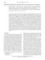
Figure 2 | Allosteric binding of FPP to FPPS. (a) Overall structure, discovery map (inset, green mesh, Fo À Fc contoured at 3s), and phosphorus anomalous
signal (inset, magenta, contoured at 3s). Only one subunit (the crystallographic asymmetric unit) is shown for clarity; the biological assembly is a
homodimer. A stereo image of the final 2Fo À Fc map around the bound ligand is shown in Supplementary Fig. 1. (b) Binding interactions by FPP
pyrophosphate. (c) Binding interactions by IPP pyrophosphate (PDB ID 4H5E). (d) FPP in space-filling representation. The surface of the binding pocket is
also represented. (e) Induced-fit conformational change accompanying FPP binding. The apo-enzyme structure is shown in grey (PDB ID 2F7M). (f–h)
Allosteric pocket in unliganded, FPP-bound and fully closed states, respectively.
NATURE COMMUNICATIONS | 8:14132 | DOI: 10.1038/ncomms14132 | www.nature.com/naturecommunications
3
ARTICLE
0
10 20 30 40 50
0.0
0
–0.2
–0.4
–0.6
10 20 30 40 50
0
Time (min)
10 20 30 40 50
0.0
0
–0.2
–0.4
0.0
–2.0
–4.0
0
1
2
[FPP]/[FPPS]
3
10 20 30 40 50
0
10 20 30 40 50
0.0
–0.4
–0.8
0.0
Q (kcal mol–1)
Q (kcal mol–1)
0.0
Q (kcal mol–1)
c
Time (min)
dQ/dt (μcal s–1)
dQ/dt (μcal s–1)
b
Time (min)
dQ/dt (μcal s–1)
a
NATURE COMMUNICATIONS | DOI: 10.1038/ncomms14132
–2.0
–4.0
0
1
2
3
4
5 0
[DMAPP]/[FPPS]
1
2
–4.0
–8.0
0
1
2
3 0
1
2
3
[GPP]/[FPPS]
Figure 3 | Ligand binding to FPPS characterized by ITC. (a) FPP binding in absence of Mg2 ỵ . The raw thermogram is shown in the upper panel, and the
binding isotherm with the fitted curve in the lower panel. (b) DMAPP binding in absence (left panels) and presence (right panels) of Mg2 ỵ .
(c) GPP binding in absence (left panels) and presence (right panels) of Mg2 ỵ .
surprising partly because such protein rearrangement could
not be predicted from the existing structures.
It has been well established that the conformational transition
between the open and closed states of FPPS dictates the
progression of its catalytic cycle. As described earlier, the closure
of the enzyme enables IPP binding and subsequent catalysis.
Opening, on the other hand, facilitates the translocation of
GPP or the release of FPP on formation of these products.
With FPP bound in the allosteric pocket, the enzyme adopts
the open conformational state. Despite the local differences
introduced by FPP binding, its overall structure is very similar
to that of the apo-enzyme form (Protein Data Bank (PDB)
ID 2F7M; Ca root mean squared deviation ¼ 0.21 Å). Of
particular significance is that closure of the enzyme brings
aH/aJ closer to aC/aG and, as a result, drastically reduces the
volume of the allosteric pocket (Fig. 2h). Therefore, allosterically
bound FPP can be considered as a molecular wedge that prohibits
this conformational transition via steric hindrance. The implication of this insight is clear. FPPS in the unliganded open state
is ready to bind a new DMAPP molecule and thus begin another
catalytic cycle; but if FPP binds to its allosteric pocket first,
the enzyme will stall in its open state and not be able to proceed
to the next catalytic step. To assess the physiological relevance
of this scenario, it was essential to determine the binding affinity
of FPP.
Thermodynamic characterization of FPP and substrate binding.
The in-solution binding of FPP to FPPS was characterized by
isothermal titration calorimetry (ITC). It was shown to be an
exothermic process driven by both favourable enthalpy and
entropy changes, in which one FPP molecule binds to a single site
on the enzyme with a dissociation constant (Kd) of 5.3 mM
(Fig. 3a; Table 2). The single site deduced here most certainly
represents the allosteric pocket based on the present crystal
structure. It is conceivable that FPP might also bind to the active
site (that is, as if just produced by the enzyme, with its head
bound to the IPP site and its tail extended into the allylic
substrate site); however, such a binding mode would be energetically unfavourable. We emphasize that this titration experiment
was done in the absence of Mg2 ỵ or other divalent metal ions,
without which FPPS cannot transition into the closed state. With
the active site of the enzyme open (and without the pyrophosphate by-product), the tail of FPP would be missing most of its
complementary packing surface and exposed to solvent. Interactions with the pyrophosphate moiety would also be suboptimal
unlike those seen with the binding of IPP (which occurs with the
4
enzyme in the closed state). This observation agrees well with the
notion that the open conformation of the enzyme facilitates
efficient release of the reaction product from the active site.
To compare with the binding affinity of FPP, we next
determined those of DMAPP and GPP. It is important to note
that while these substrates must bind to the active site
(more precisely the allylic substrate site), they should also be
able to bind to the allosteric pocket, being structural analogues
of FPP that are only shorter in the tail length. We first carried
out ITC experiments in the absence of divalent metal ions.
Without them, the substrates cannot bind to the allylic substrate
site, unable to interact with the negatively charged Asp-rich
motifs of the enzyme. The resulting data demonstrated that
DMAPP and GPP indeed bind to a single site on the enzyme
with Kd values of 43.7 and 7.6 mM, respectively (left panels,
Fig. 3b,c; Table 2). The weaker binding compared with that
of FPP is due to smaller binding enthalpies (DH, Table 2),
which likely reflect the decreased hydrophobic effect, as well as
fewer van der Waals contacts.
Binding of DMAPP and GPP to FPPS was signicantly tighter
in the presence of Mg2 ỵ (right panels, Fig. 3b,c; Table 2). The
Kd values (2.2 and 2.1 mM for DMAPP and GPP, respectively)
are in excellent agreement with a previously reported Km value
(2.07 mM for GPP)3, supporting the interpretation that the
substrates were in fact binding to the active site now.
Interestingly, the binding affinities of DMAPP and GPP are
similar here unlike at the allosteric pocket; the less favourable
enthalpy change accompanying the binding of DMAPP is
compensated by the more favourable entropic counterpart
(Table 2). The one-site-binding pattern observed here is a
consequence of the enzyme closure, which renders the allosteric
pocket inaccessible to the substrates. Analogous results
demonstrating the biased binding (that is, binding exclusively
to the allylic substrate site in the presence of Mg2 þ ) have been
confirmed crystallographically with allosteric BPs9,12. In contrast
to the binding of DMAPP and GPP, FPP binding was not affected
by Mg2 ỵ (Table 2). These results suggest that under
physiological conditions (present in millimolar concentrations,
Mg2 ỵ is the second most abundant intracellular ion), DMAPP
and GPP bind preferentially to the active site, and FPP to
the allosteric site.
Reaction progress kinetic analysis. The affinity for the active
site binding of DMAPP and GPP to FPPS is less than threefold
higher than that for the allosteric binding of FPP (Table 2).
The small difference signifies that, if the allosteric binding of
NATURE COMMUNICATIONS | 8:14132 | DOI: 10.1038/ncomms14132 | www.nature.com/naturecommunications
ARTICLE
NATURE COMMUNICATIONS | DOI: 10.1038/ncomms14132
Table 2 | Thermodynamic parameters determined by ITC.
Ligand
FPP*
DMAPP*
GPP*
DMAPPz
GPPz
FPPz
n
1.12±0.01
1w
0.80±0.03
0.87±0.01
0.79±0.01
1.17±0.02
Kd (lM)
5.3±0.4
43.7±4.3
7.6±0.9
2.2±0.2
2.1±0.3
6.0±0.6
DH (kcal mol À 1)
À 5.5±0.1
À 4.5±0.2
À 5.3±0.2
À 5.8±0.1
À 7.7±0.2
À 5.6±0.1
TDS (kcal mol À 1)
1.8
1.6
1.8
2.1
0.2
1.7
DMAPP, dimethylallyl pyrophosphate; FPP, farnesyl pyrophosphate; GPP, geranyl pyrophosphate.
The experiment was carried out in triplicate.
*Titrated in absence of Mg2 ỵ .
wThe molar binding ratio was not varied during the data fitting process due to a low c value (that is, a weak inflection point).
zTitrated in presence of Mg2 ỵ .
FPP indeed inhibits the enzyme, the rate of its reaction would be
sensitive to the change in the substrate to product concentration
ratio. To probe for time-dependent product inhibition, we
analysed FPPS reaction progress, employing the ‘same excess’
protocol13. The evolution of entire reaction was monitored
by calorimetry, where two reactions were carried out with
different initial concentrations of GPP and IPP (only the second
part of the catalytic cycle was examined for simplicity), but with
the same difference in the concentrations of the two substrates
(that is, [GPP0]–[IPP0] ¼ [excess] ¼ 24 mM; blue and red curves,
Fig. 4a). Although GPP and IPP concentrations both decrease as
the reaction continues, the change is linked to the reaction
stoichiometry (for every molecule of GPP consumed, one
molecule of IPP is consumed), and thus [excess] remains
constant throughout the full course of the reaction. Therefore,
the two reactions represent an identical reaction that started from
different time points. At any given point that gives the same
amounts of the remaining substrates, there are only
two differences between the two reactions: (i) the reaction with
the higher initial substrate concentrations has accumulated
more FPP; and (ii) the enzyme in this reaction has carried out
more turnovers. If the activity of the enzyme had not been
affected by these differences, the rate curves for the two reactions
would have overlaid onto each other. Instead, the curve for the
reaction with the higher initial substrate concentrations traced
lower (red, lower panel, Fig. 4a). This result indicates that the
enzyme was deactivated over time and/or inhibited by the
accumulating product. An additional reaction with an initial
amount of FPP produced a depressed rate curve as well
(black, Fig. 4a), thus establishing that the reduced catalytic
efficiency is due to product inhibition. The enthalpy of reaction
(DH; equation (1), Methods) was consistent between the three
separate reactions at À 22.5, À 22.1 and À 22.3 kcal mol À 1.
We proceeded to determine some of the kinetic parameters
for FPPS reaction. The Km of IPP was of special interest, since its
Kd could not be determined directly by ITC (the catalytically
relevant IPP binding is to the FPPS–GPP complex; however,
simulating this binding initiates the enzyme reaction).
The experiment was carried out with a saturating excess of
GPP (B500-fold over enzyme and 10-fold over IPP; blue curve,
Fig. 4b) to reduce its analysis to a single-substrate problem.
A general steady-state equation that accounts for product
inhibition was used (equation (4), Methods). Fitting the data
to this model (solid black line, lower panel, Fig. 4b) resulted in
a Km of 1.1 mM, which is comparable to a literature value
of 1.8 mM3. It is also close to a Kd value (0.9 mM) determined
for the binding of IPP to an N-BP-bound FPPS complex14.
The turnover number (kcat) was calculated to be 0.90 s À 1,
slightly higher than previously reported (0.42 s À 1)3. The Km and
kcat values are within the ranges of 10 À 2–103 mM and
0.05–500 s À 1, respectively, the precise determination of these
parameters, in which the calorimetric instrument used allows for
(ref. 15). Interestingly, an analogous experiment carried out in
excess of IPP demonstrated significantly reduced enzyme activity
(red curves, Fig. 4b). IPP binds also to the allylic substrate site
at high concentrations3, in which case it acts as a competitive
inhibitor with respect to DMAPP and GPP. This substrate
inhibition would not be relevant physiologically, however, due
to the action of IPP isomerase (Fig. 1b).
The new findings of this study update our understanding
of the FPPS catalytic cycle; a figure illustrating substrate
binding, product release and the conformational transition
involved, as well as the measured equilibrium and rate constants,
is presented (Fig. 5). Still unknown is the Kd values of the
products. Deduced from the crystallographic and thermodynamic
data, the Kd of FPP for the active site should be much higher than
that for the allosteric pocket. We have attempted to determine
the binding affinity of the by-product PPi both in the presence
and absence of Mg2 ỵ ; however, the results did not demonstrate
an apparent binding event, possibly indicating that the Kd of
PPi is also high.
Discussion
The significance of the mevalonate pathway has been well
established. The effectiveness of N-BPs in inducing osteoclast
death is a clear testimony to its essentialness. Overactivity of
the pathway would also be detrimental as inferred by the
many human diseases arising from hyperlipidemic conditions.
Naturally, the pathway is kept in check by multiple layers
of control mechanisms. It has been long known that the
gateway enzyme hydroxylmethylglutaryl coenzyme A reductase
(HMGCR; Fig. 1b) is feedback-regulated based on the level of
cholesterol both at the transcriptional and post-transcriptional
(via enzyme degradation) levels16. It was found more recently
that the transcription of FPPS is also regulated by the same
mechanism used for HMGCR (that is, through the actions of
sterol regulatory element binding proteins)17,18. Examples of
protein level regulation include that of mevalonate kinase
(Fig. 1b), which is inhibited by the longer-chain prenyl
pyrophosphates GPP, FPP and GGPP19.
Now, the current study provides data indicating that FPPS
is also feedback regulated at the protein level. Significantly,
the enzyme is inhibited by its own product and in an allosteric
manner. Allostery refers to the phenomenon in which binding
of an effector molecule at one site of a protein changes its affinity
for a ligand at a spatially distinct second site. FPPS inhibition
described in the present report clearly embodies this ‘action at a
distance’ principle. FPP binding at the new druggable site,
purely by altering the enzyme’s conformational ensemble,
interferes with DMAPP binding at the distantly located allylic
substrate site (Fig. 5). It is noteworthy that geranylgeranyl
NATURE COMMUNICATIONS | 8:14132 | DOI: 10.1038/ncomms14132 | www.nature.com/naturecommunications
5
ARTICLE
NATURE COMMUNICATIONS | DOI: 10.1038/ncomms14132
a
0
Time (s)
200
0
200
0.0
0.0
[GPP0] = 48 μM
[IPP0] = 24 μM
–1.0
[GPP0] = 72 μM
[IPP0] = 48 μM
[GPP0] = 48 μM
[IPP0] = 24 μM
[FPP0] = 24 μM
–2.0
dQ/dt (μcal s–1)
dQ/dt (μcal s–1)
b
Time (s)
–0.5
[GPP0] = 190 μM
[IPP0] = 19 μM
–1.0
[GPP0] = 19 μM
[IPP0] = 190 μM
0.4
v (μM s–1)
v (μM s–1)
Vmax = 0.36 ± 0.01 μM s–1
0.2
0.2
Km = 1.11 ± 0.06 μM
0.1
0.0
0.0
0
20
40
0
5
[S] (μM)
[IPP] (μM)
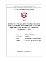
Figure 4 | Reaction progress kinetic analysis of FPPS. (a) Same excess experiment. Thermograms are shown in the upper panel, and differential rate data
generated from the thermograms in the lower panel. The initial substrate and product concentrations are indicated. (b) Determination of kinetic
parameters. The data from the excess IPP experiment (red) were not regression-analysed due to apparent substrate inhibition.
a
b
IPP
PPi
DMAPP
Kd = 2.2 μM
DMAPP
Kd =
5–6 μM
IPP
GPP
Active cycle
FPP
Inactive
state
Kd =
2.1 μM
FPP
kcat = 0.90 s–1
Km =
1.1 μM
FPP
PPi
IPP
Figure 5 | Conformational transition and catalytic cycle of FPPS. (a) Superimposition of open (FPP bound, cyan) and closed (substrate bound, green)
states. DMAPP was modelled in based on the structures of FPPS in complex with substrate analogues (PDB IDs 1RQI and 4H5E). Yellow spheres are
Mg2 ỵ ions coordinated to the Asp-rich motifs of the enzyme. (b) Schematic representation of FPPS catalytic cycle.
pyrophosphate synthase (GGPPS), the enzyme immediately
downstream of FPPS (Fig. 1b), is also inhibited by its
own product20. This inhibition, however, is not of allosteric
nature. GGPP binds in the heart of GGPPS active site and inhibits
the enzyme by directly competing with its allylic substrate20.
Enzymes that are allosterically inhibited by their own products
are uncommon. Such an inhibition mechanism allows enzymes
to have an immediately responsive feedback process (as opposed
to feedback by downstream metabolites) without compromising
their catalytic efficiency (active site product inhibition
often involves slow product release and/or backward reaction).
One of the few examples of allosteric product feedback is found
in hexokinase-1 (ref. 21), which catalyses the phosphorylation
of glucose by ATP, the first enzymatic step in glucose metabolism. This enzyme is allosterically inhibited by physiological
6
concentrations of its product, glucose-6-phosphate, and
thus controls the influx of substrate into the glycolytic pathway.
Regulation and modulation of enzyme catalytic activity should
be useful in controlling the flux of precursors and products
in any metabolic pathway. The cryptic nature of the allosteric
pocket in FPPS and the fact that its physiological effector
remained unidentified for a long time thus raise an intriguing
possibility: allosteric product inhibition might be more prevalent
than currently known amongst metabolic enzymes.
Efforts to exploit the allosteric pocket of FPPS as a therapeutic
target are actively ongoing. With the pocket dubbed
as the ‘Achilles’ heel’ of the enzyme, the enthusiasm in the
field is clearly evident. Potent allosteric inhibitors of FPPS may
have a wide range of applications, in addition to their potential
use as anticancer drugs. For example, they could serve as
NATURE COMMUNICATIONS | 8:14132 | DOI: 10.1038/ncomms14132 | www.nature.com/naturecommunications
ARTICLE
NATURE COMMUNICATIONS | DOI: 10.1038/ncomms14132
cholesterol-lowering agents; nature has already appropriated
a similar strategy at the transcriptional level. They may also
prove useful against neurodegenerative diseases; a genetic
link between elevated levels of FPPS and phosphorylated tau
protein, a key factor in neurodegeneration, has been established9.
Here we revisit the observation that the FPP pyrophosphate
bound to the allosteric pocket superimposes poorly with the
BPs of our allosteric inhibitors. On the other hand, the first series
of allosteric inhibitors discovered by Jahnke et al.8 is carboxylate
based. It is encouraging that the allosteric pocket supports diverse
binding poses of different functionalities. Indeed, discovery
of new inhibitors based on known drug scaffolds (for example,
salicylic acid)22 and those incorporating distinct functional
groups for tissue selectivity (for example, monophosphonate)23
has been reported very recently. In this light, it is pertinent that
FPP binding occurs through an induced-fit mechanism involving
expansion of the allosteric pocket. Discovery of additional
inhibitors that can exploit such a conformational change is
expected.
As an enzyme that catalyses two reactions in a sequential
manner, FPPS is a challenging enzyme to study. To make
the matter even more complex, its substrates and products
are analogues that differ only in their hydrocarbon tail length.
Perhaps because of these complications, and despite the bulk
of research done, certain aspects of the enzyme have remained
undiscovered for decades. In this work, we have demonstrated
through a modern kinetic approach that FPPS is inhibited
by FPP. Our crystal structure reveals that the product can trap
the enzyme in an unreactive state by binding to its allosteric
pocket. On the basis of the affinities of the substrates
and products measured, this binding should be sensitive to
the fluctuating levels of the prenyl pyrophosphates in vivo.
The allostery thus provides an exquisite means of regulating
and fine-tuning these levels. The consequences of our findings
on mammalian biology call for future cellular metabolomic
studies.
wavelengths of the X-ray beams used were 1.5418 and 0.97949 Å, respectively. Both
data sets were processed with the xia2 package24; however, Friedel mates were
not merged for the home-source set. Only the synchrotron data were used for
structure determination. The initial model was built by a difference Fourier
method with a solvent-omitted starting model generated from PDB entry 2F7M.
This model was improved through iterative rounds of manual and automated
refinement with Coot25 and REFMAC5 (ref. 26). Ramachandran statistics for
the final model show 97% of the residues in the favoured regions and 3% in
the allowed regions. An anomalous signal map was calculated from the
home-source data with SHELXC27 and ANODE28. The phase information used
in this calculation was obtained from the structure model refined against the
synchrotron data. Data collection and structure refinement statistics are
summarized in Table 1.
Methods
in which Q is the total heat associated with producing n moles of product, [S0] is
the starting concentration of the limiting substrate and V is the volume of the
calorimetric cell. Q was calculated by integrating the thermal power (dQ/dt)
measured over the complete course of reaction, whereas [S0] and V were known.
Once DH was determined, the substrate concentration ([S]) could be determined as
a function of time as described in the equation:
R t dQ
dt
ẵS ẳ ẵS0 0 dt :
2ị
V DH
Expression and purification of human FPPS. A pET-based plasmid encoding
human FPPS with an N-terminal His6 tag was transformed into Escherichia coli
BL21 (DE3) cells. The cells were grown in LB at 37 °C until the OD600 of
0.6–0.8 was reached. Expression of the recombinant enzyme was induced by
1 mM isopropylthiogalactoside overnight at 18 °C. To collect the enzyme, the
cells were lysed in a buffer containing 50 mM HEPES (pH 7.5), 500 mM NaCl,
2 mM b-mercaptoethanol, 5 mM imidazole and 5% glycerol. The lysate was applied
to a metal ion affinity column (Ni-nitrilotriacetic acid), from which the enzyme was
eluted with an increasing imidazole gradient. The enzyme containing fractions
were pooled and further purified by size-exclusion chromatography (Superdex
200). For storage, the purified enzyme was concentrated to B20 mg ml À 1 by
ultrafiltration.
Isoprenyl pyrophosphates. DMAPP, IPP, GPP (trans isomer) and
FPP (trans,trans isomer) were all purchased from Sigma-Aldrich. When came
as a methanol/ammonia solution, the solvent was removed from the sample by
desiccating in a centrifugal evaporator. The compounds were dissolved in
appropriate buffers for different experiments as described below.
Crystallization. FPP was prepared at 5 mM concentration in the final purification
buffer (10 mM HEPES (pH 7.5), 500 mM NaCl, 2 mM b-mercaptoethanol
and 5% glycerol). MgCl2 was prepared as a 100 mM aqueous solution. FPP and
MgCl2 were added to the purified enzyme to give the concentrations of 1 mM FPP,
2 mM MgCl2 and 10 mg ml À 1 enzyme. A single crystal was obtained at
22 °C by vapour diffusion in a sitting drop composed of 1 ml FPP/MgCl2/enzyme
mixture and 1 ml crystallization solution (80 mM TrisHCl (pH 8.5),
1.6 M ammonium phosphate and 20% glycerol).
Structure determination. Diffraction data were collected under cryogenic
conditions (100 K) first at the home lab with a MicroMax-007 HF generator
and a Saturn 944 ỵ charge-coupled device detector, and then at a synchrotron
(Beamline 08ID-1, Canadian Light Source, Saskatoon, SK, Canada). The
Binding assay. Binding experiments were carried out at 30 °C with a MicroCal
iTC200 system. The purified enzyme was dialyzed overnight against the binding
assay buffer (50 mM HEPES (pH 7.5), 150 mM NaCl, 2 mM b-mercaptoethanol
and 5% glycerol). Ligand (DMAPP, GPP and FPP) and MgCl2 solutions were
prepared in the used dialysate. Each titration experiment consisted of a first
1 ml injection followed by 18 2 ml injections of a ligand solution into the
204.1 ml calorimetric cell loaded with the enzyme solution. The concentration
of the enzyme in the cell was 100 mM (in monomers), and those of the ligands
in the titration syringe ranged from 1 to 2 mM. When added, the concentration
of MgCl2 was 5 mM. Heats of dilution were measured by injecting the ligands
into the buffer alone and subtracted from the corresponding titration data.
The data were fitted to the single-site-binding model implemented in the
Origin software package provided with the ITC instrument.
Reaction assay. Reaction calorimetry was also carried out at 30 °C with
a MicroCal iTC200 system. The enzyme and substrates were prepared in
the reaction buffer (50 mM HEPES (pH 7.5), 150 mM NaCl, 2 mM MgCl2,
2 mM b-mercaptoethanol and 5% glycerol) in the same way described for the
binding experiments. The reactions were assayed by a single injection method15,
where 10 ml substrate solution of both GPP and IPP was injected into the
calorimetric cell containing the enzyme. When added, FPP was preincubated
together with the enzyme. The concentration of the enzyme in the cell was
400–500 nM, and those of the substrates in the syringe were 0.4–4 mM. Heats of
dilution were measured and subtracted from the actual reaction data. The raw data
were processed with the Enzyme Assay module of the Origin package, and plots
of reaction rate as a function of substrate concentration were generated
(five data points were binned for each data point displayed). Briefly, The molar
reaction enthalpy (DH) was determined first based on the relationship:
Q ¼ n DH ẳ ẵS0 V DH;
1ị
The rate of reaction could be calculated from dQ/dt for any given time point:
Rate ẳ
dẵP
dẵS
1 dQ
ẳ
ẳ
:
dt
dt
V DH dt
3ị
For kinetic measurements, the temporal experimental heat ow must be monitored
accurately. To ensure that the heat flow measured was not convoluted with
the rate of heat transfer through the reactor wall, the experimental data were
mathematically corrected by the apply time constant function implemented in the
Origin software. Km and Vmax values were determined by fitting the rate data to the
following equation29:
dẵP
ẳ
dt
Vmax ẵS
1 Km =Kp ị
:
ẵS0 ỵ Kp
Kp =Km 1ị ỵ ẵS
4ị
The value of 6 mM was substituted for the product affinity term (Kp) as determined
in the binding assay. To carry out analysis with only the portion of the reaction
exhibiting steady-state behaviour, data obtained during the induction period were
omitted.
Data availability. Sequence information on human FPPS is available in the
UniProt Knowledgebase under accession code P14324. The PDB accession codes
1RQI, 2F7M, 4H5E, 4LPG and 4QXS were used in this study. Coordinates and
structure factor of the structure reported here have been deposited into the Protein
Data Bank under accession code 5JA0. All other relevant data are available from
the corresponding author upon reasonable request.
NATURE COMMUNICATIONS | 8:14132 | DOI: 10.1038/ncomms14132 | www.nature.com/naturecommunications
7
ARTICLE
NATURE COMMUNICATIONS | DOI: 10.1038/ncomms14132
References
1. McTaggart, S. J. Isoprenylated proteins. Cell. Mol. Life Sci. 63, 255–267
ð2006Þ:
2. Hosfield, D. J. et al. Structural basis for bisphosphonate-mediated inhibition of
isoprenoid biosynthesis. J. Biol. Chem. 279, 8526–8529 (2004).
3. Kavanagh, K. L. et al. The molecular mechanism of nitrogen-containing
bisphosphonates as antiosteoporosis drugs. Proc. Natl Acad. Sci. USA 103,
7829–7834 (2006).
4. Rondeau, J. M. et al. Structural basis for the exceptional in vivo efficacy of
bisphosphonate drugs. ChemMedChem. 1, 267–273 (2006).
5. Russell, R. G. Bisphosphonates: the first 40 years. Bone 49, 2–19 (2011).
6. Berndt, N., Hamilton, A. D. & Sebti, S. M. Targeting protein prenylation for
cancer therapy. Nat. Rev. Cancer 11, 775–791 (2011).
7. Rogers, M. J., Crockett, J. C., Coxon, F. P. & Monkkonen, J. Biochemical
and molecular mechanisms of action of bisphosphonates. Bone 49, 34–41
(2011).
8. Jahnke, W. et al. Allosteric non-bisphosphonate FPPS inhibitors identified by
fragment-based discovery. Nat. Chem. Biol. 6, 660–666 (2010).
9. De Schutter, J. W. et al. Multistage screening reveals chameleon ligands of the
human farnesyl pyrophosphate synthase: implications to drug discovery for
neurodegenerative diseases. J. Med. Chem. 57, 5764–5776 (2014).
10. Gritzalis, D. et al. Probing the molecular and structural elements of ligands
binding to the active site versus an allosteric pocket of the human farnesyl
pyrophosphate synthase. Bioorg. Med. Chem. Lett. 25, 1117–1123 (2015).
11. Park, J., Lin, Y. S., Tsantrizos, Y. S. & Berghuis, A. M. Structure of human
farnesyl pyrophosphate synthase in complex with an aminopyridine
bisphosphonate and two molecules of inorganic phosphate. Acta Crystallogr.
F Struct. Biol. Commun. 70, 299–304 (2014).
12. Leung, C. Y. et al. Thienopyrimidine bisphosphonate (ThPBP) inhibitors of the
human farnesyl pyrophosphate synthase: optimization and characterization of
the mode of inhibition. J. Med. Chem. 56, 7939–7950 (2013).
13. Blackmond, D. G. Reaction progress kinetic analysis: a powerful methodology
for mechanistic studies of complex catalytic reactions. Angew. Chem. Int. Ed.
Engl. 44, 4302–4320 (2005).
14. Park, J., Lin, Y. S., De Schutter, J. W., Tsantrizos, Y. S. & Berghuis, A. M.
Ternary complex structures of human farnesyl pyrophosphate synthase bound
with a novel inhibitor and secondary ligands provide insights into the
molecular details of the enzyme’s active site closure. BMC Struct. Biol. 12, 32
(2012).
15. Freyer, M. W. & Lewis, E. A. Isothermal titration calorimetry: experimental
design, data analysis, and probing macromolecule/ligand binding and kinetic
interactions. Methods Cell Biol. 84, 79–113 (2008).
16. Goldstein, J. L. & Brown, M. S. Regulation of the mevalonate pathway. Nature
343, 425–430 (1990).
17. Ericsson, J., Jackson, S. M., Lee, B. C. & Edwards, P. A. Sterol regulatory
element binding protein binds to a cis element in the promoter of the farnesyl
diphosphate synthase gene. Proc. Natl Acad. Sci. USA 93, 945–950 (1996).
18. Horton, J. D. et al. Combined analysis of oligonucleotide microarray data from
transgenic and knockout mice identifies direct SREBP target genes. Proc. Natl
Acad. Sci. USA 100, 12027–12032 (2003).
19. Hinson, D. D., Chambliss, K. L., Toth, M. J., Tanaka, R. D. & Gibson, K. M.
Post-translational regulation of mevalonate kinase by intermediates of the
cholesterol and nonsterol isoprene biosynthetic pathways. J. Lipid Res. 38,
2216–2223 (1997).
20. Kavanagh, K. L., Dunford, J. E., Bunkoczi, G., Russell, R. G. & Oppermann, U.
The crystal structure of human geranylgeranyl pyrophosphate synthase reveals
a novel hexameric arrangement and inhibitory product binding. J. Biol. Chem.
281, 22004–22012 (2006).
21. Mulichak, A. M., Wilson, J. E., Padmanabhan, K. & Garavito, R. M. The
structure of mammalian hexokinase-1. Nat. Struct. Biol. 5, 555–560 (1998).
8
22. Marzinzik, A. L. et al. Discovery of novel allosteric non-bisphosphonate
inhibitors of farnesyl pyrophosphate synthase by integrated lead finding.
ChemMedChem. 10, 1884–1891 (2015).
23. Jahnke, W. et al. A general strategy for targeting drugs to bone. Angew. Chem.
Int. Ed. Engl. 54, 14575–14579 (2015).
24. Winter, G., Lobley, C. M. & Prince, S. M. Decision making in xia2. Acta
Crystallogr. D Biol. Crystallogr. 69, 1260–1273 (2013).
25. Emsley, P., Lohkamp, B., Scott, W. G. & Cowtan, K. Features and development
of Coot. Acta Crystallogr. D Biol. Crystallogr. 66, 486–501 (2010).
26. Murshudov, G. N. et al. REFMAC5 for the refinement of macromolecular
crystal structures. Acta Crystallogr. D Biol. Crystallogr. 67, 355–367 (2011).
27. Sheldrick, G. M. Experimental phasing with SHELXC/D/E: combining chain
tracing with density modification. Acta Crystallogr. D Biol. Crystallogr. 66,
479–485 (2010).
28. Thorn, A. & Sheldrick, G. M. ANODE: anomalous and heavy-atom density
calculation. J. Appl. Crystallogr. 44, 1285–1287 (2011).
29. Koerber, S. C. & Fink, A. L. The analysis of enzyme progress curves by
numerical differentiation, including competitive product inhibition and enzyme
reactivation. Anal. Biochem. 165, 75–87 (1987).
Acknowledgements
We thank the beamline personnel at the Canadian Light Source for data collection.
This work was supported by grants from the Canadian Institute of Health Research
to Y.S.T. (CIHR-126062) and A.M.B. (MOP-114889), and the Fonds de recherche du
Que´bec—Nature et technologies to both Y.S.T. and A.M.B. (FRQ-NT PR-181227).
A.M.B. holds a Canada Research Chair in Structural Biology.
Author contributions
J.P. designed the study and performed all experiments unless noted otherwise. M.Z. and
A.M. participated as undergraduate project students: M.Z. set-up the crystallization trays,
and A.M. carried out part of the ITC experiments. J.P. analysed all experimental data and
wrote the manuscript together with Y.S.T. and A.M.B.
Additional information
Supplementary Information accompanies this paper at />naturecommunications
Competing financial interests: The authors declare no competing financial interests.
Reprints and permission information is available online at />reprintsandpermissions/
How to cite this article: Park, J. et al. Human farnesyl pyrophosphate synthase
is allosterically inhibited by its own product. Nat. Commun. 8, 14132
doi: 10.1038/ncomms14132 (2017).
Publisher’s note: Springer Nature remains neutral with regard to jurisdictional claims in
published maps and institutional affiliations.
This work is licensed under a Creative Commons Attribution 4.0
International License. The images or other third party material in this
article are included in the article’s Creative Commons license, unless indicated otherwise
in the credit line; if the material is not included under the Creative Commons license,
users will need to obtain permission from the license holder to reproduce the material.
To view a copy of this license, visit />
r The Author(s) 2017
NATURE COMMUNICATIONS | 8:14132 | DOI: 10.1038/ncomms14132 | www.nature.com/naturecommunications