hyperbaric oxygen promotes osteogenic differentiation of bone marrow stromal cells by regulating wnt3a catenin signaling an in vitro and in vivo study
Bạn đang xem bản rút gọn của tài liệu. Xem và tải ngay bản đầy đủ của tài liệu tại đây (2.3 MB, 15 trang )
Stem Cell Research (2014) 12, 260–274
Available online at www.sciencedirect.com
ScienceDirect
www.elsevier.com/locate/scr
Hyperbaric oxygen promotes osteogenic
differentiation of bone marrow stromal cells
by regulating Wnt3a/β-catenin signaling—An
in vitro and in vivo study☆
Song-Shu Lin a,c,1 , Steve W.N. Ueng c,1 , Chi-Chien Niu c , Li-Jen Yuan c ,
Chuen-Yung Yang c , Wen-Jer Chen c , Mel S. Lee d , Jan-Kan Chen b,⁎
a
Institute of Biomedical Sciences, Chang Gung University, Taoyuan, Taiwan
Department of Physiology, Chang Gung University, Taoyuan, Taiwan
c
Department of Orthopaedics, Chang Gung Memorial Hospital, Taoyuan, Taiwan
d
Department of Orthopaedics, Chang Gung Memorial Hospital, Chiayi, Taiwan
b
Received 7 June 2013; received in revised form 9 October 2013; accepted 23 October 2013
Available online 1 November 2013
Abstract We hypothesized that the effect of hyperbaric oxygen (HBO) on bone formation is increased via osteogenic
differentiation of bone marrow stromal cells (BMSCs), which is regulated by Wnt3a/β-catenin signaling. Our in vitro data
showed that HBO increased cell proliferation, Wnt3a production, LRP6 phosphorylation, and cyclin D1 expression in
osteogenically differentiated BMSCs. The mRNA and protein levels of Wnt3a, β-catenin, and Runx2 were upregulated while
those of GSK-3β were downregulated after HBO treatment. The relative density ratio (phospho-protein/protein) of Akt and
GSK-3β was both up-regulated while that of β-catenin was down-regulated after HBO treatment. We next investigated whether
HBO affects the accumulation of β-catenin. Our Western blot analysis showed increased levels of translocated β-catenin that
stimulated the expression of target genes after HBO treatment. HBO increased TCF-dependent transcription, Runx2 promoter/
Luc gene activity, and the expression of osteogenic markers of BMSCs, such as alkaline phosphatase activity, type I collagen,
osteocalcin, calcium, and the intensity of Alizarin Red staining. HBO dose dependently increased the bone morphogenetic
protein (BMP2) and osterix production. We further demonstrated that HBO increased the expression of vacuolar-ATPases,
which stimulated Wnt3a secretion from BMSCs. Finally, we showed that the beneficial effects of HBO on bone formation were
related to Wnt3a/β-catenin signaling in a rabbit model by histology, mechanical testing, and immunohistochemical assays.
Accordingly, we concluded that HBO increased the osteogenic differentiation of BMSCs by regulating Wnt3a secretion and
signaling.
© 2013 The Authors. Published by Elsevier B.V. All rights reserved.
☆ This is an open-access article distributed under the terms of the Creative Commons Attribution-NonCommercial-No Derivative Works
License, which permits non-commercial use, distribution, and reproduction in any medium, provided the original author and source are
credited.
⁎ Corresponding author at: Department of Physiology, College of Medicine, Chang Gung University, 259 Wen-Hwa 1st Road, Kweishan,
Taoyuan 333, Taiwan.
E-mail address: (J.-K. Chen).
1
Lin, S.S. and Ueng, S.W.N. contributed equally to this article.
1873-5061/$ - see front matter © 2013 The Authors. Published by Elsevier B.V. All rights reserved.
/>
Hyperbaric oxygen promotes osteogenic differentiation by regulating Wnt3a/β-catenin signaling
Introduction
Bone loss induced by hypoxia is associated with various pathophysiological conditions such as ischemia (Vogt et al., 1997).
The long-term culturing of human bone marrow stromal cells
(BMSCs) under hypoxia conditions promotes a genetic program
that maintains their undifferentiated and multi-potent status
(Basciano et al., 2011). Hypoxia induces BMSC proliferation and
enhances long-term BMSC expansion, but results in a population with impaired osteogenic differentiation potential (Fehrer
et al., 2007; Pattappa et al., 2013). Hypoxia inhibits osteogenic
differentiation in BMSCs by regulating Runx2 via the basic
helix–loop–helix (bHLH) transcription factor TWIST (Yang
et al., 2011).
Hyperbaric oxygen (HBO) therapy is a safe noninvasive
modality that increases the oxygen tension of tissues and
microvasculature (Korhonen et al., 1999). HBO increases the
expression of placental growth factor in BMSCs (Shyu et al.,
2008), fibroblast growth factor (FGF)-2 in osteoblasts (Hsieh
et al., 2010), and the Wnt-3 protein in neural stem cells
(Wang et al., 2007). The BMSC population contains a subset
comprised of skeletal stem cells, which contribute to the
regeneration of mesenchymal tissues such as bone, cartilage, muscle, ligament, tendon, and adipocyte in vivo, and
cartilage in pellet cultures in vitro (Pittenger et al., 1999).
Previous studies have suggested that Wnt signaling could be
used to stimulate bone healing (Minear et al., 2010) and
fracture repair (Komatsu et al., 2010). We first reported the
beneficial effects of HBO on bone lengthening in a rabbit
model (Ueng et al., 1998). However, little is known about
the effects of HBO on the Wnt signaling pathway in BMSCs.
Autocrine and paracrine Wnt signaling operates in stem
cell populations and regulates mesenchymal lineage specification. The target cells for the Wnt proteins expressed by
BMSCs may be either BMSCs themselves or other cell types in
the bone marrow (Etheridge et al., 2004). Wnt proteins are
secreted lipid-modified signaling molecules that influence
multiple processes during animal development (Nusse,
2003). The Wnt family of signaling proteins mediates cell–
cell communication (Lorenowicz and Korswagen, 2009; Port
and Basler, 2010). In the absence of the Wnt protein,
β-catenin is phosphorylated by glycogen synthase kinase-3β
(GSK-3β) and subsequently degraded by proteasomes (Zeng
et al., 2005). On target cells, secreted Wnt proteins interact
with the receptors Frizzled and low-density lipoprotein
receptor-related (LRP) 5/6 to activate the β-catenin
pathway (Logan and Nusse, 2004). Activation of the Frizzled
receptor complex results in the inhibition of a phosphorylation cascade that stabilizes intracellular β-catenin levels.
β-Catenin is subsequently translocated into the nucleus to
form a transcriptionally active β-catenin T-cell factor
(TCF)/lymphoid enhancer factor (LEF) DNA-binding complex that regulates the Wnt target gene. Among Wnt family
members, Wnt3a is involved in the proliferation and
differentiation of BMSCs (De Boer et al., 2004). Once
BMSCs are committed to the osteogenic lineage, canonical
Wnt signaling stimulates their differentiation (Ling et al.,
2009; Eijken et al., 2008). Canonical Wnt signaling promotes
osteogenesis by directly stimulating Runx2 gene expression
(Gaur et al., 2005). Runx2 activates osteocalcin, which is an
osteoblast-specific gene expressed by differentiated osteoblasts (Ducy, 2000).
261
Vacuolar ATPases (V-ATPases) are large multi-subunit
complexes that are organized into V0 and V1 domains, which
operate by a rotary mechanism (Forgac, 2007). V-ATPasedriven proton pumping and organellar acidification are essential for vesicular trafficking along both the exocytotic and
endocytotic pathways of eukaryotic cells. In Wnt producing
cells, vacuolar acidification is required for Wnt signaling
(Cruciat et al., 2010; Coombs et al., 2010). The secretion of
Wnt3a protein into the cell culture medium was shown to be
dependent on vacuolar pH. Moreover, acidification inhibitor
was shown to decrease secreted and increase cell-associated
Wnt3a. The inhibition of V-ATPase blocks Wnt3a secretion and
inhibits Wnt/β-catenin signaling both in cultured human cells
and in vivo (Coombs et al., 2010).
In the present study, we found that HBO increased cell
proliferation, LRP6 phosphorylation, and cyclin D1 expression in osteogenically differentiated BMSCs. HBO increased
the osteogenic differentiation of BMSCs via regulation of
Wnt3a signaling as well as increased the TCF-dependent
transcription and Runx2 promoter/Luc gene activity. Because Wnt/β-catenin signaling is an upstream activator of
BMP2 expression in osteoblasts, we found that HBO dose
dependently increased the BMP2 and osterix production.
Since endosomal acidification is an essential function of the
Wnt secretion pathway, we further demonstrated that HBO
increased the expression of V-ATPases to stimulate Wnt3a
secretion. Finally, we showed the beneficial effects of HBO
on bone formation via Wnt/β-catenin signaling regulation in
a rabbit model.
Materials and methods
In vitro study
The experimental protocol was approved by the human subjects Institutional Review Board of the Chang Gung Memorial
Hospital.
Surgical procedures
We harvested BMSCs from patients who underwent iliac bone
grafting for spine fusion. During bone graft harvesting,
10 mL of bone marrow was aspirated and collected in a
heparin-rinsed syringe.
Isolation and cultivation of BMSCs
Each marrow sample was washed with Dulbecco's phosphatebuffered saline (DPBS). Up to 2 × 10⁸ nucleated cells in 5 mL of
DPBS were loaded onto 25 mL of Percoll cushion (Pharmacia
Biotech). A density gradient was used as the isolation
procedure to eliminate unwanted cell types that were present
in the marrow aspirate. A small percentage of cells were
isolated from the density interface at 1.073 g/mL. The cells
were re-suspended and plated at 2 × 10⁵ cells in T-75 flasks.
The cells were maintained in Dulbecco's Modified Eagle's
Medium-Low Glucose (DMEM-LG; Gibco, Grand Island, NY)
that contained 20% fetal bovine serum (FBS) and antibiotics at
37 °C in a humidified atmosphere of 5% CO2 and 95% air. After
7 d of primary culturing, the non-adherent cells were removed
by changing the medium. The BMSCs grew as symmetric
colonies and were subcultured at 10 to 14 d by treatment
with 0.05% trypsin (Gibco) and seeded into fresh flasks.
262
Flow cytometric analysis of surface antigen expression
When confluent, the BMSCs were passaged 1 in 3, and a sample
was analyzed for MSC marker expression by flow cytometry. The
cells were washed in phosphate-buffered saline (PBS), and then
removed from the flask by 0.05% trypsin (Gibco). 1 × 10⁵ cells
were incubated with each mouse monoclonal primary antibody
at 4 °C for 30 min. Mouse FITC-conjugated anti-CD105 antibody
(1:100 dilution), mouse PE-conjugated anti-CD146 antibody
(1:100 dilution), and mouse FITC-conjugated anti-CD34 antibody (1:100 dilution) were purchased from Becton Dickinson
(Oxford, UK). Mouse FITC-conjugated anti-αSMA antibody (1:25
dilution) was purchased from Abcam (Cambridge, UK). Mouse
PE-conjugated anti-STRO-1 antibody (1:50 dilution) was purchased from Santa Cruz (CA, USA). After wash, the cells were
resuspended in 500 μL wash buffer and analyzed on a BD flow
cytometer (Oxford, UK).
Cell exposure to intermittent HBO
Cells were cultured in complete medium (DMEM-LG containing
10% FBS and antibiotics) and the osteogenic groups were
cultured in osteogenic induction medium (DMEM-LG containing
10% FBS, antibiotics, 100 μM ascorbate-2 phosphate, 100 nM
dexamethasone, and 10 mM β-glycerophosphate). Control cells
were maintained in 5% CO2/95% air throughout the experiment.
The hyperoxic cells were exposed to 100% O2 for 25 min and
then to 5% CO2/95% air for 5 min at 2.5 ATA (atmospheres
absolute) in a hyperbaric chamber (Huxley Corporation, Taipei,
Taiwan) for 90 min every 36 h.
Cell proliferation assay
Cell proliferation was quantified using the WST-1 cell
proliferation reagent (Roche, Penzberg, Germany) according
to the manufacturer's protocol. About 2 × 10³ BMSCs/well
were plated on 24-well cell culture plates and incubated at
37 °C in 5% CO2/95% air. After 12 h, the culture medium was
changed to complete or osteogenic induction medium with
10% FBS and the cells were exposed to HBO (day 1). Cells
were incubated for 36 h after HBO treatment, 100 μL/well
of WST-1 was added, and then incubated for 4 h. The
absorbance of each sample was determined in triplicate
using an ELISA plate-reader (MRX; Dynatech Labs) at 440 nm.
On days 4, 7, 10 and 14, the absorbance of each sample was
determined as described above.
RNA preparation and real-time quantitative polymerase
chain reaction (Q-PCR) analysis
About 2.5 × 10⁵ BMSCs were plated onto 100 mm cell culture
dishes. After culturing for 1, 4, and 7 d with or without HBO
treatment, the cultured cells were rinsed with PBS. Total RNA
was extracted using a Qiagen RT kit (Qiagen, USA) according to
the manufacturer's instructions. Each RNA sample was further
purified using an RNeasy Mini Column (Qiagen). The RNA
concentration was evaluated by A260/A280 measurement. To
detect the Wnt3a, GSK-3β, β-catenin, Runx 2, type I collagen,
osteocalcin, BMP2, osterix, and GAPDH RNA transcripts, cDNA
was analyzed using an ABI PRISM 7900 sequence detection
system and TaqMan PCR Master Mix (Applied Biosystems, Foster
City, CA). The cycle threshold (Ct) values were obtained, and
the data were normalized to GAPDH expression using the ΔΔCt
method to calculate the relative mRNA level of each target
gene.
S.-S. Lin et al.
Small interfering RNA transfection
On day 1, 2 × 10⁵ BMSCs were plated onto a 6-well tissue
culture plate in 2.5 mL of OPTI-MEM (Invitrogen, Carlsbad,
CA) medium that was free of antibiotics and serum. The
BMSCs were then transfected with human β-catenin small
interfering (si)RNA or scrambled siRNA (Stealth RNAi,
Invitrogen) using Lipofectamine RNAiMAX (Invitrogen) according to the manufacturer's instructions. After 8 h of
transfection, the culture medium was changed to osteogenic
medium with 10% FBS and the cells were exposed to HBO
treatment. On days 4 and 7, the cells were re-transfected
once and exposed to HBO. After an additional 24 h of
culturing, the BMSCs were harvested for analysis. The
silencing effect on β-catenin and downregulation of Runx 2
were detected by real-time PCR after the treatments.
Western blot analysis
About 2.5 × 10⁵ BMSCs were plated on 100 mm cell culture
dishes. After culturing for 7 d or 14 d with or without HBO
treatment, the cells were washed with PBS and extracted using
M-PER protein extraction reagent (Thermo, USA). The protein
content was quantitated using a protein assay kit (Pierce
Biotechnology, IL), separated by 7.5% SDS-PAGE, and transferred onto membranes using a transfer unit (Bio-Rad, USA).
After blocking, the membranes were incubated with 1000fold diluted rabbit antibodies against Wnt3a, phosphor-LRP6,
GSK-3β (Cell Signaling, MA, USA), LRP6 (Abcam, Cambridge,
UK), or mouse antibodies against β-catenin (Millipore, Temecula, CA), β-actin (Millipore), Runx 2 (Millipore), Wnt1 (Abcam),
Akt (Abcam), phosphor-Akt (Ser472) (Abcam), phosphor-GSK3β (Ser9) (Abcam), phosphor-β-catenin (Ser33/37, Thr41)
(Cell Signaling), BMP2 (Abcam), and osterix (Abcam). After
washing, the membranes were further incubated for 2 h with
10,000-fold goat anti-mouse IgG (Calbiochem, USA) or goat
anti-rabbit IgG (Millipore) conjugated to horseradish peroxidase. The membranes were then washed and rinsed with ECL
detection reagents (Millipore). The bands were photographed
using ECL Hyperfilm (Amersham Pharmacia Biotech, UK) and
their intensity was quantified using an image-analysis system
(Image-Pro plus 5.0).
Preparation of cytosolic and nuclear fractions for
β-catenin detection
About 2.5 × 10⁵ BMSCs were plated on 100 mm cell culture
dishes. After culturing for 7 d with or without HBO treatment,
the cells were rinsed with PBS, treated with 0.05% trypsin, and
then collected by centrifugation at 800 g. NE-PER nuclear and
cytoplasmic extraction reagents (Thermo Science, USA) were
used to isolate cytoplasmic and nuclear extracts from the cells.
The protein content was quantitated using a protein assay kit
(Pierce), and separated by 7.5% SDS-PAGE to detect β-catenin
(Millipore) and TATA binding protein (TBP; Abcam).
On days 1, 4, and 7, the BMSCs were transfected with
β-catenin siRNA or scrambled siRNA and exposed to HBO as
described above. After an additional 24 h of culturing, the
cytoplasmic and nuclear extracts were harvested for β-catenin
detection as described above.
Transcription activity of the β-catenin–TCF/LEF complex
Cells were seeded in 24-well tissue culture plates at 5 × 10⁴
cells/well in 0.5 mL of Opti-MEM (Invitrogen) at 12 h before
transfection. On the day of transfection (day 1), 900 ng of
Hyperbaric oxygen promotes osteogenic differentiation by regulating Wnt3a/β-catenin signaling
the TOPFLASH or FOPFLASH construct (Upstate, Chicago, IL)
together with 100 ng of the pGL4.74 [hRluc/TK] plasmid
(Promega, Madison, WI) was used to transfect the cells in
each well. The pGL4.74 [hRluc/TK] plasmid containing the
Renilla luciferase gene was used as an internal control for
normalizing the transfections. Transient transfections using
Lipofectamine LTX and PLUS reagent (Invitrogen) were
performed according to the manufacturer's instructions. Eight
hours after transfection, the transfection medium was changed
to osteogenic induction medium with 10% FBS, and the cells
were exposed to HBO. On days 4 and 7, the cells were
re-transfected once and exposed to HBO as described above.
After an additional 24 h of culturing, the BMSCs were washed
with PBS and harvested using 100 μL/well of passive lysis
buffer (Promega). The cell lysates (20 μL) were evaluated for
luciferase activity using a Dual-Luciferase Reporter Assay Kit
(Promega). Luciferase activity was measured according to the
manufacturer's instructions and normalized to the values for
Renilla luciferase.
Construction of Runx2 promoter-luciferase constructs and
expression vectors
Human Runx2 gene promoter fragments were generated by
direct PCR amplification from human genomic DNA. The
sequence-specific primer pairs were all designed to contain
an XhoI site and a HindIII site for subsequent cloning. Desired
DNA fragments were PCR amplified and inserted into the
luciferase reporter vector pGL4.10 [luc2] (Promega). The
inserts were positioned in the sense orientation relative to
the luciferase coding sequence between the XhoI and HindIII
sites. Proper insertion was verified by direct DNA sequencing.
The 302-bp (−317 to −16) fragment containing the human
Runx 2 promoter (Drissi et al., 2000; Zhang et al., 2009)
was amplified from human DNA using the forward primer
(5′-AGACTCGAGCCCTTAACTGCAGAGCTCTGCT-3′) and the
reverse primer (5′-TGGCTG GTAGTGACCTGCGGAGATTA-3′).
The fragment was inserted into pGL4.10 [luc2] via the XhoI and
HindIII sites to obtain the vector pGL4-Runx 2-Luc.
Dual-luciferase reporter assay
Co-transfection of luciferase reporter plasmid DNA mixture
(pGL4-Runx2-Luc: pGL4.74 [hRluc/TK] = 20:1) was performed
using Lipofectamine LTX and PLUS reagent (Invitrogen). The
cells were seeded in 6-well tissue culture plates at 2 × 10⁵
cells/per well in 2.5 mL Opti-MEM (Invitrogen) at 12 h before
transfection. On the day of transfection (day 1), the cells were
exposed to DNA-Lipofectamine LTX and PLUS mixtures containing 2.5 μg of the luciferase reporter plasmid DNA mixture.
At 8 h after transfection, the transfection medium was
changed to osteogenic induction medium with 10% FBS and
the cells were exposed to HBO. After 24 h, the cells were
washed with PBS and harvested using 500 μL/well of passive
lysis buffer (Promega). Cell lysates (20 μL) were evaluated for
luciferase activity using a Dual-luciferase reporter assay kit
(Promega). On days 4, 7, and 10, the cells were re-transfected
once and exposed to HBO as described above.
Quantitative measurement of alkaline phosphatase
activity
After culturing for 7, 14, and 21 d with or without HBO
treatment, the cultured cells were washed with PBS. A 5-mL
aliquot of the alkaline phosphatase substrate buffer (50 mM
263
glycine and 1 mM MgCl2, pH 10.5), containing soluble chromogenic alkaline phosphatase substrate (2.5 mM p-nitrophenyl
phosphate), was added at room temperature. Twenty minutes
after adding the substrate, 1 mL of the buffer was removed
from the culture and mixed with 1 mL of 1 N NaOH to halt each
reaction. The absorbance of each mixture was determined
in triplicate using an ELISA plate-reader (MRX; Dynatech
Labs) at 405 nm. Enzyme activity was expressed as n mole
p-nitrophenol/min.
Calcium level quantification
After culturing for 7, 14, and 21 d with or without HBO
treatment, the cultured cells were rinsed with PBS and
placed into 5 mL of 0.5 N HCl. Calcium was extracted from
the cells by shaking them for 24 h. Cellular debris was centrifuged and the calcium in the supernatant was measured
using a Quantichrom calcium assay kit (DICA-500, Bioassay
Systems, USA).
Alizarin Red staining
After culturing for 21 d with or without HBO treatment, the
medium was aspirated from the dish. Cells were rinsed twice
with 10 mL of PBS, and then fixed in 10% buffered formalin.
After 45 min, the formalin was carefully aspirated and the
cells were washed with distilled water. A 10-mL aliquot of
freshly prepared 2% (w/v) Alizarin Red S solution (pH 4.2)
was added, and the dishes were kept in the dark for 3 min,
then thoroughly washed with distilled water. The presence
of calcium deposit was indicated by the development of a
bright orange-red precipitate on the mineralized matrix.
Wnt secretion factor assay
ATP6V0 and ATP6V1 are 2 subunits of V-ATPase. After culturing
for 1, 4, and 7 d with or without HBO treatment, the culture
medium was collected and the cells were washed with PBS,
after which the proteins were extracted using the M-PER
protein extraction reagent (Thermo, USA). Each protein
extraction was separated by 7.5% SDS-PAGE to detect ATP6V1
(Abcam) and β-actin (Millipore). The secreted Wnt3a in the
collected medium was quantified by ELISA (USCN Life Science
Inc., Wuhan, China).
RNAi treatment against V-ATPases
BMSCs were transfected with siRNA or scrambled siRNA against
ATP6V1 (Santa Cruz) on days 1, 4, and 7 using the same protocol
as previously described. Silencing was detected by Western
blot analysis after the treatments. The secreted Wnt3a protein
in the collected medium was quantified by ELISA (USCN).
Statistical analysis
Data are given as mean ± standard deviation of the results from
3 or 4 independent experiments. Data were analyzed using SPSS
software. A p value less than 0.05 was defined as statistically
significant.
In vivo study
All rabbits were cared for in accordance with the regulations
of the National Institutes of Health of the Republic of China,
under the supervision of a licensed veterinarian.
264
S.-S. Lin et al.
Surgical procedures
Eight 14-week-old male New Zealand white rabbits were
randomly divided into 2 groups. The first group (n = 4) went
through intermittent 2.5 ATA HBO therapy, the second group
(n = 4) was used as a control. Under sterile conditions and
general anesthesia with ketamine hydrochloride (Ketalar,
Parke-Davis, Taiwan) and Rompun (Bayer, Leverkusen, Germany) intravenous injection, a 5-cm incision was made over the
medial aspect of the right tibia, and 4 stainless-steel screws
were inserted. A uniplanar lengthening device (Traumafix, NY)
was fixed with the 4 screws. The tibia was osteotomized at
the tibiofibular junction between two inner screws using an
airtome under saline irrigation. After a waiting period of 7 d,
during which the interrupted blood circulation and endosteum
in the marrow space were thought to recover, distraction was
started at a rate of 0.5 mm every 12 h for 5 d (this produced a
gap of 5 mm).
Mechanical testing
All of the animals were sacrificed at 6 weeks after surgery and
underwent mechanical testing. The tibiae bone segments
containing the lengthening sites and their corresponding
controls were aligned along their longitudinal axes and potted
in holding tubes with methylmethacrylate. The potted samples
were then mounted on a Material Testing System (MTS) machine
(Bionix MTS, Minneapolis, MN). Specimens were tested until
ultimate failure occurred during external rotation along their
longitudinal axes at 1°/s. The percentage of maximal torque
(maximal torque of lengthened bone / maximal torque of
control bone) was calculated using the non-operated contralateral tibiae as an internal control. Differences between the 2
groups were analyzed by 2-tailed Student's t-test to determine
the statistical significance. The fracture samples were microscopically and immunohistochemically examined to assess the
failure site.
Animal exposure to intermittent HBO
All of the animals were housed in a hyperbaric chamber (Perry
Baromedical Corporation, Riviera Beach, FL). When they were
in the chamber, the HBO group was exposed to 2.5 ATA of 100%
O2 for 25 min and then to normal air for 5 min at 2.5 ATA. The
steps outlined above were repeated 3 times daily. The control
group was exposed to 1 ATA of normal air. All the animals were
allowed to freely move in their cages when they were not in the
chamber.
Tissue processing, hematoxylin–eosin (H&E) staining,
and histologically quantifying
After decalcification, the tissue blocks were cut in half
through the defect area and embedded in paraffin.
Five-micron sections were cut and stained with H&E.
The changes of area in the fracture callus were
quantified by using an image-analysis system (Image-Pro
Plus 5.0).
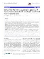
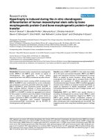
Figure 1 Flow cytometry analysis of passage 1 cells from 1 patient. The filled areas represent the distribution of cells stained by
the respective antibodies; the open areas are control cells without staining. Percentages in parentheses indicate the percentages of
cells positively stained by the respective antibodies in the flow cytometry analysis.
Hyperbaric oxygen promotes osteogenic differentiation by regulating Wnt3a/β-catenin signaling
265
Immunohistochemical detection of Wnt3a, GSK-3β,
β-catenin, Runx 2, and V-ATPase
The tissue sections were deparaffinized, dehydrated, and
treated with proteinase K (25 μg/mL, Sigma, MO) for 60 min.
Endogenous peroxidase activity was blocked with 3% H2O2. The
presence and distribution of Wnt3a, GSK-3β, β-catenin, Runx
2, and V-ATPase were determined using 5 μg/mL of anti-Wnt3a
(Santa Cruz, CA), anti-Runx 2 (Santa Cruz), anti-β-catenin (BD
Bioscience, CA), anti-V-ATPase (Santa Cruz), and anti-GSK-3β
antibodies (Enzo Life Science, PA) at 4 °C overnight. Subsequently, a biotinylated linking 2° Ab was used for 15 min.
Bound immunoglobulin was detected using a LSAB peroxidase
substrate kit (Dako, Carpinteria, CA) and 0.1% methyl green
(Dako) was used for counterstaining.
Results
In vitro study
Flow cytometry analysis
Primary adherent human BMSCs from 3 donors were cultured in
control medium, and cells were analyzed for expression of BMSC
markers using flow cytometry at passage 1. The percentage of
cells expressing the BMSC markers CD146, CD105, Stro-1, α-SMA
and CD34 were shown in Fig. 1. The mean percentages of
CD146+, CD105+, Stro-1+, α-SMA+, and CD34+ cells in the cell
preparations from 3 patients were calculated to be 27.6% ±
1.3%, 85.7% ± 5.8%, 32.7% ± 1.3%, 53.3% ± 2.1%, and 0.21% ±
0.09%, respectively.
Effect of HBO on cell proliferation rate of BMSCs
A decrease in cell proliferation following HBO treatment was
observed when the BMSCs were cultured in complete medium
for 7, 10, and 14 d. No significant differences were detected
in alkaline phosphatase activity between control and HBO
group at each time point (Fig. 2A, *p N 0.05, **p b 0.05,
***p b 0.01, n = 3). However, an increase in cell proliferation
following HBO treatment was noted when BMSCs were already
committed to the osteoblast lineage which was confirmed by
the evaluated expression of alkaline phosphatase activity after
culturing for 7, 10, and 14 d in osteogenic conditions (Fig. 2B,
*p N 0.05, **p b 0.05, ***p b 0.01, n = 3).
Effect of HBO on LRP6 phosphorylation and activation of
the Wnt3a/β-catenin pathway
The Western blot data showed that the protein levels of Wnt3a
(1.54 ± 0.12-fold, *p b 0.05, n = 3), total LRP6 (2.03 ±
0.27-fold, *p b 0.05, n = 3), and phosphorylated LRP6 (2.59 ±
0.51-fold, **p b 0.01, n = 3) were upregulated after culturing
for 7 d with HBO treatment. In addition, the activation of the
Wnt3a pathway resulted in an enhanced expression of Wnt3a
target gene, the protein cyclin D1 (1.90 ± 0.25-fold, *p b 0.05,
n = 3) (Fig. 3A).
The real-time Q-PCR data showed that the mRNA levels of
Wnt3a (2.59 ± 0.57-fold, **p b 0.01 on D1; 2.21 ± 0.49-fold,
**p b 0.01 on D4; 3.13 ± 0.75-fold, **p b 0.01 on D7, n = 3),
β-catenin (1.41 ± 0.21-fold, p N 0.05 on D1; 1.68 ± 0.20-fold,
*p b 0.05 on D4; 1.78 ± 0.12-fold, *p b 0.05 on D7, n = 3), and
Runx2 (1.08 ± 0.11-fold, p N 0.05 on D1, 1.69 ± 0.18-fold, *p b
0.05 on D4, 1.72 ± 0.16-fold, *p b 0.05 on D7, n = 3) were
upregulated, while that of GSK-3β (1.02 ± 0.03-fold, p N 0.05
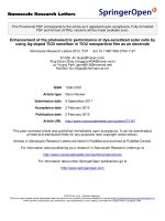
Figure 2 Hyperbaric oxygenation alters the proliferation
of undifferentiated and osteogenically differentiated BMSCs.
(A) Decreased cell proliferation by HBO treatment was seen when
BMSCs were cultured in complete medium. No significant differences were detected in alkaline phosphatase activity between
control and HBO group at each time point (*p N 0.05, **p b 0.05,
***p b 0.01, n = 3). (B) Increased cell proliferation following HBO
was observed when BMSCs were committed to the osteoblast
lineage, which was confirmed by the alkaline phosphatase activity.
The results of the control and HBO groups were compared by
Student's t-tests. Each bar represents the mean ± standard
deviation (*p N 0.05, **p b 0.05, ***p b 0.01; n = 3).
on D1, 0.67 ± 0.11-fold, *p b 0.05 on D4, 0.54 ± 0.09-fold,
*p b 0.05 on D7, n = 3) was downregulated after HBO treatment
(Fig. 3B). The silencing effect on β-catenin (Induction + HBO vs.
Induction + HBO + siRNA, ***p b 0.01, Fig. 3C) and downregulating effect for Runx2 (Induction + HBO vs. Induction + HBO +
siRNA, **p b 0.05, Fig. 3D) by β-catenin siRNA were detected by
real-time PCR after the treatments. In Fig. 3, the data shown
are from cells culturing in osteogenic medium for 7 d. These
cells are beginning to differentiate down to the osteoblastic
pathway which was confirmed by the up-regulation of Runx 2
expressions.
The Western blot data showed that the protein levels of
Wnt3a (1.54 ± 0.12-fold, p* b 0.05, n = 3), β-catenin (1.85 ±
0.13-fold, p** b 0.01, n = 3) and Runx2 (1.61 ± 0.11-fold,
p** b 0.01, n = 3) were upregulated but that of GSK-3β
(0.78 ± 0.05-fold, p* b 0.05, n = 3) was downregulated after
HBO treatment (Fig. 4A). HBO increased the osteogenic
differentiation of the BMSCs as well as its effect on Wnt3a
266
S.-S. Lin et al.
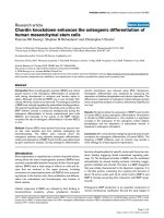
Figure 3 Hyperbaric oxygenation promotes LRP6 phosphorylation to activate Wnt3a signaling and osteogeneic differentiation of
BMSCs. (A) Western blot analysis revealed that the protein levels of Wnt3a (1.54 ± 0.12-fold, p b 0.05, n = 3), total LRP6 (2.03 ±
0.27-fold, p b 0.05, n = 3), and phosphorylated LRP6 (2.59 ± 0.51-fold, p b 0.01, n = 3) were upregulated after culturing for 7 d with
HBO treatment. In addition, the activation of the Wnt3a pathway resulted in enhanced expression of cyclin D1 (1.90 ± 0.25-fold,
p b 0.05, n = 3). (B) mRNA levels of Wnt3a (**p b 0.01 on D1, D4, and D7, n = 3), β-catenin (p N 0.05 on D1; *p b 0.05 on D4; *p b 0.05
on D7, n = 3), and Runx2 (p N 0.05 on D1; *p b 0.05 on D4; *p b 0.05 on D7, n = 3) were up-regulated, whereas that of GSK-3β
(p N 0.05 on D1; *p b 0.05 on D4; *p b 0.05 on D7, n = 3) was downregulated after HBO treatment. (C) Silencing effect for β-catenin
(Induction + HBO vs. Induction + HBO + siRNA, ***p b 0.01, n = 3) and (D) downregulating effect for Runx2 (Induction + HBO vs.
Induction + HBO + siRNA, **p b 0.05, n = 3) by β-catenin siRNA were detected by real-time PCR after the treatments. Abbreviations:
Ind, induction medium; I + H, induction medium + HBO, S-siRNA, scrambled siRNA.
signaling. However, there was no significant effect of HBO on
the Wnt 1 production.
The protein levels of β-catenin in the nuclear fractions were
up-regulated after HBO treatment (2.44 ± 0.17-fold, p b 0.01,
n = 3, Fig. 4B). HBO increased the translocation of β-catenin
from the cytosol into the nucleus. To confirm the effect of
HBO on Runx2 expression via translocation of β-catenin, the
increased protein levels of β-catenin and Runx2 by HBO
treatment were all down-regulated through β-catenin siRNA
treatment (β-catenin:0.32 ± 0.05-fold, p b0.01, n = 3; Runx2:
0.39 ± 0.15-fold, p b 0.05, n = 3; Fig. 4C).
To further investigate the effects of HBO on the activation
of Wnt3a and PI3K–Akt pathways, the levels of phospho-Akt
(Ser 473), phospho-GSK-3β (Ser 9), and phospho-β catenin (Ser
33/37) have been examined and the results are shown in Fig. 5.
The relative optical density ratio (phospho-protein/protein)
for Akt (41.7% ± 9% vs. 88.4% ±21.8%, *p b 0.05, n = 3) and
GSK-3β (41.1% ± 5.1% vs. 64.84% ± 12%, *p b 0.05, n = 3) were
both shown to be up-regulated while that of β-catenin
(77.4% ± 9.5% vs. 29.8% ± 3.4%, **p b 0.01, n = 3) was
down-regulated after HBO treatment.
Effect of HBO on the transcriptional activity of the
β-catenin–TCF/LEF complex and Runx2 promoter/Luc
gene activity
In the nucleus, β-catenin interacts with TCF/LEF transcription
factors and upregulates Wnt3a target genes. To further
evaluate the activation of the β-catenin–TCF/LEF complex,
we measured the activity of both TOP flash (containing the
wild-type TCF binding sites) and FOP flash (mutant TOP flash) in
BMSCs cultured in osteogenic medium after HBO treatment.
Fig. 6A shows that there was increased TOP flash activity
following HBO stimulation (1.58 ± 0.02-fold, **p b 0.01, n = 3),
whereas the FOP flash activity (1.07 ± 0.05-fold, p N 0.05,
n = 3) was not affected. These results demonstrate that HBO is
able to enhance the transcription of genes that are targeted by
the TCF transcription factor. To elucidate the mechanisms that
underlie the effects of HBO on Runx2 gene expression in BMSCs
Hyperbaric oxygen promotes osteogenic differentiation by regulating Wnt3a/β-catenin signaling
267
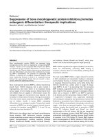
Figure 4 Hyperbaric oxygenation activates Wnt3a/β-catenin signaling via increased translocation of β-catenin of BMSCs. (A) Protein
levels of Wnt3a (p b 0.05), β-catenin (p b 0.01), and Runx2 (p b 0.01) were upregulated but that of GSK-3β (p b 0.05) was downregulated
after HBO treatment. No significant effect of HBO on the Wnt 1 production. (B) Protein levels of β-catenin in the nuclear fractions were
upregulated after HBO treatment (p b 0.01). (C) The increased protein levels of β-catenin and Runx 2 induced by HBO treatment were all
downregulated following β-catenin siRNA treatment. Data are shown as mean ± standard deviation and analyzed by Student's t-test.
Abbreviations: I, induction medium; I + H, induction medium + HBO; S-siRNA, scrambled siRNA; TBP, TATA binding protein.
cultured in osteogenic medium, we examined its effect on the
transcriptional regulation of cloned human Runx2/Luc reporter
constructs. Our data showed that HBO upregulated Runx2/Luc
gene transcription to 2.5-fold greater than that of the control
using the Runx2 construct containing the −317 to −16 promoter
regions (control vs. HBO: 4.09 ± 1.19-fold vs. 7.82 ± 2.13-fold,
Figure 5 Effects of HBO on the activation of Wnt3a/β-catenin and PI3K–Akt pathways. The protein levels of Akt, GSK-3β, β catenin,
phospho-Akt (Ser 473), phospho-GSK-3β (Ser 9), and phospho-β catenin (Ser 33/37) were examined. The relative optical density ratio
(phospho-protein/protein) for Akt (41.7% ± 9% vs. 88.4% ± 21.8%, *p b 0.05, n = 3) and GSK-3β (41.1% ± 5.1% vs. 64.84% ± 12%, *p b 0.05,
n = 3) was both up-regulated while that of β-catenin (77.4% ± 9.5% vs. 29.8% ± 3.4%, **p b 0.01, n = 3) was down-regulated after HBO
treatment.
268
S.-S. Lin et al.
compared to the control group (24.2% ± 2.7% vs. 63.9% ±
7.7%, **p b 0.01, n = 3, Fig. 7D).
Effects of HBO on BMP-2 and osterix production
HBO dose dependently increased the mRNA levels of BMP2
(1.31 ± 0.15 fold on D7, p N 0.05; 2.72 ± 0.52 fold on D14,
*p b 0.05) and osterix (1.23 ± 0.12 fold on D7, p N 0.05;
4.52 ± 0.63 fold on D14, *p b 0.05). HBO also increased the
protein levels of BMP2 (1.75 ± 0.25 fold, *p b 0.05) and
osterix (2.57 ± 0.37 fold, *p b 0.05) on D14 (Fig. 8).
Effect of HBO on ATP6V1 and Wnt3a secretion
Protein levels of ATP6V1 were upregulated after HBO treatment
in the cell lysates (Induction + HBO/Induction: 2.67 ±0.32-fold,
*p b 0.05, n = 3) and the effect of HBO was reduced following
ATP6V1 siRNA treatment (Induction +HBO + siRNA/Induction:
1.28 ± 0.13-fold, *p b 0.05, n = 3; Fig. 9A). No significant effect
on the ATP6V1 level was shown after scrambled siRNA
treatment. The amount of Wnt3a in the collected culture
medium was up-regulated after HBO treatment (Induction vs.
Induction + HBO: 92.7 ± 6.3 vs. 143.7 ±16.5, *p b 0.05, n = 3)
and the effect of HBO on Wnt3a secretion was reduced following
ATP6V1 siRNA treatment (Induction + HBO vs. Induction +
HBO + siRNA: 143.7 ± 16.5 vs. 87.1 ± 6.1, **p b 0.01, n = 3;
Fig. 9B). No significant effect on the Wnt3a levels was shown
after scrambled siRNA treatment.
Figure 6 Hyperbaric oxygenation enhances transcriptional
activity of β-catenin–TCF/LEF complex and Runx2 promoter
activity. (A) HBO enhances the TCF-dependent transcription.
Ratio of the relative luciferase activity between the control and
HBO was calculated. Each bar represents the value of mean ± SD
and analyzed by Student's t-test (**p b 0.01; n = 3). (B) HBO
increases Runx2 promoter activity. Empty pGL4 vector served as
a negative control. The ratio of the relative luciferase activity
between the control and HBO was calculated and analyzed by
Student's t-test (*p b 0.05, **p b 0.01; n = 4).
*p b 0.05 on D4, 5.24 ± 2.43-fold vs. 12.00 ± 0.69-fold, **p b
0.01 on D7, 6.73 ± 0.93-fold vs. 12.58 ± 1.37-fold, **p b 0.01 on
D10, n = 4; Fig. 6B).
Effect of long term exposure to HBO on mRNA and protein
expression
To deposit calcium, osteogenically induced BMSCs must
enter the late stage of osteogenesis. We further investigated
the long-term effects of HBO on BMSCs. The mRNA levels of
type I collagen (2.99 ± 0.4-fold, *p b 0.05 on D14, n = 3) and
osteocalcin (3.09 ± 0.28-fold, **p b 0.01 on D14, n = 3) were
upregulated after HBO treatment (Fig. 7A). In addition, HBO
significantly increased the alkaline phosphatase activity
after 7 d (35.8 ± 1.8 vs. 46.0 ± 3.5, *p b 0.05, n = 3), 14 d
(54.4 ± 4.5 vs. 83.1 ± 4.1, **p b 0.01, n = 3), and 21 d
(43.8 ± 3.1 vs. 55.4 ± 3.2, *p b 0.05, n = 3) of culturing
(Fig. 7B) along with calcium levels after 14 d (126.8 ± 25.9
vs. 231.4 ± 22.2, *p b 0.05, n = 3) and 21 d (343.2 ± 36.8 vs.
507.4 ± 20.8, *p b 0.05, n = 3) of culturing (Fig. 7C) in the
osteogenic induction medium. The deposition of a calcified
matrix on the surface of the culture dish became evident by
Alizarin Red staining. Greater positive staining of the matrix
at the surface layer of the HBO group was observed
In vivo study
Surgery was successful in all 8 rabbits. Distraction was
started at a rate of 0.5 mm every 12 h for 5 d and produced
a gap of 5 mm. The manual evaluation before mechanical
testing showed that at the sixth week, all the specimens
from both groups were immobile.
Histology and mechanical testing
The distraction sites were filled with hard calluses in the
tissue sections of the HBO group (Fig. 10B). However, more
fibrous tissue and cartilage were present in the control
group (Fig. 10A). The lengthened right tibiae exhibited
spiral fractures across the regenerate site. The mechanical
properties were shown in Table 1. The mean percentage of
maximal torque was 96.8% ± 5.6% in the HBO group (n = 4)
and 73.7% ± 4.2% in the non-HBO group (n = 4). The data
indicated that the mechanical properties of the HBO group
were superior to those of the non-HBO group (*p b 0.01).
Immunohistochemistry
The callus is composed of calcified cartilage and newly
formed woven bone. The callus area is larger in HBO group
than in control group (1.71 ± 0.23 fold, *p b 0.01). Immunohistochemical analysis of the protein expression of Wnt3a
(Figs. 10C,D), GSK-3β (Figs. 10E,F), β-catenin (Figs. 10G,H),
Runx2 (Figs. 10I,J), and V-ATPase (Figs. 10K,L) was performed. The levels of Wnt3a (Fig. 10D), β-catenin (Fig. 10H),
Runx2 (Fig. 10J), and V-ATPase (Fig. 10L) were upregulated,
while that of GSK-3β (Fig. 10F) was downregulated after
HBO treatment. The elevated V-ATPase levels (Fig. 10L)
were associated with increased Wnt3a expression (Fig. 10D)
and the elevated β-catenin levels (Fig. 10H) were associated
with increased Runx2 (Fig. 10J) in the HBO treated rabbits.
Hyperbaric oxygen promotes osteogenic differentiation by regulating Wnt3a/β-catenin signaling
269
Figure 7 Long-term hyperbaric oxygenation increases osteogenesis of BMSCs. (A) HBO increased mRNA levels of type I collagen and
osteocalcin after 14 d of culturing. (B) HBO increased alkaline phosphatase activity after 7 d, 14 d, and 21 d of culturing. (C) HBO
increased calcium levels after 14 d and 21 d of culturing. (D) Positive Alizarin Red staining through the matrix at the surface layer of
the HBO group was greater than that of the control group (100 ×). The differences between the control and HBO were calculated
(**p b 0.01). Each bar represents the value of the mean ± standard deviation and analyzed by Student's t-test (*p b 0.05, **p b 0.01;
n = 3). Abbreviations: Ind, induction medium; I + H, induction medium + HBO.
The expression data related to Wnt3a/β-catenin signaling
are consistent with our in vitro findings. The staining
intensity and distribution of Runx2 expression were greater
in the HBO treated rabbits compared with the controls,
which reflects increased bone formation in the HBO group.
Discussion
Human BMSCs cultured in hypoxia show greater proliferation
than those cultured in normoxic conditions (Grayson et al.,
2006; Fehrer et al., 2007). However, both inhibitory and
enhancing effects of hypoxia on osteogenic differentiation
have been reported (Grayson et al., 2006; Fehrer et al., 2007;
Pattappa et al., 2011). Because HBO increases the oxygen
tension in vivo (Ueng et al., 1998; Korhonen et al., 1999) and in
vitro (Ueng et al., 2013; Niu et al., 2013), we used HBO to
alter the hypoxic microenvironment for cell proliferation and
differentiation and activate the oxygen sensitive pathways.
Our findings support those of previous studies, which suggest
that undifferentiated BMSCs and committed BMSCs could
respond differently to oxygen signals (Fehrer et al., 2007).
HBO decreases cell proliferation when undifferentiated BMSCs
are cultured in complete medium (Fig. 2A). However, increased levels of cell proliferation were induced by HBO
treatment when the BMSCs were committed to the osteoblast
lineage (Fig. 2B). These findings were further validated by the
evaluated expression levels of cyclin D1 after HBO treatment
(Fig. 3A). Although the responses of osteoblasts to HBO have
been documented (Wu et al., 2007; Hsieh et al., 2010), the
direct effects of HBO on human BMSCs that are induced to
differentiate down the osteoblastic pathway have, to the best
of our knowledge, not been previously investigated.
Oxygen availability regulates stem cells via Wnt/β-catenin
signaling (Mazumdar et al., 2010). Because HBO has stimulatory
effects on cell growth (Fig. 2B), we wanted to identify the
molecular mechanisms involved by assessing the Wnt/β-catenin
pathway. Our data showed that the protein levels of Wnt3a,
phosphorylated LRP6, and cyclin D1 were upregulated after
culturing for 7 d with HBO treatment (Fig. 3A). A key step after
Wnt stimulation is the phosphorylation of the LRP6 intracellular
domain. This phosphorylation event stabilizes the Wnt signaling
transducer β-catenin (Bilic et al., 2007). Activation of the
Wnt3a pathway results in enhanced expression of the Wnt3a
target gene, cyclin D1, which is required for G1/S phase
traversal (Xiong et al., 1997). Osteoblasts were induced to enter
the S and G2/M phases of the cell cycle after HBO treatment
(Hsieh et al., 2010). HBO increases the proliferation of BMSCs
that are beginning to differentiate down the osteoblastic
pathway via Wnt3a signaling (Fig. 3), which was in contrast to
270
S.-S. Lin et al.
Figure 8 Effects of HBO on BMP-2 and osterix production. HBO
dose dependently increased the mRNA expression of BMP2 (D7,
p N 0.05; D14, *p b 0.05) and osterix (D7, p N 0.05; D14, *p b0.05).
HBO also increased the protein levels of BMP2 (1.75 ± 0.25 fold,
*p b 0.05) and osterix (2.57 ± 0.37 fold, *p b 0.05) on D14.
previous observations that hypoxia selectively activates Wnt/
β-catenin signaling in undifferentiated neural stem cells but not
in differentiated neurons (Mazumdar et al., 2010).
When BMSCs act as target cells, the canonical β-catenin
signaling pathway can be stimulated in response to Wnt1,
Wnt3a, and Wnt8 or by inhibition of GSK-3 (Westendorf
et al., 2004). Wnt/β-catenin directly stimulates Runx2 gene
expression via the TCF-binding site (Gaur et al., 2005). In the
present study, the mRNA (Fig. 3B) and protein (Fig. 4A)
levels of Wnt3a, β-catenin, and Runx2 were upregulated,
while that of GSK-3β was downregulated after HBO treatment. HBO increased β-catenin mRNA production to stimulate Runx2 mRNA expression and this was confirmed by
β-catenin siRNA treatment (Figs. 3C–D). In addition, accumulated β-catenin was subsequently translocated into the
nucleus (Fig. 4B) where it upregulated Runx2 protein
expression and this was also confirmed by β-catenin siRNA
treatment (Fig. 4C).
In the Wnt signaling pathway, β-catenin is phosphorylated by GSK-3β, which leads to its degradation via the
ubiquitin/proteasome pathway (Zeng et al., 2005). The
activity of GSK-3β is reduced by phosphorylation of its
N terminus at the Serine 9 residue by Akt (Cross et al.,
1995). Lithium, a pharmacological GSK-3 inhibitor, has been
shown to enhance GSK-3 serine phosphorylation by activation of phosphatidylinositol 3-kinase (PI3-kinase)-dependent
Akt (Chalecka-Franaszek and Chuang, 1999). In the present
study, HBO has similar effects which can increase the Serine
9 phosphorylation of GSK-3β through PI3-kinase-mediated
phosphorylation of Akt (at the Serine 473 residue), thus
decreases the activity of GSK-3β (Fig. 5).
Fig. 6A showed that there was increased TOP flash activity following HBO stimulation. The activation of the TOP
flash reporter was specific to the Wnt3a genes (Gazit et al.,
Figure 9 Hyperbaric oxygenation increases Wnt3a secretion via
ATP6V1 production. (A) Protein levels of ATP6V1 in the cell lysates
were upregulated after HBO treatment (*p b 0.05) and the effect
of HBO was reduced following ATP6V1 siRNA treatment (*p b 0.05).
(B) The amount of Wnt3a in the collected culture medium was
upregulated after HBO treatment (*p b 0.05) and the effect of HBO
on Wnt3a secretion was reduced by ATP6V1 siRNA treatment
(**p b 0.01). Abbreviations: I, induction medium; I + H, induction
medium + HBO; S-siRNA, scrambled siRNA.
1999; Lu et al., 2004). HBO increased Wnt3a expression,
which enhanced the β-catenin–TCF transcriptional activity
in this study.
The major isoforms of Runx2 involved in osteogenesis are
type1 (T1) and type2 (T2) Runx2. T1 Runx2 is regulated by
the proximal promoter P2; whereas T2 Runx2 is regulated by
the distal promoter P1 (Sudhakar et al., 2001). T2 Runx2 (P1
promoter) is induced upon stimulation with BMP2 or activation of the canonical Wnt and β-catenin/TCF1 pathways
(Gaur et al., 2005). Hypoxia or TWIST did not inhibit P1 but
it did inhibit P2 promoter activity in BMSCs undergoing
osteogenic differentiation (Yang et al., 2011). In the present
study, HBO activated the canonical Wnt and β-catenin/TCF1
pathways to increase Runx2/Leu promoter activity (Fig. 6B).
However, the effects of HBO on the individual P1 and P2
promoter activities need to be further investigated.
Hyperbaric oxygen promotes osteogenic differentiation by regulating Wnt3a/β-catenin signaling
271
Figure 10 Beneficial effects of HBO on bone formation via regulation of Wnt3a/β-catenin signaling. The distraction sites were
filled with calcified cartilage and newly formed woven bone in the tissue sections of the HBO group (B, 40 ×). However, more fibrous
tissue and cartilage were present in the control group (A, 40 ×). The levels of Wnt3a (D, 100 ×), β-catenin (H, 100 ×), Runx2 (J, 100 ×),
and V-ATPase (L, 100 ×) were upregulated, whereas that of GSK-3β (F, 100 ×) was downregulated after HBO treatment. The staining
intensity and distribution of the Runx2 expression levels were greater in the HBO treated rabbits compared with the controls, which
reflects greater bone formation in the HBO group. Control group: A, C, E, G, I, and K. HBO group: B, D, F, H, J, and L.
Wnt signaling activates the endogenous BMP2 gene through
a TCF response enhancer region (Zhang et al., 2013). BMP-2
stimulates the expression of osterix which is required for
osteoblast differentiation and bone formation (Nakashima
et al., 2002; Lee et al., 2003). Because HBO activated the
Wnt/β-catenin/TCF pathways (Fig. 6B), we further investigated the effects of HBO on BMP-2 production and found that HBO
dose dependently increased the mRNA expression of BMP2 and
osterix. In addition, HBO also increased the protein levels of
BMP2 and osterix (Fig. 8). Wnt/β-catenin signaling is an
upstream activator of BMP2 expression in osteoblasts (Zhang
et al., 2013). Our results provided novel insights into the nature
of functional cross talk integrating the BMP and Wnt/β-catenin
pathways in osteoblastic differentiation after HBO treatment.
Osteoblasts originate from BMSCs via a stepwise maturation
process. During the early stages of osteogenesis, the cell
cannot deposit calcium to form mineralized bone (Ducy et al.,
1997). To deposit calcium, the cells must enter the late stage
272
of osteogenesis (Nakashima et al., 2002). We further investigated the long-term effects of HBO (14 and 21 d) on the
osteogenic differentiation of BMSCs and found that HBO
significantly increased the expression of osteogenic markers,
including type I collagen, osteocalcin (Fig. 7A), alkaline phosphatase activity (Fig. 7B), and calcium (Fig. 7C). Enhanced
positive Alizarin Red staining through the matrix at the surface
layer of the HBO group was also seen compared to the control
group (Fig. 7D).
V-ATPases is a pH regulator in acidic subcellular compartments including the Golgi complex, vesicles, and lysosomes.
Wnt3a secretion requires its binding to the carrier protein
wntless (WLS) and Wls-dependent secretion of Wnt3a was
shown to require vacuolar acidification (Coombs et al, 2010).
In the presence of acidification inhibitors, the Wnt3a–Wls
complex is able to reach the cell surface but the release of
Wnt3a from Wls is hindered (Coombs et al, 2010). Treatment of
cells with siRNA targeting 2 subunits of V-ATPase (ATP6V1 and
ATP6V0) inhibited Wnt signaling (Cruciat et al., 2010). When
osteogenically differentiated BMSCs act as Wnt producing cells,
increased V-ATPase expression (Fig. 9A) and Wnt3a secretion
(Fig. 9B) were induced by HBO treatment. Secretion of Wnt3a is
impaired upon inhibition of V-ATPase. Wnt3a is retained in the
producing cells, and is therefore, unable to move into the
culture medium during ATP6V1 siRNA treatment (Fig. 9B).
Bone repair requires the mobilization of adult skeletal stem
cells to allow deposition of cartilage and bone at the injury
site. These stem cells are believed to come from multiple
sources including the bone marrow and periosteum (Colnot,
2009). HBO treatment increases the number of circulating
hematopoietic stem cells (Thom et al., 2006) and endothelial
precursor cells (Liu and Velazquez, 2008). However, there is no
convincing evidence that BMSCs can be liberated from the bone
marrow. HBO effects on circulating BMSCs have not been
elucidated. Previously, we showed that HBO increased bone
mineral density and torsional strength of lengthened tibia in a
rabbit model (Ueng et al., 1998). In the present study, we
further demonstrated that Wnt3a/β-catenin signaling plays a
crucial role in bone healing after HBO treatment. The levels of
Wnt3a (Fig. 10D), β-catenin (Fig. 10H), Runx2 (Fig. 10J), and
V-ATPase (Fig. 10L) were upregulated, whereas that of GSK-3β
(Fig. 10F) was downregulated after HBO treatment. Therefore,
HBO increased β-catenin production or decreased β-catenin
degradation by upregulating Wnt3a or down-regulating GSK-3β
expression. Expression of stabilized β-catenin in cells committed to the osteoblast lineage improves osteogenesis, thereby
leading to enhanced bone healing.
Both canonical Wnt pathway (Wnt3a/β-catenin) and noncanonical Wnt pathway (Wnt5a, which signals mainly through
the Wnt/calcium) have been shown to regulate the differentiation state of BMSCs. In addition, several microRNAs (miRNAs)
have recently been discovered as important regulators of
osteoblast gene expression, such as Mir-31 (Baglìo et al., 2013),
Mir-93 (Yang et al., 2012), Mir-141, Mir-200a, Mir-133a,
Mir-204, and Mir-211 (Chen et al., 2013). It is currently not
clear which pathway is at work regulating the differentiation
state of the cells.
Osteoblast maturation requires the phenotype promoting
activity of the transcription factor Runx2, which controls both
cell growth and differentiation. Runx2 is hyper-phosphorylated
by CDK1/cyclin B during mitosis and dynamically converted
into a hypo-phosphorylated form by PP1/PP2A-dependent
S.-S. Lin et al.
dephosphorylation after mitosis to support the post-mitotic
regulation of Runx2 target genes (Rajgopal et al., 2007). In the
present study, the activation of the Wnt3a pathway by HBO
treatment resulted in an enhanced expression of Wnt3a target
gene, the protein cyclin D1 (Fig. 2A), which is required for cell
cycle G1/S transition (Sherr and Roberts, 1999). Further studies
are required to investigate the expression of CDK1/cyclin B and
PP1/PP2A in osteoblasts after HBO treatment.
Several studies have concluded that HBO has different
effects on osteoblast proliferation in vitro. Wong et al. showed
that 100% O2 at 2 ATA once daily inhibited growth of primary
osteoblasts and resulted in a significant increase in apoptosis
(Wong et al., 2008). Comparatively, Hsieh et al. found that
providing 50% O2 at 2.5 ATA twice daily increased growth of an
osteoblast cell line (Hsieh et al., 2010). In the present study,
100% O2 at 2.5 ATA once every 36 h promoted proliferation of
committed BMSCs (Fig. 2B). The differences among the present
results and those reported by Wong et al. and Hsieh et al. may
stem from the use of different cells (committed BMSCs,
primary culture osteoblasts, and an osteoblast cell line),
different levels of pressure (1 ATA, 2 ATA, and 2.5 ATA),
different O2 concentrations (21%, 50%, and 100%), and different
treatment durations (once daily, twice daily, and once every
36 h). Because 100% O2 at 2 ATA once daily inhibited growth of
primary osteoblasts (Wong et al., 2008), the duration was
modified from once daily to once every 36 h and found to have
a positive effect on cell proliferation in this study (Fig. 2B).
HBO treatment not only increased cell proliferation of committed BMSCs, but also suppressed the apoptosis in degenerated intervertebral disc cells (Niu et al., 2013) and
osteoarthritic chondrocytes (Ueng et al., 1998) in previous
studies. Our data support the notion that different cell types
have distinct growth responses after exposure to HBO in vitro.
Environmental oxygen levels affect tissue vascularization
and fracture healing. A previous study suggested that hyperoxia
(50% O2 and 1 ATA) increased tissue vascularization but did
not significantly alter osteogenesis during the early stages
of fracture healing (Lu et al., 2013). Most of the inspired
atmospheric oxygen was carried by hemoglobin (Hb) and
delivery to the fracture site. After Hb saturation, the oxygen
level may not be high enough to increase the osteogenesis in
the avascular fracture site. In this study, HBO (hyperbaric
oxygen, a combination of 100% O2 and 2.5 ATA) increased
osteogenesis of bone healing. After Hb saturation, HBO may
result in greater amounts of O2 dissolved in plasma to improve
the environmental oxygen levels at the avascular fracture
site than hyperoxia treatment only. The additive effects of
increased pressure and increased O2 were demonstrated in this
study. Long-term and repeated HBO treatments may increase
oxidative stress (Korhonen et al., 1999); however, tolerance to
HBO treatment can be extended by intermittent exposure. The
authors used a clinical HBO protocol in this study. Because
exposure to HBO in clinical protocols is rather brief (typically
b 2 h/d), studies show that antioxidant defenses are adequate
so that biochemical stresses related to increases in ROS are
reversible (Korhonen et al., 1999).
Conclusions
Considering the previous studies and our findings, we propose
the following model: When osteogenically differentiated
Hyperbaric oxygen promotes osteogenic differentiation by regulating Wnt3a/β-catenin signaling
Table 1
Results of mechanical testing.
Rabbit
Control group
1 Lengthened
Non-lengthened
2 Lengthened
Non-lengthened
3 Lengthened
Non-lengthened
4 Lengthened
Non-lengthened
Mean
S.D.
HBO group
1 Lengthened
Non-lengthened
2 Lengthened
Non-lengthened
3 Lengthened
Non-lengthened
4 Lengthened
Non-lengthened
Mean
S.D.
Rotation
angle
(degree)
Maximum
torque
(N-mm)
7.9
13.6
8.1
15.7
10.4
15.4
8.2
14.9
2378
3338
2171
3147
2368
3036
2280
2998
Percentage of
maximum torque
(lengthened/
non-lengthened)
71.2%
69.0%
78.0%
76.1%
73.6%
4.2%
12.6
12.4
9.6
10.8
8.8
11.2
10.2
12.0
3375
3229
2861
2946
2905
3178
3009
3195
104.5%
97.1%
91.4%
94.2%
96.8%
5.6%
BMSCs act as Wnt3a-producing cells, the level of Wnt3a is
upregulated after HBO treatment. In addition, protein levels of
ATP6V1 and Wnt3a secretion were also upregulated after HBO
treatment. When osteogenically differentiated BMSCs act as
Wnt3a targeting cells, phosphorylated LRP6, β-catenin,
TCF-dependent transcription, Runx2 promoter/Luc gene activity, and the expression of osteogenic markers were upregulated after HBO treatment. Because Wnt/β-catenin signaling is an
upstream activator of BMP2 expression in osteoblasts, we
further found that HBO dose dependently increased the BMP2
and osterix production. Finally, we showed that the beneficial
effects of HBO on bone formation were related to Wnt3a/
β-catenin signaling in a rabbit model. After understanding the
regulatory factors and molecular mechanisms, HBO may serve
as a therapeutic approach to increase bone healing in clinical
studies.
Acknowledgments
This research was supported in part by grants from the National
Science Council and Chang Gung Memorial Hospital, Taiwan,
Republic of China.
References
Baglìo, S.R., Devescovi, V., Granchi, D., Baldini, N., 2013. MicroRNA
expression profiling of human bone marrow mesenchymal stem
273
cells during osteogenic differentiation reveals osterix regulation
by miR-31. Gene 527, 321–331.
Basciano, L., Nemos, C., Foliguet, B., de Isla, N., de Carvalho, M.,
Tran, N., Dalloul, A., 2011. Long term culture of mesenchymal
stem cells in hypoxia promotes a genetic program maintaining their
undifferentiated and multipotent status. BMC Cell Biol. 12, 1–12.
Bilic, J., Huang, Y.L., Davidson, G., Zimmermann, T., Cruciat, C.M.,
Bienz, M., Niehrs, C., 2007. Wnt induces LRP6 signalosomes and
promotes dishevelled-dependent LRP6 phosphorylation. Science
316, 1619–1622.
Chalecka-Franaszek, E., Chuang, D.M., 1999. Lithium activates the
serine/threonine kinase Akt-1 and suppresses glutamate-induced
inhibition of Akt-1 activity in neurons. Proc. Natl. Acad. Sci. U. S.
A. 96, 8745–8750.
Chen, Q., Liu, W., Sinha, K.M., Yasuda, H., de Crombrugghe, B.,
2013. Identification and characterization of microRNAs controlled by the osteoblast-specific transcription factor osterix.
PLoS ONE 8, e58104.
Coombs, G.S., Yu, J., Canning, C.A., Veltri, C.A., Covey, T.M.,
Cheong, J.K., Utomo, V., Banerjee, N., Zhang, Z.H., Jadulco, R.C.,
Concepcion, G.P., Bugni, T.S., Harper, M.K., Mihalek, I., Jones,
C.M., Ireland, C.M., Virshup, D.M., 2010. WLS-dependent secretion
of WNT3A requires Ser209 acylation and vacuolar acidification.
J. Cell Sci. 123, 3357–3367.
Colnot, C., 2009. Skeletal cell fate decisions within periosteum and
bone marrow during bone regeneration. J. Bone Miner. Res. 24,
274–282.
Cross, D.A., Alessi, D.R., Cohen, P., Andjelkovich, M., Hemmings,
B.A., 1995. Inhibition of glycogen synthase kinase-3 by insulin
mediated by protein kinase B. Nature 378, 785–789.
Cruciat, C.M., Ohkawara, B., Acebron, S.P., Karaulanov, E., Reinhard,
C., Ingelfinger, D., Boutros, M., Niehrs, C., 2010. Requirement of
prorenin receptor and vacuolar H+-ATPase-mediated acidification
for Wnt signaling. Science 327, 459–463.
De Boer, J., Wang, H.J., Van Blitterswijk, C., 2004. Effects of Wnt
signaling on proliferation and differentiation of human mesenchymal stem cells. Tissue Eng. 10, 393.
Drissi, H., Luc, Q., Shakoori, R., Chuva De Sousa Lopes, S., Choi, J.Y.,
Terry, A., Hu, M., Jones, S., Neil, J.C., Lian, J.B., Stein, J.L., Van
Wijnen, A.J., Stein, G.S., 2000. Transcriptional autoregulation of
the bone related cbfa1/Runx 2 gene. J. Cell. Physiol. 184, 341–350.
Ducy, P., 2000. Cbfa1: a molecular switch in osteoblast biology.
Dev. Dyn. 219, 461–471.
Ducy, P., Zhang, R., Geoffroy, V., Ridall, A.L., Karsenty, G., 1997.
Osf2/Cbfa1: a transcriptional activator of osteoblast differentiation. Cell 89, 747–754.
Eijken, M., Meijer, I.M., Westbroek, I., Koedam, M., Chiba, H.,
Uitterlinden, A.G., Pols, H.A., van Leeuwen, J.P., 2008. Wnt
signaling acts and is regulated in a human osteoblast differentiation dependent manner. J. Cell. Biochem. 104, 568–579.
Etheridge, S.L., Spencer, G.J., Heath, D.J., Genever, P.G., 2004.
Expression profiling and functional analysis of Wnt signaling
mechanisms in mesenchymal stem cells. Stem Cells 22,
849–860.
Fehrer, C., Brunauer, R., Laschober, G., Unterluggauer, H., Reitinger,
S., Kloss, F., Gully, C., Gassner, R., Lepperdinger, G., 2007.
Reduced oxygen tension attenuates differentiation capacity of
human mesenchymal stem cells and prolongs their lifespan. Aging
Cell 6, 745–757.
Forgac, M., 2007. Vacuolar ATPases: rotary proton pumps in physiology
and pathophysiology. Nat. Rev. Mol. Cell Biol. 8, 917–929.
Gaur, T., Lengner, C.J., Hovhannisyan, H., Bhat, R.A., Bodine, P.V.,
Komm, B.S., Javed, A., van Wijnen, A.J., Stein, J.L., Stein, G.S.,
Lian, J.B., 2005. Canonical WNT signaling promotes osteogenesis
by directly stimulating Runx2 gene expression. J. Biol. Chem.
280, 33132–33140.
Gazit, A., Yaniv, A., Bafico, A., Pramila, T., Igarashi, M., Kitajewski,
J., Aaronson, S.A., 1999. Human frizzled 1 interacts with
274
transforming Wnts to transduce a TCF dependent transcriptional
response. Oncogene 18, 5959–5966.
Grayson, W.L., Zhao, F., Izadpanah, R., Bunnell, B., Ma, T., 2006.
Effects of hypoxia on human mesenchymal stem cell expansion
and plasticity in 3D constructs. J. Cell. Physiol. 207, 331–339.
Hsieh, C.P., Chiou, Y.L., Lin, C.Y., 2010. Hyperbaric oxygenstimulated proliferation and growth of osteoblasts may be
mediated through the FGF-2/MEK/ERK 1/2/NF-kB and PKC/JNK
pathways. Connect. Tissue Res. 51, 497–509.
Korhonen, K., Kuttila, K., Niinikoski, J., 1999. Subcutaneous
tissue oxygen and carbon dioxide tensions during hyperbaric
oxygenation: an experimental study in rats. Eur. J. Surg. 165,
885–890.
Komatsu, D.E., Mary, M.N., Schroeder, R.J., Robling, A.G., Turner,
C.H., Warden, S.J., 2010. Modulation of Wnt signaling influences
fracture repair. J. Orthop. Res. 28, 928–936.
Lee, M.H., Kwon, T.G., Park, H.S., Wozney, J.M., Ryoo, H.M., 2003.
BMP-2-induced Osterix expression is mediated by Dlx5 but is
independent of Runx2. Biochem. Biophys. Res. Commun. 309,
689–694.
Ling, L., Nurcombe, V., Cool, S.M., 2009. Wnt signaling controls the
fate of mesenchymal stem cells. Gene 433, 1–7.
Liu, Z.J., Velazquez, O.C., 2008. Hyperoxia, endothelial progenitor
cell mobilization, and diabetic wound healing. Antioxid. Redox
Signal. 10, 1869–1882.
Logan, C.Y., Nusse, R., 2004. The Wnt signaling pathway in
development and disease. Annu. Rev. Cell Dev. Biol. 20, 781–810.
Lorenowicz, M.J., Korswagen, H.C., 2009. Sailing with the Wnt:
charting the Wnt processing and secretion route. Exp. Cell Res.
315, 2683–2689.
Lu, D., Zhao, Y., Tawatao, R., Cottam, H.B., Sen, M., Leoni, L.M., Kipps,
T.J., Corr, M., Carson, D.A., 2004. Activation of the Wnt signaling
pathway in chronic lymphocytic leukemia. PNAS 101, 3118–3123.
Lu, C., Saless, N., Wang, X., Sinha, A., Decker, S., Kazakia, G., Hou,
H., Williams, B., Swartz, H.M., Hunt, T.K., Miclau, T., Marcucio,
R.S., 2013. The role of oxygen during fracture healing. Bone 52,
220–229.
Mazumdar, J., O'Brien, W.T., Johnson, R.S., LaManna, J.C., Chavez,
J.C., Klein, P.S., Simon, M.C., 2010. O2 regulates stem cells
through Wnt/β-catenin signalling. Nat. Cell Biol. 12, 1007–1013.
Minear, S., Leucht, P., Jiang, J., Liu, B., Zeng, A., Fuerer, C., Nusse,
R., Helms, J.A., 2010. Wnt proteins promote bone regeneration.
Sci. Transl. Med. 2, 29ra30.
Nakashima, K., Zhou, X., Kunkel, G., Zhang, Z., Deng, J.M., Behringer,
R.R., de Crombrugghe, B., 2002. The novel zinc finger-containing
transcription factor osterix is required for osteoblast differentiation and bone formation. Cell 108, 17–29.
Niu, C.C., Lin, S.S., Yuan, L.J., Chen, L.H., Wang, I.C., Tsai, T.T.,
Lai, P.L., Chen, W.J., 2013. Hyperbaric oxygen treatment
suppresses MAPK signaling and mitochondrial apoptotic pathway
in degenerated human intervertebral disc cells. J. Orthop. Res.
31, 204–209.
Nusse, R., 2003. Wnts and hedgehogs: lipid-modified proteins and
similarities in signaling mechanisms at the cell surface. Development 130, 5297.
Pattappa, G., Thorpe, S.D., Jegard, N.C., Heywood, H.K., de Bruijn,
J.D., Lee, D.A., 2013. Continuous and uninterrupted oxygen
tension influences the colony formation and oxidative metabolism of human mesenchymal stem cells. Tissue Eng. C Methods
19, 68–79.
Pittenger, M.F., Mackay, A.M., Beck, S.C., Jaiswal, R.K., Douglas,
R., Mosca, J.D., Moorman, M.A., Simonetti, D.W., Craig, S.,
Marshak, D.R., 1999. Multilineage potential of adult human
mesenchymal stem cells. Science 284, 143–147.
Port, F., Basler, K., 2010. Wnt trafficking: new insights into Wnt
maturation, secretion and spreading. Traffic 11, 1265–1271.
Rajgopal, A., Young, D.W., Mujeeb, K.A., Stein, J.L., Lian, J.B., van
Wijnen, A.J., Stein, G.S., 2007. Mitotic control of RUNX2
S.-S. Lin et al.
phosphorylation by both CDK1/cyclin B kinase and PP1/PP2A
phosphatase in osteoblastic cells. J. Cell. Biochem. 100,
1509–1517.
Sherr, C.J., Roberts, J.M., 1999. Cdk inhibitors: positive and
negative regulators of G1-phase progression. Genes Dev. 13,
1501–1512.
Shyu, K.G., Hung, H.F., Wang, B.W., Chang, H., 2008. Hyperbaric
oxygen induces placental growth factor expression in bone
marrow-derived mesenchymal stem cells. Life Sci. 83, 65–73.
Sudhakar, S., Katz, M.S., Elango, N., 2001. Analysis of type-I and
type-II RUNX2 protein expression in osteoblasts. Biochem.
Biophys. Res. Commun. 286, 74–79.
Thom, S.R., Bhopale, V.M., Velazquez, O.C., Goldstein, L.J., Thom,
L.H., Buerk, D.G., 2006. Stem cell mobilization by hyperbaric
oxygen. Am. J. Physiol. Heart Circ. Physiol. 290, H1378–H1386.
Ueng, S.W.N., Lee, S.S., Lin, S.S., Wang, C.R., Liu, S.J., Yang, H.F.,
Tai, C.L., Shih, C.H., 1998. Bone healing of tibial lengthening
is enhanced by hyperbaric oxygen therapy: a study of bone
mineral density and torsional strength on rabbits. J. Trauma 44,
676–681.
Ueng, S.W.N., Yuan, L.J., Lin, S.S., Niu, C.C., Chan, Y.S., Wang,
I.C., Yang, C.Y., Chen, W.J., 2013. Hyperbaric oxygen treatment
prevents nitric oxide-induced apoptosis in articular cartilage
injury via enhancement of the expression of heat shock protein
70. J. Orthop. Res. 31, 376–384.
Vogt, M.T., Cauley, J.A., Kuller, L.H., Nevitt, M.C., 1997. Bone
mineral density and blood flow to the lower extremities: the
study of osteoporotic fractures. J. Bone Miner. Res. 12, 283–289.
Wang, X.L., Yang, Y.J., Xie, M., Yu, X.H., Liu, C.T., Wang, X., 2007.
Proliferation of neural stem cells correlates with Wnt-3 protein
in hypoxic–ischemic neonate rats after hyperbaric oxygen
therapy. NeuroReport 18, 1753–1756.
Westendorf, J.J., Kahler, R.A., Schroeder, T.M., 2004. Wnt signaling
in osteoblasts and bone diseases. Gene 341, 19–39.
Wong, A.K., Schönmeyr, B.H., Soares, M.A., Li, S., Mehrara, B.J.,
2008. Hyperbaric oxygen inhibits growth but not differentiation
of normal and irradiated osteoblasts. J. Craniofac. Surg. 19,
757–765.
Wu, D., Malda, J., Crawford, R., Xiao, Y., 2007. Effects of
hyperbaric oxygen on proliferation and differentiation of
osteoblasts from human alveolar bone. Connect. Tissue Res.
48, 206–213.
Xiong, W., Pestell, R.G., Watanabe, G., Li, J., Rosner, M.R.,
Hershenson, M.B., 1997. Cyclin D1 is required for S phase
traversal in bovine tracheal myocytes. Am. J. Physiol. 272,
L1205–L1210 (6 Pt1).
Yang, D.C., Yang, M.H., Tsai, C.C., Huang, T.F., Chen, Y.H., Hung,
S.C., 2011. Hypoxia inhibits osteogenesis in human mesenchymal
stem cells through direct regulation of RUNX2 by TWIST. PLoS ONE
6, e23965.
Yang, L., Cheng, P., Chen, C., He, H.B., Xie, G.Q., Zhou, H.D., Xie,
H., Wu, X.P., Luo, X.H., 2012. MiR-93/Sp7 function loop
mediates osteoblast mineralization. J. Bone Miner. Res. 27,
1598–1606.
Zeng, X., Tamai, K., Doble, B., Li, S., Huang, H., Habas, R.,
Okamura, H., Woodgett, J., He, X., 2005. A dual-kinase
mechanism for Wnt co-receptor phosphorylation and activation.
Nature 438, 873–877.
Zhang, Y., Hassan, M.Q., Xie, R.L., Hawse, J.R., Spelsberg, T.C.,
Montecino, M., Stein, J.L., Lian, J.B., van Wijnen, A.J., Stein, G.S.,
2009. Co-stimulation of the bone-related Runx2 P1 promoter in
mesenchymal cells by SP1 and ETS transcription factors at
polymorphic purine-rich DNA sequences (Y-repeats). J. Biol.
Chem. 284, 3125–3135.
Zhang, R., Oyajobi, B.O., Harris, S.E., Chen, D., Tsao, C., Deng,
H.W., Zhao, M., 2013. Wnt/β-catenin signaling activates bone
morphogenetic protein 2 expression in osteoblasts. Bone 52,
45–56.