diagnostic role of magnetic resonance angiography in swyer james syndrome case series of two cases
Bạn đang xem bản rút gọn của tài liệu. Xem và tải ngay bản đầy đủ của tài liệu tại đây (679.6 KB, 4 trang )
Case Report
www.lungindia.com
Diagnostic role of magnetic resonance angiography in
Swyer James syndrome: Case series of two cases
Umesh C. Parashari, Ragini Singh, Anit Parihar, Pallavi Aga, Rajesh Yadav
Department of Radiodiagnosis, CSM Medical University (Upgraded K.G. Medical University), Lucknow, India
ABSTRACT
Swyer James syndrome is a rare syndrome which occurs due to viral illness in early childhood. The post infective
obliterative bronchiolitis results in arrest of lung growth and alveolarization with reduced vascularity resulting in classical
radiological features. We describe two cases of patients fulfilling all the criteria of the syndrome - 1) Unilateral hyperlucent
small lung in chest radiograph with air trapping on expiration, small ipsilateral hila and pulmonary artery. 2) Diffuse
decrease in attenuation of lung parenchyma with bronchiectasis and reduction in vascularity. 3) Unilateral pruned tree
appearance on angiography (MRA). The clinical presentation was recurrent chest infection in a child and infrequent
bouts of hemoptysis in a middle aged female. The study demonstrates the role of magnetic resonance angiography in
diagnosing the condition.
KEY WORDS: Bronchiolitis obliterans, MR angiography, pruned tree appearance, unilateral hyperlucent lung
Address for correspondence: Dr. Umesh Chandra Parashari, Department of Radio Diagnosis, C.S.M.M.U. Lucknow, India. E-mail:
DOI: 10.4103/0970-2113.68326
INTRODUCTION
We report a case series of two cases of Swyer James
syndrome. [1] The syndrome is rare and occurs following
viral insult in infancy or early childhood. The infective
insult leads to acute obliterative bronchiolitis resulting
in arrest of progressive alveolarization and proper
development of lung. The syndrome results in reduced
vascularity with paucity of bronchial subdivisions (cut off
at 4th to 5th generation). The study was performed to rule out
the cause of recurrent respiratory infection in first patient
and infrequent bouts of hemoptysis in another patient.
CASE REPORTS
Case 1
A 13-year-old female child presented with recurrent
attacks of respiratory tract infection for last few years.
Physical examination revealed presence of crepts in almost
whole of left lung. Arterial blood gas analysis showed
reduced oxygenation. The pulmonary function test showed
diminution of flow with reduced FEV1.
The radiological evaluation started with inspiratory
chest radiograph, which showed small hyperluscent left
hemithorax with evidence of air trapping on expiratory
Lung India • Vol 27 • Issue 3 • Jul - Sep 2010
radiograph. The left hilum was small. Slight hyperinflation
of right lung was also noted [Figure 1 a and b]. High
resolution CT (HRCT) was done following chest radiograph,
which showed small left hemithorax with diffuse decrease
in attenuation. Paucity of broncho vascular markings with
proximal bronchiectasis was noted in left hemithorax. The
pulmonary artery was smaller on the left side [Figure 1c].
Magnetic resonance (MR) angiography was performed
after HRCT revealed typical pruned tree appearance on left
side confirming the diagnosis of Swyer James syndrome
[Figure 1d].
Case 2
A 51-year-old woman presented with infrequent bouts of
hemoptysis for approximately 10 years. Hemoptysis was
streaky in nature. Patient did not have any other complaint.
Physical examination revealed presence of crepts in some
areas in left hemithorax. The arterial blood gas analysis
showed slightly reduced oxygenation. On pulmonary
function test, diminution of flow with reduction of FEV1
was noted. The PA view chest radiograph showed small
hyperlucent left hemithorax with evidence of air trapping
on expiration. Small left hilum with few bronchiectasis
changes were also noted along with slight hyperinflation
of right lung [Figure 2a and b]. HRCT lung showed small
left hemithorax with diffuse decrease in attenuation,
161
Parashari, et al.: Diagnostic role of MR angiography in Swyer James syndrome
a
c
b
c
d
b
d
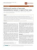
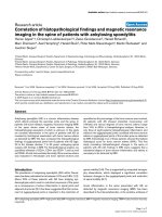
Figure 1: Inspiratory (a) and expiratory (b) chest radiograph of 13
year old girl showing small hyperluscent left hemithorax with mild
mediastinal swing towards normal (right) side(thin white arrow). HRCT
(c) demonstrates decrease in volume of left lung with decrease in
bronchovascular markings on left side with bronchiectatic changes
(white arrow head). MR angiography (d) confirming the diagnosis by
revealing smaller pulmonary artery on left side with typical pruned tree
appearance (thick white arrow)
proximal bronchiectasis with paucity of broncho vascular
markings. The pulmonary artery was smaller on the left
side [Figure 2c]. MR angiography showed typical pruned
tree appearance on left side confirming the diagnosis
[Figure 2d].
DISCUSSION
The Swyer James syndrome is also called Macleod
syndrome/ Bret’s syndrome / Janus syndrome in honor of
workers who initially described this rare entity.[2,3] They
also demonstrated its association with Fallot’s tetrology.
The condition arises as a result of some viral insult in
infancy or childhood. The agents which are implicated
are adenovirus, respiratory syncital virus, influenza virus,
Mycoplasma pneumoniae, Streptococcus pneumoniae and
Staphylococcus. In the initial eight years of life the lung
growth occurs by progressive alveolarization and later on
the growth occurs by expansion of preexisting bronchi.
The infective insult during infancy or childhood results
in post infectious acute obliterative bronchiolitis (usually
developing after six months to three years), which causes
arrest of growth and alveolarization leading to hypoplasia
of affected lung with reduced vascularity with paucity of
bronchial subdivisions (cut off at 4th to 5th generation).
Proteases released by phagocytes may be causative
for elastolysis. Increase in CD8+ cells is also noted in
broncho alveolar lavage of patients suffering from Swyer
162
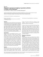
a
Figure 2: Inspiratory (a) and expiratory (b) chest radiographs of
51-year-old female showing small hyperluscent left hemithorax with
air trapping on expiration. HRCT (c) shows reduced volume of left
lung with decrease in parenchymal attenuation and bronchovascular
markings on left side. Bronchiectatic areas are seen in left hemithorax
(white arrow head). MR angiography (d) shows typical pruned tree
appearance on left side (thick white arrow)
James syndrome. The disease usually affects one lung;
however, both lungs and part of the lung may be involved.
The affected lung or portion of the lung will not develop
properly and will be smaller than its counterpart along
with evidence of air trapping, resulting in unilateral
hyperlucency. Finally, fibrous obliteration of airway lumen
occurs.
The usual presentation of the patient is recurrent bouts of
cough, fever, dyspnoea, which may be exertional. Some
times the patients may present with history of hemoptysis,
as in one of our cases. Weight loss may also be present. The
syndrome may be complicated by spontaneous multi vessel
coronary dissection.[4] The Swyer James syndrome results
in chronic lung illness with abnormal lung dynamics
during inspiration and expiration (demonstrated as
abnormal time attenuation curves during inspiration and
expiration) with air trapping which increases on expiration
with bronchial and bronchiolar abnormalities. Placental
transmogrification of lung has been described recently in
patients of Swyer James syndrome,[5] structures resembling
placental villi in lung parenchyma were described.
After the initial infective insult radiographic findings
appear after months to years. Usual investigations
performed are chest radiograph, high resolution computed
tomography (both of these investigation should be done in
inspiration and expiration), MRI, angiography, ventilation
perfusion scanning.
Chest X-ray is the initial modality of investigation which
demonstrates small hyperlucent affected lung/ region
with compensatory hyperinflation of contra lateral lung.
Evidence of air trapping may be noted in expiration which
is a sine qua non for making diagnosis of Swyer James
Lung India • Vol 27 • Issue 3 • July - Sep 2010
Parashari, et al.: Diagnostic role of MR angiography in Swyer James syndrome
syndrome. Swing of mediastinal may be noted towards the
normal lung on expiration. The hila on the affected side
will be smaller. Evidence of bronchiectasis, scarring, and
irregular pulmonary vasculature may be noted. Excursions
of hemi diaphragm will also be markedly asymmetrical.
Brochography may show dilated bronchi with sharply
terminated segments. It is rarely used now a days.
HRCT is done with thin collimation in both phases
of respiration to demonstrate air trapping. For proper
demonstration of mosaic pattern prone position may be
required. There will be evidence of diffuse decrease in
attenuation of lung parenchyma on the affected side which
will be smaller in size with reduction in the broncho
vascular markings and smaller ipsilateral pulmonary artery.
Paucity of bronchial subdivisions, proximal bronchiectasis
and expiratory air trapping are best appreciated on HRCT. [6]
MRI itself does not contribute to the final diagnosis;
however, MR angiography demonstrates smaller pulmonary
artery and its branches on the affected side. The narrowed
attenuated arteries coursing through the radiolucent lung
will produce “pruned tree appearance”.
Matched ventilation perfusion defects may be demonstrated
by ventilation perfusion scanning due to abnormal
development of pulmonary vasculature and lung
parenchyma. Marked air trapping may also be noted
in the washout phase.[7] Scintigraphy may demonstrate
findings of Swyer James syndrome in absence of significant
radiological signs with demonstration of additional areas
Lung India • Vol 27 • Issue 3 • Jul - Sep 2010
of involvement which will be normal on radiograph.
The important differential diagnosis of the Swyer James
syndrome are congenital lobar emphysema, congenital
hypoplasia of the lung, hypoplastic pulmonary artery,
compensatory unilateral emphysema secondary to
lobectomy, pulmonary embolic disease, pneumothorax,
foreign body in air way.[8]
REFERENCES
1.
2.
3.
4.
5.
6.
7.
8.
Macís Robles MD, Martínez Mengual BM, Amador Tejón MJ, López
Fonticiella MP. Swyer-MacLeod syndrome or unilateral hyperlucent
lung. Ann Med Intern 2006;23:557-8.
Swyer PR, James GC. A case of unilateral pulmonary emphysema.
Thorax 1953;8:133-6.
Macleod WM. Abnormal transradiancy of one lung. Thorax 1954;9:147-53.
Davutoglu V, Ege I, Kucukdurmaz Z, Oylumlu M, Akdemir I. SwyerJames-MacLeod syndrome complicated by spontaneous multivessel
coronary dissection. Int J Cardiol 2005;99:359-60.
Marchevsky AM, Guintu R, Koss M, Fuller C, Houck W, McKenna RJ.
Swyer-James (MacLeod) syndrome with placental transmogrification of
the lung: A case report and review of the literature. Arch Pathol Lab
Med 2005;129:686-9.
Gómez Belda AB, Martínez-Moragón E, Fernández Fabrellas E. SwyerJames syndrome: Diagnostic contributions of helical computerized
tomography. Arch Bronconeumol 2000;36:421.
Arslan N, Ilgan S, Ozkan M, Yuksekol I, Bulakbasi N, Pabuscu Y,
et al. Utility of ventilation and perfusion scan in the diagnosis of young
military recruits with an incidental finding of hyperlucent lung. Nucl
Med Commun 2001;22:525-30.
Müller NL. Unilateral hyperlucent lung: MacLeod versus Swyer-James.
Clin Radiol 2004;59:1048.
Source of Support: Nil, Conflict of Interest: None declared.
163
Copyright of Lung India is the property of Medknow Publications & Media Pvt. Ltd. and its content may not be
copied or emailed to multiple sites or posted to a listserv without the copyright holder's express written
permission. However, users may print, download, or email articles for individual use.