characterization of murine macrophages from bone marrow spleen and peritoneum
Bạn đang xem bản rút gọn của tài liệu. Xem và tải ngay bản đầy đủ của tài liệu tại đây (1.5 MB, 10 trang )
Wang et al. BMC Immunology 2013, 14:6
/>
RESEARCH ARTICLE
Open Access
Characterization of murine macrophages from
bone marrow, spleen and peritoneum
Changqi Wang1*†, Xiao Yu1,2†, Qi Cao1, Ya Wang1, Guoping Zheng1, Thian Kui Tan1, Hong Zhao1,3, Ye Zhao1,
Yiping Wang1 and David CH Harris1
Abstract
Background: Macrophages have heterogeneous phenotypes and complex functions within both innate and
adaptive immune responses. To date, most experimental studies have been performed on macrophages derived
from bone marrow, spleen and peritoneum. However, differences among macrophages from these particular
sources remain unclear. In this study, the features of murine macrophages from bone marrow, spleen and
peritoneum were compared.
Results: We found that peritoneal macrophages (PMs) appear to be more mature than bone marrow derived
macrophages (BMs) and splenic macrophages (SPMs) based on their morphology and surface molecular
characteristics. BMs showed the strongest capacity for both proliferation and phagocytosis among the three
populations of macrophage. Under resting conditions, SPMs maintained high levels of pro-inflammatory cytokines
expression (IL-6, IL-12 and TNF-α), whereas BMs produced high levels of suppressive cytokines (IL-10 and TGF-β).
However, SPMs activated with LPS not only maintained higher levels of (IL-6, IL-12 and TNF-α) than BMs or PMs, but
also maintained higher levels of IL-10 and TGF-β.
Conclusions: Our results show that BMs, SPMs and PMs are distinct populations with different biological functions,
providing clues to guide their further experimental or therapeutic use.
Keywords: Macrophage, Bone marrow, Spleen, Peritoneum
Background
Macrophages play an essential role in both innate and
adaptive immunity [1]. Macrophages are the indispensable part of the host defense system because of their
presence in virtually every type of tissue, their capacity
to contain the majority of infections in the early phase
of their development, and their ability to mount specific
immunological responses.
Macrophages are distributed in all tissues and organs
after birth. The distribution patterns of macrophages
have been shown by labeling the colony-stimulated factor 1 receptor (Csf1r) promoter with green fluorescent
protein (GFP) [2] or by specific F4/80 antibody (Ab)
staining of macrophages [3]. It has been found that distinctive morphological differences within and among
* Correspondence:
†
Equal contributors
1
Centre for Transplant and Renal Research at Westmead, Sydney, NSW,
Australia
Full list of author information is available at the end of the article
macrophage populations could be attributed to their heterogeneity [4]. The heterogeneity of macrophages may
be important for their diverse and flexible participation
in immune responses. Therefore, it is important to
examine the phenotypic and functional differences
amongst macrophages from different origins, such as
spleen, bone marrow and peritoneum.
Peritoneal macrophages (PMs) have been widely used
as a macrophage source in mice since the 1960s [5,6].
Possibly due to the low organ tension within the peritoneal cavity, PMs are remarkably distinct from macrophages of other tissues [7]. For example, PMs have
higher expression of inducible nitric oxide synthase and
IL-12 than do splenic macrophages (SPMs) [8].
SPMs were originally located in the cords of Billroth
in splenic red pulp and termed red pulp macrophages,
which show a high acid phosphatase activity and several
detectable macrophage markers, such as F4/80, Mac-1
and MOMA-2 [9-12]. Previous studies have found that
SPMs differ significantly from PMs in their requirements
© 2013 Wang et al.; licensee BioMed Central Ltd. This is an Open Access article distributed under the terms of the Creative
Commons Attribution License ( which permits unrestricted use, distribution, and
reproduction in any medium, provided the original work is properly cited.
Wang et al. BMC Immunology 2013, 14:6
/>
for activation [13], and exhibit different levels of CD40L,
IL-1 and scavenger receptors [14,15]. It has been
reported in a tumor-bearing mouse study, that cytotoxicity was significantly decreased in PMs,while markedly
increased in SPMs [16]. However, the differences of
SPMs with other resident macrophages have not been
fully addressed.
Another source for commonly used macrophages is
the bone marrow. The growth of bone marrow macrophages (BM) requires macrophage colony-stimulating
factor (M-CSF). In the past, studies of macrophages have
had a bias towards macrophages derived from one specific organ. For instance, BMs have been commonly used
due to their homogeneity, ability to be transfected, proliferation capacity and longer lifespan. However, the application of BMs in experimental studies also has
difficulty due to the instability of their phenotype and
functions in vivo [17]. BMs are relatively flexible in their
response to modification; for example, their proliferation
can be regulated by changing the concentration of
growth factor M-CSF [18].
For those reasons, it is important to define differences
among macrophages derived from spleen, bone marrow
and peritoneal cavity. The aim of this study was to explore differences in morphology, phenotype, proliferation, phagocytosis, antigen presentation and cytokine
expression of murine SPMs, BMs and PMs.
Page 2 of 10
Results
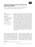
Morphological difference of SPMs, BMs and PMs
PMs displayed a larger cell size (Figure 1G) and higher
lysosomal content than both SPMs and BMs (Figure 1D,
E and F). SPMs had a more elongated spindle shape than
PMs and BMs (Figure 1A, B and C), and lower lysosomal content. BMs contained less cytoplasm than PMs
or SPMs.
Phenotype differences of SPMs, PMs and BMs
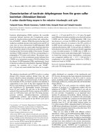
The expression of CD115, CD206, GR-1, CD80, CD86,
MHCII, B7-H1, B7-H2, B7-H3 and B7-H4 was examined
by flow cytometry analysis. CD115 was expressed frequently on BMs (65.4 ± 3.0%), and significantly less on
SPMs (2.4 ± 0.4%) and PMs (3.6 ± 0.2%). Similarly, Gr-1
exhibited a much more frequent expression on BMs
(56.2 ± 2.3%) than on SPMs (6.6 ± 0.7%) or PMs (8.3 ±
1.1%) (Figure 2A, D).
CD80, CD86 and MHC II are important costimulatory
molecules for T cell stimulation. PMs demonstrated high
frequent expression of MHC II (25.5 ± 3.2%) and CD86
(45.3 ± 2.7%), whereas, BMs had high expression of
CD80 (34.6 ± 2.6%). SPMs showed relatively low expression of CD80 (5.5 ± 0.8%) and CD86 (36.1 ± 1.9%)
(Figure 2B, D).
Expression of other costimulatory ligands including
B7-H1, B7-H2, B7-H3 and B7-H4 was examined by flow
Figure 1 Morphological characteristics of cultured macrophages derived from spleen (A, D), bone marrow (B, E) and peritoneal cavity
(C, F), and their cell size assessment (G). All cells were cultured in complete RPMI1640 on 6-well plates, and after removal of supernatant, cells
were then stained with Giemsa-wright dye (A, B, C) and to demonstrate lysosome, anti-LAMP1 (D, E, F) (original magnification x400). Cell size
was assessed by flow cytometry analysis (G).
Wang et al. BMC Immunology 2013, 14:6
/>
Page 3 of 10
Figure 2 Expression of surface molecules on resting SPM, BM and PM was determined by flow cytometry. Red solid lines, staining with
(A) anti-CD115, anti-CD206, anti-Gr-1, (B) anti-CD80, anti-CD86, anti-MHC II, (C) anti-B7-H1, anti-B7-H2, anti-B7-H3 and anti-B7-H4; grey filled ,
staining with the relevant isotype controls. The percentage positivity is shown at the upper right of each histogram. Data are representative of 5
separate experiments of each macrophage preparation. D: summary data of surface molecules expression. Data are mean ± SEM. *p < 0.05,
**p < 0.01.
cytometry. The expression of B7-H1 was much more
frequent on PMs (66.7 ± 0.8%) than SPMs (32.5 ± 2.5%)
or BMs (30.7 ± 1.3%). Low expression level of B7-H2,
B7-H3 and B7-H4 was shown for all three macrophage
types (Figure 2C, D).
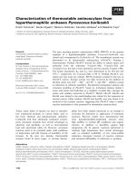
showed similar patterns to those with 2 ng/ml M-CSF.
The proliferation of BMs and SPMs was much greater
than that in low concentration M-CSF (Figure 3B).
However, an increase of M-CSF concentration up to 10
ng/ml did not enhance proliferation capability of PMs.
Proliferative capability of SPMs, BMs and PMs
The proliferative capability of SPMs, BMs and PMs was
assessed. Under culture with 2 ng/ml M-CSF (Figure 3A),
BMs showed a much stronger proliferative capability than
SPMs and PMs. BM numbers increased from day 4, and
continued until to day14 when there was a 60 fold
increase over baseline. However, SPMs showed less proliferation with only a 7 fold increase. In contrast, there was
no proliferation in PMs during the 14 day culture
(Figure 3B).
In response to 10 ng/ml of M-CSF (Figure 3B), the
proliferation of the three macrophage populations
Capacity of phagocytosis
Phagocytic capacity of these three populations of macrophages was examined. A substantial amount of FITCdextran was taken up by the macrophages derived from
the three different sources. BMs (97.9 ± 1.2% of cells)
exhibited the highest phagocytotic ability compared to
SPMs (64.7 ± 3.1%) and PMs (78.9 ± 2.6%) (Figure 4A).
The mean fluorescence intensity (MFI) of BMs, SPMs
and PMs was 1980 ± 145, 645 ± 29 and 1232 ± 77 respectively (Figure 4B), indicating the higher phagocytotic
ability of individual BMs. The MFI value of PMs was
Wang et al. BMC Immunology 2013, 14:6
/>
Page 4 of 10
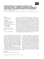
Cytokine expression profile of SPMs, BMs and PMs
Cytokine mRNA expression profiles were examined.
Under resting conditions, BMs produced significantly
higher levels of IL-10 and TGF-β than SPMs and PMs.
SPMs produced significantly higher levels of IL-6, IL-12
and TNF-α than BMs and PMs. However, following LPS
activation, SPMs still expressed high levels of proinflammatory cytokines (IL-6, IL-12 and TNF-α) in comparison to BMs or PMs. SPMs expressed significantly
higher level of suppressive cytokine IL-10 and TGF-β
than PMs. SPMs also expressed significantly higher level
of TGF-β than BMs (Figure 6).
Figure 3 Macrophage growth rate treated with different M-CSF
concentrations. BM, SPM and PM were cultured with M-CSF in
concentrations of 2 ng/ml (A) or 10 ng/ml (B) for 0, 4, 7 and 14
days. The numbers of macrophages were quantified. Images are
representative of 3 separate experiments. Data are mean ± SEM.
*p < 0.05, **p < 0.01.
higher than SPMs indicating the higher phagocytotic
capability of PMs.
Antigen presenting capacity
SPMs, BMs and PMs were analyzed for their ability to
present OVA antigen to OVA-specific DO11.10 CD4+ T
cells by [3H]-thymidine incorporation assay. DCs generated from bone marrow were used as positive control.
Each of these types of macrophage exhibited a much
lower OVA-specific antigen presenting ability than DCs,
and there was no significant difference in the ability of
presenting OVA-specific antigen among the three types
of macrophage (Figure 5).
Discussion
Macrophages have heterogeneous phenotypes and complex functions within both innate and adaptive immune
responses [19]. To date, most experimental studies have
been performed on BMs, isolated SPMs and PMs [1].
However, differences among macrophages from these
particular sources remain unclear. In this study, the features of macrophages from spleen, bone marrow and
peritoneal cavity were compared. We found that PMs
appear to be more mature than SPMs and BMs, based
on their morphology and surface molecular characterizatics. BMs showed the strongest capacity in both proliferation and phagocytosis among the three populations of
macrophage; under resting conditions, SPMs maintained
high level pro-inflammatory cytokine expression (IL-6,
IL-12 and TNF-α), whereas, BMs had high level expression of suppressive cytokines (IL-10 and TGF-β); after
LPS activation, SPMs expressed relatively high levels of
all those cytokines.
In macrophage studies, macrophage cell lines including J774A.1, RAW264.7, P388D1 and U937 [20,21] can
be used, however, continuous subculture of these cell
lines may cause gene loss and impair macrophage immune functions. Therefore, macrophages from bone
marrow, spleen and peritoneum in primary culture are
more commonly used. To date, macrophage studies have
been performed and validated extensively using BMs
[22-24], but less so with SPMs and PMs. Unlike macrophages obtained directly from spleen and peritoneum,
BMs can be fully differentiated in vitro from macrophage
dendritic cell precursors [25]. Although there are many
advantages in using BMs in immunological studies, such
as their high yield, homogeneity and long lifespan [23],
the features of BM macrophages are not fully characterized. Morphological changes of macrophages from three
sources were examined to compare their maturation.
Consistent with the previous studies [26], there are some
similarities among SPMs, BMs and PMs with regard to
their sphere and deeply stained nuclei, but SPMs and
PMs contained much more cytoplasm than BMs, suggesting that BMs may be less mature then SPMs and
Wang et al. BMC Immunology 2013, 14:6
/>
Page 5 of 10
Figure 4 FITC-dextran uptake assay of macrophages from the three different sources. (A) Purified macrophages were incubated with FITCdextran at 37°C for 45 min, and then washed extensively to remove excess FITC-dextran, followed by FACS analysis. Representative histograms
are shown. Solid grey histograms represent control groups; solid red lines represent the percentage of phagocytic macrophages. (B) Group
histograms showing both population and median fluorescence intensity (MFI) values. Data are the mean ± SEM from five separate experiments.
*p < 0.05.
PMs. When comparing cytoplasm of SPMs with PMs,
PMs exhibited a larger size and lysosomal content than
SPMs, suggesting that PMs may be more mature than
SPMs. In addition to morphological analysis, surface
molecular expression could also be used, at least in part,
to indicate the maturity of the three populations. A
study from Alatery showed that both SPMs and BMs
were not fully mature and needed to undergo a further
maturation in vitro in culture [26]. Our study detected
Figure 5 Stimulation of CD4+ T cells by macrophages
presenting OVA in [3H]-thymidine incorporation assays. Isolated
macrophages and dendritic cells (DCs) were loaded with OVA
(10 μg/ml) and irradiated; then co-cultured with DO11.10 CD4+ T
cells for 48 hours. Cultures were then pulsed with [3H]-thymidine,
and incorporated counts determined. DCs were used as positive
control. Data are the mean ± SEM from three separate experiments.
surface molecular expression that related to macrophage
maturation and function. PMs had high level MHC II
and CD86 expression, whereas BMs had high level
CD115 and GR-1 expression. MHC II and CD86 are
expressed highly on fully functional macrophages, which
also indicates their maturity [27,28]. CD115 and Gr1 are
usually expressed on precursors of monocytes and
macrophages, indicating that the cells are less differentiated and more immature [29]. Therefore, our study
showed that PMs appear to be the most mature macrophage, followed by SPMs, then BMs. These differences
are likely important considerations in the experimental
use of macrophages from different sources.
Following great technical improvements in the in vitro
generation of macrophages, they are now considered as
candidates for cell therapy [17,30-32]. Currently, there is
a much variation in the preparation of macrophages
from different sources for therapeutic use. A recent
study of muscle regeneration demonstrated the therapeutic potential of macrophages derived from bone marrow [33]. However, both the experimental and clinical
use of regulatory macrophages (M2) for treating central
nervous system injury relied on generation of macrophages from peripheral blood. Previously we have
demonstrated the therapeutic efficacy of M2 macrophages derived from spleen, but not bone marrow, to resolve inflammation and repair the kidney injury [34-37].
We have shown a similar efficacy of M2 macrophages
derived from peritoneum as from spleen (unpublished
data). This demonstrates the importance of the origin of
Wang et al. BMC Immunology 2013, 14:6
/>
Page 6 of 10
Figure 6 Cytokine mRNA expression profiles of the three populations (SPMs, BMs and PMs) with and without activation with LPS.
mRNA levels of IL-10, TGF-β, IL-6, IL-12 and TNF-α in SPMs, BMs and PMs were measured by real time PCR with β-actin as the housekeeping
gene; (n = 5). Values are expressed as 10x (gene of interest vs β-actin). *p < 0.05, **p < 0.01, ***p < 0.001.
macrophages used for treating disease. In this present
study, the proliferative, phagocytotic and antigen presenting ability of BMs, SPMs, and PMs were assessed. It
was found that BMs exhibited the strongest proliferative
capability among the three populations, with SPMs demonstrating slight and PMs no proliferative capability,
suggesting that macrophages derived from spleen and
peritoneum might be more functionally and phenotypically stable. This observation is consistent with our previous report that M2 macrophage generated from bone
marrow rather than spleen showed strong proliferation
in vivo and failed to protect against renal disease, apparently due to the loss of function and phenotype of
macrophages linked to their proliferation ability [35]. In
addition to proliferative ability, phagocytotic capacity of
macrophages was assessed. BMs have been shown to
maintain the highest capability of phagocytosis [38,39],
which was confirmed in our study and may be an important consideration in regards to their therapeutic efficacy.
T-cell activation and proliferation is associated with
many chronic inflammatory diseases, including chronic
kidney disease, rheumatoid arthritis and atherosclerosis
[17,40,41]. Inhibition of T-cell activation is important in
effectively suppressing inflammatory responses. A previous study showed that B7-H1 binding to its receptor, PD1, results in inhibition of antigen-induced T-cell activation
Wang et al. BMC Immunology 2013, 14:6
/>
[42]. High expression of B7-H1 on PMs suggests PMs
might inhibit T cell activation more effectively than SPMs
or BMs. Such a property of PMs indicates a greater potential for treating chronic inflammatory diseases.
Although SPMs, BMs and PMs exhibited different
levels of expression of molecules involved in antigen
presentation, such as MHCII, CD80 and CD86, they
showed similar antigen presenting ability. Many PMs are
recruited into peritoneal cavity in response to bacterial
infection, in greater amount than other cell types
[43,44]. In spleen, several subpopulations of macrophage
have been characterized in vivo, including F4/80+ red
pulp macrophages, MOMA-1+ marginal metallophilic
macrophages, ER-TR9+ marginal zone macrophages and
MOMA-2+ white pulp macrophages in mice [7]. F4/80
is prodominantly expressed on red pulp macrophages,
but not on others such as marginal metallophilic macrophages, marginal zone macrophages and white pulp
macrophages. Therefore, F4/80 stained cells might be
less diverse and could be considered as a relative uniform population. However, other subpopulations of
splenic macrophages require further study.
Comparison of cytokine expression profile of SPMs,
BMs and PMs might contribute to the understanding of
their distinct properties and provide a valuable reference
for further macrophage related studies. The significantly
higher expression of TGF-β and IL-10 by resting BMs in
comparison to SPMs and PMs suggests that in vitro generated BMs might be potentially more likely to have a
M2 phenotype. M-CSF has been shown to induce differentiation of BMs from bone marrow progenitors [45]
and also to induce human macrophages into a M2
phenotype [46]. Compared to an only 1 day in vitro incubation time of SPMs and PMs, the requirement of 7
days stimulation of bone marrow cells with M-CSF may
push them towards M2 differentiation. Combined with
high proliferation and phagocytosis ability of BM, thus
suggests that BMs might be less mature and phenotypically stable than SPMs and PMs, giving caution to the use
of BMs in cell therapy. Alternatively, pro-inflammatory
cytokines including IL-6, IL-12 and TNF-α were significantly more highly expressed on SPMs with and without
activation than BMs or PMs, which may be relevant to
the specific microenvironment of spleen. In spleen, SPMs
play an important role in removal of red cells, which may
require SPMs to produce abundant cytotoxicity-associated
cytokines such as IL-12, TNF-α and IL-6 [47,48]. Therefore, cytokine expression of BMs, SPMs and PMs reflect
their biological function.
Conclusions
In summary, we report a side-by-side comparison study
of macrophages derived from spleen, bone marrow and
peritoneum. This study demonstrates their distinct
Page 7 of 10
characteristics which are likely relevant to their respective roles in immune response. It also provides a powerful
reference for choosing macrophages of specific origins
not only for experimental study but also for therapeutic use.
Methods
Animals
Six- to eight-week-old male BALB/c mice purchased
from the Animal Resources Centre (Perth, Australia)
were used in this study. DO11.10 mice were obtained
from Animal House of Westmead Hospital (Animal
Care Facility, Westmead Hospital, NSW, Australia). All
animal experiments were approved by the Animal Ethics
Committee of the Sydney West Area Health Service. All
mice were housed in a specific pathogen-free environment and were maintained under constant temperature
(22°C) and humidity, on a 12-hour light/dark cycle in
the Animal House of Westmead Hospital. Then, mice
were fed with acidified water and commercial mouse
chow (protein 18.9%; Glen Forrest Stockfeeders, Glen
Forrest, WA, Australia) ad libitum. Mice were sacrificed
by CO2 inhalation.
Preparation of SPMs, BMs, PMs, DCs and CD4+ T cells
Mice were sacrificed by CO2 inhalation. Spleens were
dissected from abdominal cavity and filtered through a
40-μm nylon strainer. Red cell lysis buffer was used to
remove red cells. A single splenic cell suspension then
was obtained. FACS sorting was performed to obtain F4/
80 positive and CD11c negative cells; then the harvested
cells (0.5-1x10⁶) were cultured for 24 hours with
complete RPMI1640 supplemented with 10% FBS, 2 mM
L-glutamine, 50 U/ml penicillin, 50 μg/ml streptomycin,
10 mM HEPES (N-2-hydroxyethylpiperazine-N’-2ethanesulfoinc acid) and 0.1 mM nonessential amino
acids (all from Life Technologies) and 10 ng/ml M-CSF
(R&D Systems) in 6-well plates (BD Bioscience), at 37°C.
For macrophage activation, cells were stimulated with
100 ng/ml LPS (Sigma) for 24 hours. The cells were harvested by trypsin (0.5%) (Invitrogen).
Pelvic and femoral bones were dissected; and all the
remaining tissue on the bones was removed. Each bone
end was cut off, and bone marrow was expelled.
Cells from bone marrow were cultured for 7 days with
10 ng/ml M-CSF; medium was changed every two days.
Adherent cells were detached by trypsin (0.5%) digestion. FACS sorting (BD Bioscience) was performed to
obtain F4/80 positive and CD11c negative cells; then the
harvested cells (0.5-1×10⁶) were cultured for 24 hours in
complete RPMI1640 with 10% FBS in 6-well plates, at
37°C. For macrophage activation, cells were stimulated
with 100ng/ml LPS (Sigma) for 24 hours. The cells were
harvested by trypsin (0.5%).
Wang et al. BMC Immunology 2013, 14:6
/>
Page 8 of 10
Table 1 Antibodies for flow cytometry analysis
Gimesa-Wright staining and lysosome staining
Antibody
Flurochrome
anti-mouse F4/80
PE-conjugated
Cells were cultured in 6-well plates and fixed by 100%
methanol for 10 min at -20°C; and after air-drying, 1 ml
of Gimesa-Wright dye (Sigma) was added into each well
for 3 min at room temperature and then washed with
PBS completely. Stained cells were examined under microscopy (Nikon) with magnification x400. For lysosome
staining, cells were fixed by 100% methanol for 10 min
at -20°C. Anti-mouse lysosome associated membrane
protein 1 (LAMP1) (1/400; Abcam) was used as primary
antibody and Alexa FluorW 488 (green) goat anti-rabbit
IgG (1/1000; eBioscience) was used as second antibody.
DAPI was used to stain the cell nuclei (blue). Images
were captured by fluorescent microscope (Olympus)
with magnification x400.
anti-mouse CD11c
APC-conjugated
anti-mouse CD80
PE-conjugated
anti-mouse CD86
PE-conjugated
anti-mouse CD115
PE-conjugated
anti-mouse CD206
APC-conjugated
anti-mouse Gr-1
PECy7-conjugated
anti-mouse MHC II
PE-conjugated
anti-mouse B7-H1
PE-conjugated
anti-mouse B7-H2
PE-conjugated
anti-mouse B7-H3
PE-conjugated
anti-mouse B7-H4
PE-conjugated
Flow cytometry analysis
All antibodies were from eBioscience.
Peritoneal membrane was separated from under the
abdominal musculature. 5-7 ml ice cold PBS was
injected into peritoneal cavity; peritoneum was gently
and completely massaged; PBS was then aspirated from
peritoneal cavity. Peritoneal cells were enriched by centrifugation, then purified by FACS sorting by selecting
F4/80 positive and CD11c negative population, then the
harvested cells (0.5-1×10⁶) were cultured for 24 hours in
complete RPMI1640 with 10% FBS in 6-well plates, at
37°C. For macrophage activation, cells were stimulated
with 100ng/ml LPS (Sigma) for 24 hours. The cells were
harvested by trypsin (0.5%). The purity of detached cells
was assessed by Flow analysis (Additional file 1).
To obtain dendritic cells, cells from bone marrow were
cultured for 7 days with 10 ng/ml GM-CSF and 10 ng/ml
IL-4; medium was changed every two days. Floating cells
were removed by PBS washing, adherent cells were considered as DCs.
OVA-specific CD4+ T cells were isolated from DO11.10
mice. DO11.10 mice were sacrificed by CO2 inhalation.
Spleen were dissected from abdominal cavity and filtered
through a 40-μm nylon strainer. Red cell lysis buffer was
used to remove red cells. A single splenic cell suspension
then was obtained and incubated with mouse CD4
MicroBeads (Miltenyi Biotec) for 15 min on ice. MACSbead separation was performed to obtain CD4+ T cells.
The macrophages were resuspended in PBS containing
2% fetal bovine serum (FBS). Non-specific Ab binding
was blocked with addition of Fc block Ab, then
fluorochrome-labelled Abs against macrophage surface
markers were added in a concentration of 1:200; cells
were stained for 20 min on ice and washed 3 times with
cold PBS. Unstained samples were prepared for cell size
assessment. Data were collected with Flow Cytometer
LSRII and analyzed with Flow Jo software. Abs used in
this study are listed in Table 1.
Proliferation assay
Macrophages derived from spleen, bone marrow and
peritoneal cavity were purified. Then, purified macrophages were cultured in separate 6-well plates at the
concentration of 1 × 10⁴ cells per well. Medium used
was complete RPMI 1640 with M-CSF in two concentrations (10 ng/ml and 2 ng/ml). Medium was changed
every two days. The cell number was measured by
counting under microscope, on days 4, 7 and 14.
FITC-dextran uptake assay
In order to measure macrophage phagocytic ability, the
FITC-dextran uptake assay was set up by incubating cells
with FITC-dextran in triplicate plates. Briefly, purified
macrophages were cultured on 12-well plates at a concentration of 0.5 × 10⁵ cells/well. FITC-dextran was added into
Table 2 Primers for real time PCR
Gene
Sequences of primers
β-actin
Left:50-GATTACTGCTCTGGCTCCTAG-3’
Right:50-GCCACCGATCCACACAGAGT-3’
IL-10
Left:50-CCAGTACAGCCGGGAGACA-3’
Right:50-CAGCTGGTCCTTTGTTTGAAAG-3’
0
TGF-β
Left:5 -TTAGGAAGGACCTGGGTTGG-3’
Right:50-AGGGCAAGGACCTTGCTGTA-3’
IL-6
Left:50-CACAAGTCCGGAGAGGAGAC-3’
Right:50-TTGCCATTGCACAACTCTTT-3’
0
IL-12
Left:5 -GACATCACACGGGACCAAAC-3’
Right:50-TACCAAGGCACAGGGTCATC-3’
TNF-α
Left:50-TGCCTATGTCTCAGCCTCTTC-3'
Right: 50-GAGGCCATTTGGGAACTTCT-3'
Wang et al. BMC Immunology 2013, 14:6
/>
each well at a final concentration of 0.5 mg/ml, and the
culture plates was incubated at 4°C and 37°C for 45min.
After incubation, wells were washed extensively to remove
excess FITC-dextran. Macrophages were detached by
digestion with 5% trypsin. FACS analysis was performed;
median fluorescence intensity (MFI) was calculated.
[3H] thymidine incorporation assay
For analysis of in vitro T cell proliferation, isolated macrophages and dendritic cells (DCs) were incubated with 10
μg/ml ovalbumin (OVA) peptide 323-339 (Genscript,
USA) for 60 min at 37°C. OVA-loaded cells were washed
3 times with RPMI 1640. A total number of 50,000 naive
CD4+ T cells were cultured in 96-well plates with 50,000
OVA-loaded macrophages or DCs for 48 hours; then 3HThymidine (1 μCr/well) was added and the incubation
continued for a further 16 hours. Cells were harvested
using a Packard Filtermate Harvester 96 and counted by
Microbeta counter (PerkinElmer, Beaconsfield, UK).
Real time PCR analysis
RNA was extracted using the Qiagen (MD, USA) RNeasy
mini kit according to the manufacturer’s instructions; For
reverse transcription, first strand cDNA was transcribed
from total RNA using a First Strand cDNA Synthesis Kit
((Fermantas, Australia) by following the manufacturer’s
instructions. Then the SBYR Green qPCR Detection
System (Invitrogen) was employed for real-time PCR.
Real-time PCR amplification was carried out in Corbett
Rotorgene 6000 real-time Thermo cycler using a PCR
mixture containing primers, cDNA and SYBR green
mastermix. Levels of mRNA expression were normalized
to housekeeping gene β-actin mRNA levels. GraphPad
Prism 5.0 was used for statistical analysis. The primers
used in this study are listed on Table 2.
Statistical methods
The Student’s T-test was used for 2-group comparisons,
and ANOVA was used for comparisons involving 3 or
more groups. A P value of less than 0.05 was considered
statistically significant. Values are expressed as means ±
standard error (SEM).
Additional file
Additional file 1: SPMs, BMs and PMs were generated respectively,
and then stained with anti-F4/80 and anti-CD11c, the gating of F4/80+
and CD11c-cells was based their isotype controls. Data are representive
of 5 separate experiments.
Authors’ contributions
CW and XY performed all the experiments under the supervision of D C.H. H
and YW. QC and all other authors contributed to the experimental design.
CW wrote the manuscript; D C.H. H and YW revised the manuscript. All
authors approved the manuscript.
Page 9 of 10
Acknowledgements
This study was supported by the National Health & Medical Research Council
of Australia (NHMRC, grant 457345 to Yiping Wang & David Harris).
Author details
1
Centre for Transplant and Renal Research at Westmead, Sydney, NSW,
Australia. 2Department of Urology, Tongji Hospital, Tongji Medical College,
Huazhong University of Science and Technology, Wuhan, PR China.
3
Department of Biochemistry and Molecular Biology, Shanxi Medical
University, Shanxi, PR China.
Received: 4 September 2012 Accepted: 25 January 2013
Published: 5 February 2013
References
1. Gordon S: The macrophage: past, present and future. Eur J Immunol 2007,
37(Suppl 1):S9–S17.
2. Sasmono RT, Oceandy D, Pollard JW, Tong W, Pavli P, Wainwright BJ,
Ostrowski MC, Himes SR, Hume DA: A macrophage colony-stimulating
factor receptor-green fluorescent protein transgene is expressed
throughout the mononuclear phagocyte system of the mouse. Blood
2003, 101:1155–1163.
3. Hume DA, Ross IL, Himes SR, Sasmono RT, Wells CA, Ravasi T: The
mononuclear phagocyte system revisited. J Leukoc Biol 2002, 72:621–627.
4. Shortman K, Wu L: Are dendritic cells end cells? Nat Immunol 2004,
5:1105–1106.
5. Rous P: The Relative Reaction within Living Mammalian-Tissues: Ii. On
the Mobilization of Acid Material within Cells, and the Reaction as
Influenced by the Cell State. J Exp Med 1925, 41:399–411.
6. Kaufmann SH, Schaible UE: Antigen presentation and recognition in
bacterial infections. Curr Opin Immunol 2005, 17:79–87.
7. Liu G, Xia XP, Gong SL, Zhao Y: The macrophage heterogeneity:
difference between mouse peritoneal exudate and splenic F4/80+
macrophages. J Cell Physiol 2006, 209:341–352.
8. Zhu YN, Yang YF, Ono S, Zhong XG, Feng YH, Ren YX, Ni J, Fu YF, Tang W,
Zuo JP: Differential expression of inducible nitric oxide synthase and IL12 between peritoneal and splenic macrophages stimulated with LPS
plus IFN-gamma is associated with the activation of extracellular signalrelated kinase. Int Immunol 2006, 18:981–990.
9. Springer T, Galfre G, Secher DS, Milstein C: Mac-1: a macrophage
differentiation antigen identified by monoclonal antibody. Eur J Immunol
1979, 9:301–306.
10. Austyn JM, Gordon S: F4/80, a monoclonal antibody directed specifically
against the mouse macrophage. Eur J Immunol 1981, 11:805–815.
11. Hume DA, Robinson AP, MacPherson GG, Gordon S: The mononuclear
phagocyte system of the mouse defined by immunohistochemical
localization of antigen F4/80. Relationship between macrophages,
Langerhans cells, reticular cells, and dendritic cells in lymphoid and
hematopoietic organs. J Exp Med 1983, 158:1522–1536.
12. Kraal G, Rep M, Janse M: Macrophages in T and B cell compartments and
other tissue macrophages recognized by monoclonal antibody MOMA-2.
An immunohistochemical study. Scand J Immunol 1987, 26:653–661.
13. Lane TE, Wu-Hsieh BA, Howard DH: Gamma interferon cooperates with
lipopolysaccharide to activate mouse splenic macrophages to an
antihistoplasma state. Infect Immun 1993, 61:1468–1473.
14. Kim JG, Keshava C, Murphy AA, Pitas RE, Parthasarathy S: Fresh mouse
peritoneal macrophages have low scavenger receptor activity. J Lipid Res
1997, 38:2207–2215.
15. Yang X, Ye RG, Kong QY, Yang QQ, Gao Y, Zhong JH, Wang T: CD40 ligand
expression on macrophages during peritonitis in continuous ambulatory
peritoneal dialysis patients. Adv Perit Dial 2000, 16:213–215.
16. Gordon S: Pathogen recognition or homeostasis? APC receptor functions
in innate immunity. C R Biol 2004, 327:603–607.
17. Wang Y, Harris DC: Macrophages in renal disease. J Am Soc Nephrol 2011,
22:21–27.
18. Hume DA, Gordon S: Regulation of bone-marrow macrophage
proliferation. Adv Exp Med Biol 1982, 155:261–266.
19. Gordon S: The role of the macrophage in immune regulation. Res
Immunol 1998, 149:685–688.
20. Goodrum KJ: Complement component C3 secretion by mouse
macrophage-like cell lines. J Leukoc Biol 1987, 41:295–301.
Wang et al. BMC Immunology 2013, 14:6
/>
21. Baek YS, Haas S, Hackstein H, Bein G, Hernandez-Santana M, Lehrach H,
Sauer S, Seitz H: Identification of novel transcriptional regulators involved
in macrophage differentiation and activation in U937 cells. BMC Immunol
2009, 10:18.
22. Wells CA, Ravasi T, Faulkner GJ, Carninci P, Okazaki Y, Hayashizaki Y, Sweet
M, Wainwright BJ, Hume DA: Genetic control of the innate immune
response. BMC Immunol 2003, 4:5.
23. Weischenfeldt J, Porse B: Bone Marrow-Derived Macrophages (BMM):
Isolation and Applications. CSH Protoc 2008, 2008:pdb prot5080.
24. Marim FM, Silveira TN, Lima DS Jr, Zamboni DS: A method for generation
of bone marrow-derived macrophages from cryopreserved mouse bone
marrow cells. PLoS One 2010, 5:e15263.
25. Geissmann F, Manz MG, Jung S, Sieweke MH, Merad M, Ley K: Development of
monocytes, macrophages, and dendritic cells. Science 2010, 327:656–661.
26. Alatery A, Basta S: An efficient culture method for generating large quantities
of mature mouse splenic macrophages. J Immunol Methods 2008, 338:47–57.
27. Andreesen R, Brugger W, Scheibenbogen C, Kreutz M, Leser HG, Rehm A,
Lohr GW: Surface phenotype analysis of human monocyte to
macrophage maturation. J Leukoc Biol 1990, 47:490–497.
28. Wang Y, Cui X, Tai G, Ge J, Li N, Chen F, Yu F, Liu Z: A critical role of activin
A in maturation of mouse peritoneal macrophages in vitro and in vivo.
Cell Mol Immunol 2009, 6:387–392.
29. Huang B, Pan PY, Li Q, Sato AI, Levy DE, Bromberg J, Divino CM, Chen SH:
Gr-1 + CD115+ immature myeloid suppressor cells mediate the
development of tumor-induced T regulatory cells and T-cell anergy in
tumor-bearing host. Cancer Res 2006, 66:1123–1131.
30. Arnold L, Henry A, Poron F, Baba-Amer Y, van Rooijen N, Plonquet A,
Gherardi RK, Chazaud B: Inflammatory monocytes recruited after skeletal
muscle injury switch into antiinflammatory macrophages to support
myogenesis. J Exp Med 2007, 204:1057–1069.
31. Brown BN, Ratner BD, Goodman SB, Amar S, Badylak SF: Macrophage
polarization: an opportunity for improved outcomes in biomaterials and
regenerative medicine. Biomaterials 2012, 33:3792–3802.
32. Nelson PJ, Rees AJ, Griffin MD, Hughes J, Kurts C, Duffield J: The renal
mononuclear phagocytic system. J Am Soc Nephrol 2012, 23:194–203.
33. Ruffell D, Mourkioti F, Gambardella A, Kirstetter P, Lopez RG, Rosenthal N,
Nerlov C: A CREB-C/EBPbeta cascade induces M2 macrophage-specific
gene expression and promotes muscle injury repair. Proc Natl Acad Sci U
S A 2009, 106:17475–17480.
34. Wang Y, Cao Q, Zheng G, Lee VW, Zheng D, Li X, Tan TK, Harris DC: By
homing to the kidney, activated macrophages potently exacerbate renal
injury. Am J Pathol 2008, 172:1491–1499.
35. Cao Q, Zheng D, Sun Y, Wang Y, Lee VW, Zheng G, Alexander SI, Harris DC:
Impaired alternative activated macrophages from bone marrow in treatment
of CKD: Associations with their proliferation in inflamed kidney. Presented in
abstract form at the annual meeting of the American Society of Nephrology;
October 27 through November 1, 2009. San Diego, CA.
36. Cao Q, Wang C, Zheng D, Wang Y, Lee VW, Wang YM, Zheng G, Tan TK, Yu D,
Alexander SI, Harris DC: IL-25 Induces M2 Macrophages and Reduces Renal
Injury in Proteinuric Kidney Disease. J Am Soc Nephrol 2011, 22:1229–1239.
37. Zheng D, Wang Y, Cao Q, Lee VW, Zheng G, Sun Y, Tan TK, Alexander SI,
Harris DC: Transfused macrophages ameliorate pancreatic and renal
injury in murine diabetes mellitus. Nephron Exp Nephrol 2011, 118:e87–99.
38. Mitchison NA: The immunogenic capacity of antigen taken up by
peritoneal exudate cells. Immunology 1969, 16:1–14.
39. Cannon GJ, Swanson JA: The macrophage capacity for phagocytosis. J Cell
Sci 1992, 101(Pt 4):907–913.
40. Zhou X, Nicoletti A, Elhage R, Hansson GK: Transfer of CD4(+) T cells
aggravates atherosclerosis in immunodeficient apolipoprotein E
knockout mice. Circulation 2000, 102:2919–2922.
41. Isomaki P, Clark JM, Panesar M, Cope AP: Pathways of T cell activation and
terminal differentiation in chronic inflammation. Curr Drug Targets
Inflamm Allergy 2005, 4:287–293.
42. Petroff MG, Kharatyan E, Torry DS, Holets L: The immunomodulatory
proteins B7-DC, B7-H2, and B7-H3 are differentially expressed across
gestation in the human placenta. Am J Pathol 2005, 167:465–473.
43. Anderson CF, Mosser DM: A novel phenotype for an activated macrophage:
the type 2 activated macrophage. J Leukoc Biol 2002, 72:101–106.
44. Millard AL, Mertes PM, Ittelet D, Villard F, Jeannesson P, Bernard J: Butyrate
affects differentiation, maturation and function of human monocyte-derived
dendritic cells and macrophages. Clin Exp Immunol 2002, 130:245–255.
Page 10 of 10
45. Warren MK, Vogel SN: Bone marrow-derived macrophages: development
and regulation of differentiation markers by colony-stimulating factor
and interferons. J Immunol 1985, 134:982–989.
46. Xu W, Schlagwein N, Roos A, van den Berg TK, Daha MR, van Kooten C:
Human peritoneal macrophages show functional characteristics of MCSF-driven anti-inflammatory type 2 macrophages. Eur J Immunol 2007,
37:1594–1599.
47. Gately MK, Wolitzky AG, Quinn PM, Chizzonite R: Regulation of human
cytolytic lymphocyte responses by interleukin-12. Cell Immunol 1992,
143:127–142.
48. Galligioni E, Favaro D, Santarosa M, Quaia M, Spada A, Freschi A, Alberti D:
Induction and maintenance of monocyte cytotoxicity during treatment
with liposomes containing muramyl tripeptide despite tachyphylaxis to
the cytokine response. Clin Cancer Res 1995, 1:493–499.
doi:10.1186/1471-2172-14-6
Cite this article as: Wang et al.: Characterization of murine macrophages
from bone marrow, spleen and peritoneum. BMC Immunology 2013 14:6.
Submit your next manuscript to BioMed Central
and take full advantage of:
• Convenient online submission
• Thorough peer review
• No space constraints or color figure charges
• Immediate publication on acceptance
• Inclusion in PubMed, CAS, Scopus and Google Scholar
• Research which is freely available for redistribution
Submit your manuscript at
www.biomedcentral.com/submit