a mutation in the gene encoding mitochondrial mg2 channel mrs2 results in demyelination in the rat
Bạn đang xem bản rút gọn của tài liệu. Xem và tải ngay bản đầy đủ của tài liệu tại đây (4.79 MB, 11 trang )
A Mutation in the Gene Encoding Mitochondrial Mg2+
Channel MRS2 Results in Demyelination in the Rat
Takashi Kuramoto1*, Mitsuru Kuwamura2, Satoko Tokuda1,2, Takeshi Izawa2, Yoshifumi Nakane1,
Kazuhiro Kitada1,3, Masaharu Akao4, Jean-Louis Gue´net5, Tadao Serikawa1
1 Institute of Laboratory Animals, Graduate School of Medicine, Kyoto University, Kyoto, Japan, 2 Laboratory of Veterinary Pathology, Osaka Prefecture University, Osaka,
Japan, 3 Laboratory of Mammalian Genetics, Genome Dynamics Research Center, Graduate School of Science, Hokkaido University, Sapporo, Japan, 4 Department of
Cardiovascular Medicine, Graduate School of Medicine, Kyoto University, Kyoto, Japan, 5 De´partement de Biologie du De´veloppement, Institut Pasteur, Paris, France
Abstract
The rat demyelination (dmy) mutation serves as a unique model system to investigate the maintenance of myelin, because it
provokes severe myelin breakdown in the central nervous system (CNS) after normal postnatal completion of myelination.
Here, we report the molecular characterization of this mutation and discuss the possible pathomechanisms underlying
demyelination. By positional cloning, we found that a G-to-A transition, 177 bp downstream of exon 3 of the Mrs2 (MRS2
magnesium homeostasis factor (Saccharomyces cerevisiae)) gene, generated a novel splice acceptor site which resulted in
functional inactivation of the mutant allele. Transgenic rescue with wild-type Mrs2-cDNA validated our findings. Mrs2
encodes an essential component of the major Mg2+ influx system in mitochondria of yeast as well as human cells. We
showed that the dmy/dmy rats have major mitochondrial deficits with a markedly elevated lactic acid concentration in the
cerebrospinal fluid, a 60% reduction in ATP, and increased numbers of mitochondria in the swollen cytoplasm of
oligodendrocytes. MRS2-GFP recombinant BAC transgenic rats showed that MRS2 was dominantly expressed in neurons
rather than oligodendrocytes and was ultrastructurally observed in the inner membrane of mitochondria. Our observations
led to the conclusion that dmy/dmy rats suffer from a mitochondrial disease and that the maintenance of myelin has a
different mechanism from its initial production. They also established that Mg2+ homeostasis in CNS mitochondria is
essential for the maintenance of myelin.
Citation: Kuramoto T, Kuwamura M, Tokuda S, Izawa T, Nakane Y, et al. (2011) A Mutation in the Gene Encoding Mitochondrial Mg2+ Channel MRS2 Results in
Demyelination in the Rat. PLoS Genet 7(1): e1001262. doi:10.1371/journal.pgen.1001262
Editor: Gregory S. Barsh, Stanford University, United States of America
Received June 5, 2010; Accepted November 29, 2010; Published January 6, 2011
Copyright: ß 2011 Kuramoto et al. This is an open-access article distributed under the terms of the Creative Commons Attribution License, which permits
unrestricted use, distribution, and reproduction in any medium, provided the original author and source are credited.
Funding: This work was supported by grants-in-aid for Scientific Research from the Japan Society for the Promotion of Science [21300153 to TK] and a grant-inaid for Cancer Research from the Ministry of Health, Labour, and Welfare. The funders had no role in study design, data collection and analysis, decision to publish,
or preparation of the manuscript.
Competing Interests: The authors have declared that no competing interests exist.
* E-mail:
the many defects of myelination or myelin turnover. It has also
benefited from researches carried out on animal models, mostly
mutant mice and rats, including those that have been induced by
transgenesis or genetic engineering in ES cell lines [1,2]. Some of
these models have even allowed therapies to be developed in a
preclinical setting [3]. Unfortunately, only a small number of the
many genes that are directly or indirectly involved in the myelination
process have been identified and only a few of these genes have been
functionally annotated, for example, by the characterization of one or
more mutant alleles. For this reason, any new mutation occurring
spontaneously or after mutagenesis is of potential interest for
unraveling the molecular mechanisms involved in myelin assembly.
In an earlier paper we reported the discovery and pathology of a
rat mutation designated demyelination (symbol dmy), which is
characterized by severe and progressive myelin breakdown in
the CNS. We mapped the locus responsible for this myelin
disorder to rat chromosome (Chr) 17, very close to the prolactin
(Prl) locus, in a region homologous to human Chr 6p21.1-22.3 and
mouse Chr 13 [4,5]. Based on its pathological features, as well as
its genetic localization, this demyelination syndrome appeared to
be unique, with no homologue so far reported in any other
mammalian species, including humans.
Introduction
Myelin is an essential component of the nervous tissue of higher
vertebrates. It acts as a natural insulator of axonal segments
allowing, at the same time, the maintenance of axonal integrity
and the fast conduction of action potentials. It also reduces ionic
currents across the axonal membrane and stabilizes the extracellular milieu within rapidly-firing axon bundles.
In the central nervous system (CNS), myelin is produced by
oligodendrocytes, while in the peripheral nervous system (PNS),
this function is achieved by Schwann cells. Myelination is
completed within a relatively short period of time during
mammalian development and requires a high rate of production
and transport of different kinds of molecules, mostly proteins and
lipids. In adult life, myelin is constantly remodeled and the
maintenance of functional myelin sheaths requires a careful
balance of de novo synthesis and turnover. It is quite clear that any
event generating an imbalance in the myelination or remyelination
process has the greatest chance of inducing dys- or demyelination
of either the central or peripheral nervous system.
Our knowledge of the myelination process has benefited from
careful observations conducted on human patients affected by one of
PLoS Genetics | www.plosgenetics.org
1
January 2011 | Volume 7 | Issue 1 | e1001262
Demyelination in the Mrs2-Mutant Rat
down to 0.22 cM, between markers D17Kur17 and D17Got45.
Within this critical section, we found no recombination between
the dmy locus and either Aldh5a1 (aldhehyde dehydrogenase family
5, subfamily A1) or Mrs2 (mitochondrial 118 RNA splicing2) loci,
among 68762 = 1,374 meioses. The rat genome databases
revealed that D17Kur17 and D17Got45 were at position 46.78Mb and 47.26-Mb, respectively, on rat Chr 17, yielding a physical
size of 0.48 Mb of DNA for the interval containing the dmy locus.
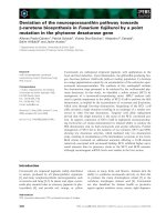
This stretch of DNA contained 6 genes (Figure 2A).
Analysis by RT-PCR of the transcription products of these 6
genes revealed that the cDNA transcribed from the Mrs2 gene was
larger in dmy/dmy mutants than in the controls (Figure 2B). After
sequencing, we found that the larger size of the dmy cDNA was due
to the insertion of an 83 bp intronic sequence between exons 3
and 4. Comparison of the two genomic sequences revealed a G-toA transition, 177 bp downstream of the end of exon 3 (Figure 2C,
Figure S1), generating a novel splice acceptor site, which
accounted for the addition of the 83bp stretch of intronic sequence
to the mutant transcript. In addition, while the Mrs2 gene
normally encodes a 434 amino-acid protein, the intronic insertion
leads to a shorter protein (106 amino acids) due to the occurrence
of a stop codon as a consequence of frame shifting within the novel
pseudo-exon X. The new protein consisted of the first 91 amino
acids of normal (wild-type) MRS2 protein followed by an
additional 15 amino acids transcribed from the intronic stretch
(Figure 2D) [10]. No nucleotide alteration was observed between
normal and mutant haplotypes in the cDNA transcribed from the
other 5 genes (Vmp, Dcdc2, Gpld1, Aldh5a1, and KIAA0319). These
findings strongly suggested that the G-to-A mutation in intron 3 of
Mrs2 in dmy/dmy rats was very likely causative of the neurological
phenotype.
Author Summary
The myelin sheath that surrounds the axon of a neuron
acts as a biological insulator. Its major function is to
increase the speed at which impulses propagate along
myelinated fibers in the central nervous system, as well as
the peripheral nervous system. Alterations or damage
affecting this structure (demyelination) result in the
disruption of signals between the brain and other parts
of the body. In the rat, mutations producing demyelination
have been frequently identified and characterized and
have contributed to a better understanding of the genetics
of myelin development, physiology, and pathology. This
paper reports the molecular characterization of a recessive
allele responsible for the progressive disruption of myelin
that was initially observed in mutant rats, previously
named demyelination (dmy). This mutation generates an
additional splicing acceptor site in an intron of the
mitochondrial Mg2+ transporter gene (Mrs2), resulting in
the insertion of a 83-bp genomic DNA segment into the
Mrs2 transcript and complete functional inactivation of the
mutant allele. We firstly defined the biological function of
MRS2 in mammals and demonstrated the crucial and
unexpected role of MRS2 in myelin physiology. Our
findings might be helpful in the development of new
therapeutic strategies for demyelinating syndromes.
In this report we demonstrate that the causative gene (Mrs2)
encodes a protein that is an essential component of the major
electrophoretic Mg2+ influx system in mitochondria [6]. This gene
has orthologues in other organisms, including lower eukaryotes
and plants [7,8]. The protein shares many of the properties of
bacterial CorA and yeast Alr1 proteins but its specific involvement
in the myelination process was not known or even suspected.
dmy/dmy rats exhibit morphological and biochemical
features characteristic of mitochondrial deficiencies
Results
The MRS2 protein functions as a major transporter protein
(Mg2+, Ni2+ and Co2+) in yeast as well as in human cells [10,11].
When this protein is functionally defective this leads to the ‘‘petite’’
phenotype in yeast and to cell death in human HEK 293 cells
[11,12]. Because mitochondrial diseases in mammals are often
accompanied by elevated lactic acid, reduced ATP, increased
cytochrome oxidase (COX) activity, and the morphological
alteration of mitochondria [13–15], we measured lactic acid levels
and ATP contents in the CNS and performed morphological
analyses of the CNS of dmy/dmy rats.
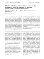
Lactic acid concentration in the cerebrospinal fluids was
significantly elevated in dmy/dmy rats when compared with normal
littermates: 126643.7 mg/dL vs 2569.6 mg/dL (average 6 SD),
P,0.002 (Figure 3A). The ATP concentration was markedly
reduced in dmy/dmy rats: 265679 mM/mg vs 99646 mM/mg
(average 6 SD), P,0.005 (Figure 3B). In the affected dmy/dmy
rats, swollen oligodendrocytes were often observed in the white
matter, showing the increased COX reaction products (Figure 3C).
Ultrastructurally, their cytoplasm contained many mitochondria
and Golgi apparatus-like membrane structures (Figure 3D). These
findings indicated that the mitochondria of dmy/dmy rats were
functionally defective.
dmy/dmy rats exhibit a phenotype with typical
demyelination
The pathology of homozygous dmy/dmy rats has been reported
in detail previously [4]. Mutant rats exhibit no significant
differences from their control littermates until 4 weeks of age.
From 5 weeks on, flaccidity of the hind limbs becomes noticeable
and evolves towards complete paralysis around 7–8 weeks of age.
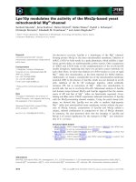
Progressive demyelination is observed in several parts of the CNS
(Figure 1), namely the corpus callosum, the capsula interna, the
striatum and the cerebellar peduncle, with major effects on the
ventral and lateral parts of the spinal cord. Astrogliosis, which is a
major feature of myelin disorder, is observed in demyelinated
areas but motor neurons remain normal and there is no sign of
associated inflammation in the white matter. The dmy mutation
can then be regarded as a mutation affecting the maintenance and
turnover of myelin rather than its initial production: this is typical
demyelination [9].
The dmy syndrome is associated with a mutation in a
splicing site of Mrs2, a gene encoding a mitochondrial
Mg2+ channel
Out of 687 dmy/dmy mutant rats, collected from the 3,252
offspring of an intercross segregating for the dmy mutation, 23
individuals were found to carry a recombinant haplotype between
the two loci that were used for the initial genetic mapping, namely;
Prl (prolactin) and Hh1tts (testis-specific histone, H1t). Further
investigation of these animals, using three novel informative SSLP
markers, allowed us to narrow the genetic interval containing dmy
PLoS Genetics | www.plosgenetics.org
Rescue of dmy/dmy mutant phenotypes by transgenic
complementation
To ascertain that the molecular defect (i.e. G-to-A transition)
observed in the dmy mutant haplotype was causative of the
abnormal phenotype observed in dmy/dmy rats, we attempted to
rescue the mutant phenotype by transgenic complementation. We
2
January 2011 | Volume 7 | Issue 1 | e1001262
Demyelination in the Mrs2-Mutant Rat
Figure 1. Demyelination in dmy/dmy rats. A. Histopathology of the cervical part of the spinal cord of dmy/+ (left) and dmy/dmy (right) rats aged
10 weeks. Luxol fast blue-HE staining. Original magnification: 6100. B. Electron microscopy of the cervical part of spinal cord of dmy/dmy rats (10
weeks). Naked axons with demyelination (arrowheads) are indicated by asterisks. Inset: control image of the spinal cord from the age-matched wild
type rat. Axons are normally myelinated. Bar = 1 mm.
doi:10.1371/journal.pgen.1001262.g001
established two independent WTC.DMY-dmy lines, expressing
each Mrs2 wild-type cDNA under the control of a cytomegalovirus
(CMV) promoter (Figure S2A), and found that all dmy/dmy
transgenic rats exhibited a completely normal phenotype, with no
paralysis of the hind limbs. Histopathological analyses demonstrated that both transgenic lines no longer exhibited any sign of
demyelination of the CNS (Figure S2B). In addition, lactic acid
levels of the cerebrospinal fluid of transgenic dmy/dmy rats had
returned to the normal range (Figure S2C). Electron microscopic
observations revealed that mitochondria of the oligodendrocytes in
transgenic rats were normal in their morphology and number
(Figure S2D). These findings confirmed that the molecular
changes reported above and observed in the Mrs2 gene were
PLoS Genetics | www.plosgenetics.org
indeed causative of the dmy-mutant phenotypes. For this reason we
decided that the symbol of the mutant allele should, from now on,
be changed to Mrs2dmy.
MRS2-GFP recombinant protein is expressed in the
mitochondria
To characterize the tissues and cell types expressing MRS2 as
well as the subcellular localization of this protein in the CNS, we
generated a strain of rats transgenic for a recombinant MRS2GFP BAC clone. These transgenic rats were expected to express
recombinant protein under the control of the endogenous, normal
Mrs2 promoter. We found that cytoplasmic dot-like MRS2-GFP
3
January 2011 | Volume 7 | Issue 1 | e1001262
Demyelination in the Mrs2-Mutant Rat
PLoS Genetics | www.plosgenetics.org
4
January 2011 | Volume 7 | Issue 1 | e1001262
Demyelination in the Mrs2-Mutant Rat
Figure 2. Positional cloning of the dmy mutation. A. The dmy locus was localized within a 0.22-cM region of chromosome 17 between D17Kur17
and D17Got45 and no recombination was observed with SSLP markers designed from Aldh5a1 and Mrs2 genomic sequences in 1,374 informative
meioses. Within the 0.48-Mb physical interval between D17Kur17 and D17Got45, harboring the dmy locus, 6 genes: Vmp (vesicular membrane protein
p24), Dcdc2 (doublecortin domain containing 2), Mrs2 (MRS2 magnesium homeostasis factor (S. cerevisiae), Gpld1 (glycosylphosphatidylinositol
specific phospholipase D1), Aldh5a1 (aldehyde dehydrogenase family 5, subfamily A1), and KIAA0319, were previously mapped. B. A larger RT-PCR
product was obtained when amplifying the 59 region of Mrs2 cDNAs from dmy/dmy rats with a primer set of rMrs2l-3&4 (59TGTACTGATCTACCCGAGTTCC-39 and 59-TCTGGAGTTATCACAGCCTTCA-39). M: molecular marker, WX174-HaeIII digest. C. Upper: Genomic
organization in the vicinity of intron 3 of the Mrs2 wild-type allele. Lower: Genomic rearrangements in the same intron 3 of the Mrs2dmy mutant
allele. In the Mrs2dmy mutant allele, a novel splice acceptor site was generated as a consequence of a G-to-A transition at 177 bp downstream of the
end of exon 3. An 83-bp genomic sequence (boxed in gray), downstream of the recently generated acceptor site (tggcag), is then inserted into the
Mrs2 mutant transcript. This sequence contains a premature stop codon (vertical arrow), which truncates the protein almost immediately
downstream of exon 3. D. Schematic representations of the wild-type and dmy MRS2 proteins. Conserved amino acid residues and transmembrane
domains are indicated by grey and purple boxes, respectively. Coiled-coil regions are indicated by horizontal orange lines. The position of the dmy
mutation is indicated by an arrowhead, and the additional 15 residues (GATWTPRILEECLES), indicated by a black box, are deduced to be added
subsequently. Bottom: Schematic representation of the topology of MRS2. Purple: transmembrane domains, Orange: coiled-coil regions. The position
of the dmy mutation is indicated by an arrowhead.
doi:10.1371/journal.pgen.1001262.g002
essential domains and accordingly its function of an Mg2+
transmembrane transporter. In other words, Mrs2dmy is a null
allele, which is totally consistent with its recessive allelic
interaction.
An MRS2 is a major transport for Mg2+ uptake into
mitochondria, its function would be expected to be important, if
not essential, for the maintenance of respiratory complex I and
accordingly for cell viability [6,11]. This assumption was
supported by the analysis of MRS2 knock-down, mediated by
shRNA in a human HEK-293 cell line, which resulted in a series
of physiological changes ranging from transient reduction of Mg2+
uptake to the complete loss of mitochondrial respiratory complex
I, with decreased mitochondrial membrane potential and cell
death, depending on the duration of knock-down treatment [11].
However, if we consider the phenotype of our mutant rat, which is
apparently limited to the myelination process with a rather long
lifespan, the role of MRS2 in the maintenance of cell integrity
should be reconsidered.
Considering the pathological features that appear to be
characteristics of the Mrs2dmy allele on the one hand, and
MRS2-specific functions, as described above on the other, it is
logical to consider that the demyelinating syndrome in mutant rats
results from a mitochondrial disease. This assertion is supported by
the observation of an elevated rate of lactic acid in the
cerebrospinal fluid, reduced ATP in the brain, increased COX
activity, and the morphological alteration of mitochondria, which
is generally considered a major characteristic of mitochondrial
diseases [13–15]. An increase in mitochondria is characteristic of
cells with reduced respiratory capacity [19]. The association of
mitochondrial dysfunction with demyelination (or leukodystrophy)
has been already reported in Leigh syndrome and mitochondrial
DNA depletion syndrome [20–23]. The tissues most frequently
affected in these mitochondrial diseases are the cerebrum,
peripheral nerves, and skeletal muscles, presumably because cells
of these tissues require more energy than any other cells in the
body. Unfortunately, the detailed pathophysiological mechanism(s)
leading to demyelination in these diseases has not yet been
unraveled. We consider that our mutant rat could be an interesting
tool for investigating this matter.
Mitochondrial dysfunction has also been observed in multiple
sclerosis (MS), one of the most common demyelination diseases,
but here again many aspects of the pathophysiology require
further investigation [24,25]. This difficulty of linking gene
functions with a specific syndrome is not so surprising if we
consider that, according to the most recent estimates, there may be
as many as 1,500 nuclear-encoded mitochondrial proteins [26]
and that less than half have been identified with experimental
support. Clearly, a complete protein inventory of this organelle
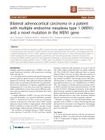
signals were observed in neurons throughout the CNS. To a lesser
extent, astrocytes and oligodendrocytes also exhibited occasional
expression of MRS2 (Figure S3). Confocal microscopy demonstrated that MRS2 is located in the mitochondria (Figure 4A–4C).
Moreover, immunoelectron microsopic examinations with antiGFP antibody revealed that MRS2 is localized in the inner
membrane of the mitochondria (Figure 4D). MRS2 expression was
also observed in the myocardium, liver, testis and skeletal muscles
(Figure S4).
Microglia activation and high expression of inflammatory
cytokines were observed in Mrs2dmy/Mrs2dmy rats
Microglial activation, characterized by cellular hypertrophy, has
been reported in various dysmyelinating and demyelinating
pathologies. To assess microglial activation, we performed
immunohistochemistry for IBA1, a specific marker of microglia.
In Mrs2dmy/Mrs2dmy rats, prolonged activation of microglia was
prominently observed at 6–7 weeks of age (Figure 5A and 5B), the
stage at which clinical symptoms such as flaccid paralysis were
commonly observed. Expression levels of proinflammatory
cytokines, such as Il1b and Il6, were also significantly higher in
Mrs2dmy/Mrs2dmy rats than in wild-type littermates at 6 weeks of age
(Figure 5C).
Discussion
Characterization, by positional cloning, of the molecular defect
responsible for the demyelinating phenotype observed in adult
dmy/dmy rats led us to incriminate a mutation in the Mrs2 gene. No
mutant allele before Mrs2dmy, which we report here, has ever been
reported at this locus in any mammalian species.
Mrs2 encodes an inner membrane Mg2+ channel in mitochondria and belongs to a family with orthologous copies in a wide
range of species [10,12]. Mrs2 was originally identified in yeast,
and orthologous copies of this gene have been identified in a
variety of organisms, including bacteria (CorA), fungi (Alr1), and
plants (AtMrs2). All proteins in the family have the same substrate
selectivity: they transport Mg2+, Co2+ and some other divalent
cations across the mitochondrial membrane. Even if these proteins
exhibit relatively low sequence similarities, they all have a few
important domains at the same relative position and can
functionally complement each other over a wide range of
phylogenetic distances [16,17]. In mammals, the normal protein
MRS2 has two universally conserved transmembrane domains
(TMs) and a conserved Gly-Met-Asn (GMN) motif close to the first
TM domain that forms part of the pore and is essential for Mg2+
transport [18] (Figure 2D, Figure S5). As we demonstrated, the
protein is truncated in dmy/dmy mutant rats, having lost both of its
PLoS Genetics | www.plosgenetics.org
5
January 2011 | Volume 7 | Issue 1 | e1001262
Demyelination in the Mrs2-Mutant Rat
Figure 3. Biochemical and morphological abnormalities in the mitochondria of dmy/dmy mutant rats. A. Lactic acid concentration in
cerebrospinal fluid of 6–7-week-old dmy/dmy rats and age-matched wild-type rats. **, P,0.002. B. ATP levels in the brain of 6–7-week-old dmy/dmy
rats and age-matched wild-type rats. **, P,0.005. C. Cytochrome oxidase staining of the spinal cords of 6–7-week-old dmy/dmy (right) and agematched wild-type (left) rats. Swollen oligodendrocytes were often seen they showed increased COX reaction product. Bar = 50 mm. D. Electron
microphotographs of a swollen oligodendrocyte in a dmy/dmy rat (right) and an oligodendrocyte in a control wild-type rat. White matter of thoracic
spine at 6 weeks of age. N: Nucleus of the oligodendrocyte. Axons adjacent to the oligodendrocyte are indicated by asterisks. Bar = 2 mm.
doi:10.1371/journal.pgen.1001262.g003
across tissues would provide a molecular framework to relate
mitochondrial biology and pathogenesis [27].
A point concerning Mrs2 gene expression in the CNS that is
worth noting after our experiments and observations is that the
PLoS Genetics | www.plosgenetics.org
gene in question is expressed at a higher rate in neurons than in
oligodendrocytes (Figure 4, Figure S3). This was rather unexpected if we consider that oligodendrocytes are the cells actually
responsible for myelination of the CNS. At this time, it remains
6
January 2011 | Volume 7 | Issue 1 | e1001262
Demyelination in the Mrs2-Mutant Rat
Figure 4. Expression of MRS2 protein in the mitochondria. MRS2-GFP recombinant protein (Green) was seen in the cytoplasm of pyramidal
cells (A). MRS2-GFP signals were colocalized with the mitochondria (B), as shown in the confocal image of GFP and mitochondrial
immunohistochemistry (C). Nuclei were stained with DAPI (Blue). Bar: 5 mm. Immunoelectron microscopy using anti-GFP antibody revealed that
MRS2-GFP signals were localized in the inner membrane of the mitochondria (arrows) (D). Bar: 200 nm.
doi:10.1371/journal.pgen.1001262.g004
molecules [29,30]. Additionally, Kuwamura and co-workers
reported prominent astrogliosis and many ED-1-positive macrophages in myelin-destroyed areas [9]. When considered together,
these morphological observations led us to believe that the
demyelination observed in dmy/dmy rats is probably enhanced by
activated microglia and astroglia.
In summary, we identified Mrs2dmy as a loss-of-function
mutation of the Mrs2 gene that normally encodes Mg2+
transporter protein of the mitochondrial inner membrane. Our
observations also demonstrate that the mechanisms underlying the
unclear whether the demyelination in dmy/dmy rats is triggered
cell-autonomously or cell-nonautonomously. Instead, it is likely
that demyelination is enhanced by the surrounding cells, such as
activated microglia and astroglia. At 6 weeks of age, when dmy/dmy
rats began to exhibit ataxia [9], cytokine levels were elevated and
microglia were activated (Figure 5), and it is considered that
activated microglia cause neuronal damage through the release of
potentially cytotoxic molecules, such as proinflammatory cytokines, reactive oxygen intermediates, proteinases, and complement
proteins [28]. Oligodendrocytes show greater vulnerability to such
PLoS Genetics | www.plosgenetics.org
7
January 2011 | Volume 7 | Issue 1 | e1001262
Demyelination in the Mrs2-Mutant Rat
Figure 5. Activation of microglia in the central nervous system of Mrs2dmy/Mrs2dmy rats. Immunohistochemistry for Iba1 in the lumbar part
of the spinal cord of wild-type (A) and Mrs2dmy/Mrs2dmy rats (B) at 6 weeks of age. Signals of Iba1 (AlexaFluor 546 nm; red), which is upregulated
during the activation of microglia, are seen in Mrs2dmy/Mrs2dmy rats much more the wild-type control. Nucleus is stained with DAPI (blue). C,
Inflammatory cytokine mRNA expression in the CNS of wild-type (%) and Mrs2dmy/Mrs2dmy (&) rats. IL1b expression was elevated in Mrs2dmy/Mrs2dmy
rats at 4 and 6 weeks of age. IL6 was elevated in Mrs2dmy/Mrs2dmy rats at 6 weeks of age. * P,0.05, ** P,0.005.
doi:10.1371/journal.pgen.1001262.g005
dmy/dmy homozygotes were identified at 7–8 weeks of age, when
paralysis of the hind limbs was obvious. 687 dmy/dmy rats were
collected out of 3,252 F2 animals (,21%) and used for fine
mapping of the dmy locus. Simple sequence length polymorphisms
(SSLPs) from the Prl (prolactin) and Hh1tts (Testis-specific histone,
H1t and H4t) genes were used for genotyping as described [31].
To refine the limits of the recombinant interval between Prl and
Hh1tts, two gene-specific and one anonymous SSLP markers were
used: Mrs2 (59-TCTCCCTTGCCTCTATCTCTCGTCT-39,59CCTGCAGTACTGGGTAAGCCTGATG-39), Aldh5a1 (59-GTTAACTGCACAAGAGCAAGCCAGT-39, 59-GCTAATGTTAAGTCATGGGGTGAGG-39), and D17Kur17 59-ACCTCTTTTTGCCAGCATTG-39, 59-CCCTGGGATTGGTCCATA-39).
All animal experiments were approved by the Animal Research
Committee of Kyoto University and were conducted according to
the Regulations on Animal Experimentation of Kyoto University.
initial development of myelin (myelination) are different from
those that are involved in its maintenance and turnover since, in
Mrs2dmy/Mrs2dmy rats, myelin development is normal while its
maintenance is defective. Our mutant rats also appear to be an
excellent animal model, not only to evaluate the causal
relationships between primary mitochondrial dysfunction and
subsequent demyelination, but also for the development of
therapies making use, for example, of cell transplantation.
Materials and Methods
Genetic fine mapping of dmy
Congenic strains WTC (NBRP#0020) and WTC.DMY-dmy
(NBRP#0021) were both from the National BioResource Project Rat, Kyoto University (Kyoto, Japan). (WTC.DMY-dmy 6 BN/
SsNSlc)F1(+/dmy) rats were intercrossed to produce F2 progeny.
PLoS Genetics | www.plosgenetics.org
8
January 2011 | Volume 7 | Issue 1 | e1001262
Demyelination in the Mrs2-Mutant Rat
conjugated anti-mouse IgG (1:500; Jackson Laboratories) or Alexa
588-conjugated anti-rabbit IgG (1:500; Molecular Probes) antibody was reacted. Nuclei were counterstained with DAPI (Vactor
Laboratories). Signals were detected with a fluorescence microscopy (Olympus, Tokyo, Japan) or a confocal imaging system
(C1Si; Nikon, Tokyo, Japan).
For immunoelectron microscopy, PFA-perfused frozen sections
were incubated with rabbit antibody against fluorescent protein
(1:2,000; Molecular Probes) at 4 uC overnight. After washing in
PBS, peroxidase-conjugated anti-rabbit IgG Fab fraction (Jackson
Laboratories, 1:1,000) and immunoreactions were reacted 3,3diaminobenzidine substrate kit (Vector Laboratories), postfixed in
1% osmium tetraoxide, dehydrated in graded ethanol, and then
embedded in epoxy resin. Ultrathin sections were examined by
electron microscopy (Hitachi, Tokyo, Japan).
RT-PCR and direct sequencing
Total RNA was isolated from the brain of 5-week-old animals
using ISOGEN (NIPPON GENE, Tokyo, Japan). RT-PCR and
direct sequencing of the PCR products were carried out as
described previously [32].
Transgenic rescue and recombinant BAC transgenics
A construct containing the CMV promoter, 1.45-kb of the Mrs2
coding sequence, and SV40 polyA signal was excised from the
vector (pCMV-Script; Agilent Technologies, CA, USA) and used
as a transgene, which was microinjected into the pronuclei of
fertilized oocytes collected from Crj:Wistar rats. Transgenic
offspring founder rats were then crossed with WTC- +/dmy rats
and then backcrossed again to WTC- +/dmy rats to obtain dmy/
dmy homozygous and also hemizygous for the transgene (dmy/dmy,
tg/-). Expression of the transgene was confirmed by RT-PCR with
primers (59-GCGAATGGAGATCCAATTTT-39, 59-GGGAGGTGTGGGAGGTTTT-39) to detect SV40 polyA sequence.
Brain RNA was treated with DNase I (New England BioLabs) to
remove contaminating genomic DNA and then subjected to
cDNA synthesis.
A rat BAC clone, CHORI-230-9K13, including the rat Mrs2
gene was modified to express MR2SL-EGFP fusion protein under
the endogenous promoter by ET recombination technology [33].
Modified genomic DNA was excised from the vector and then
used for in ovo transgenesis.
Lactic acid measurements
Cerebrospinal fluid was collected from dmy/dmy, wild-type
littermates, and dmy/dmy with the normal Mrs2 transgene at 6–7
weeks of age under isoflurane anesthesia. They were then mixed
with 0.8N perchloric acid to inactivate proteins. After centrifugation, lactic acid concentrations of the supernatants were measured
by Determiner LA (KYOWA MEDEX Co., Ltd., Tokyo, Japan).
Cytochrome oxidase histochemistry
Frozen spinal cord sections were prepared. Then, 100 ml of
freshly prepared reaction buffer [50 mM Tris/HCl (pH 7.4),
0.5 mg/ml diaminobenzidine, 20 mg/ml catalase and 0.50 mg/ml
cytochrome C] was added to each section and slides were
incubated for 30 min at 37uC.
Quantitative PCR
Real-time PCR was performed using the Thermal Cycler Dice
Real Time System (Takara Bio Inc., Otsu, Japan) with SYBR
Premix Ex Taq II (Takara Bio Inc., Otsu, Japan). By monitoring
amplification curves of a test sample and reference samples that
contained 101–106 molecules of the gene of interest, the number
of target molecules in the test sample was analyzed. The number
of target molecules was normalized to that of glyceraldehyde-3phosphate dehydrogenase (Gapdh) as an internal control. The
primers used are as follows: 59-GCTGTGGCAGCTACCTATGTCTTG-39 and 59-AGGTCGTCATCATCCCACGAG39 for the rat Interleukin-1b (Il1b), 59-CCACTTCACAAGTCGGAGGCTTA-39 and 59-GTGCATCATCGCTGTTCATACAATC-39 for the rat interleukin-6 (Il6), 59-GGCACAGTCAAGGCTGAGAATG-39 and 59-ATGGTGGTGAAGACGCCAGTA-39 for rat Gapdh.
ATP measurements
Rats were sacrificed by cervical dislocation and the brains were
immediately excised, frozen in liquid nitrogen, and stored at
280uC until measurement. In order to release cellular ATP,
frozen tissue (25 mg) was boiled for 2 min after the addition of
300 ml water containing 100 mM Tris/HCl (pH 7.75) and 4 mM
EDTA. Samples were placed on ice and homogenized by
sonification (micro tip, 1 s 610 pulse). ATP concentrations were
determined using the ATP bioluminescence assay kit HS II
(Roche) according to the manufacturer’s protocol. Data were
standardized to the protein concentration which was determined
by Coomassie Plus – the better Bradford assay kit (Pierce).
Electron microscopy
Statistical analysis
Perfusion fixation through the left ventricle was conducted with
4% paraformaldehyde in 0.1 M phosphate buffer (PB). Brains and
spinal cords were dissected and stored in 2% paraformaldehyde
and 2.5% glutaraldehyde in 0.1 M PB, then post-fixed with 2%
osmic acid for 2 hours and embedded in epoxy resin. Ultrathin
sections were double-stained with uranyl acetate and lead citrate
and examined by a Hitachi H-7500 electron microscope (Hitachi,
Tokyo, Japan).
Statistical differences in lactic acid, ATP and mRNA expressions between wild-type and dmy/dmy rats were evaluated using the
Mann-Whitney U test.
Supporting Information
Detection of the Mrs2dmy mutation. A. Chromatograms showing the Mrs2dmy G-to-A mutation. Upper: wild-type
genome. Lower: Mrs2dmy/Mrs2dmy genome. The Mrs2dmy mutation
disrupted AciI restriction site (GGCG) in the Mrs2dmy/Mrs2dmy
genome. B. Molecular diagnosis of the Mrs2dmy mutation. In the
wild type, the 349-bp PCR product amplified with primers rMrs231&32 (59-AAAGTTTGACAAAGAAGGAAACG-39 and 59GGGGATGGAGGGCTATGTAA-39) is digested with AciI but
not in Mrs2dmy/Mrs2dmy mutant rats. M: WX174-HaeIII digests.
Found at: doi:10.1371/journal.pgen.1001262.s001 (1.15 MB TIF)
Figure S1
Immunohistochemistry
Immunohistochemistry was performed as described previously
[9]. The following primary antibodies were used: monoclonal anti29, 39-cyclic nucleotide-39-phosphodiesterase (CNPase) for oligodendrocytes (1:1,000; Sigma, St. Louis, MO, USA), monoclonal
anti-mitochondria (1:100; Abcam, Cambridge, MA, USA),
polyclonal anti-GFAP for astrocytes (1:1,000; Dako, Carpinteria,
CA, USA), polyclonal anti-Iba1 for microglia/macrophages
(1:200; Wako Pure Chemical Industries, Osaka, Japan). Cy3PLoS Genetics | www.plosgenetics.org
Figure S2 Transgenic rescue experiment. A. Expression of the
transgene in the brain of a transgenic rat. Brain cDNA from Tg9
January 2011 | Volume 7 | Issue 1 | e1001262
Demyelination in the Mrs2-Mutant Rat
positive rats (Lanes 2 and 3) and Tg-negetive rats (Lanes 1and 4)
was used as templates. Brain RNA was treated with DNaseI to
remove contaminating genomic DNA. M: WX174 HaeIII digests.
B. Histopathology of the cervical part of the spinal cord of dmy/dmy
transgene-negative rats (left) and dmy/dmy transgene-positive (right)
rats aged 10 weeks. Luxol fast blue-HE staining. Original
magnification: 6100. C. Lactic acid concentration in cerebrospinal fluid of 6-7-week-old dmy/dmy rats and age-matched dmy/dmy
Mrs2 cDNA-transgenic rats. Elevated lactic acid (126 6 43.7 mg/
dL) was reduced to normal level (22 6 3.1 mg/dL). **, P , 0.002.
D. Electron microphotograph of an oligodendrocyte in a dmy/dmy
transgene-positive rat. Densely packed mitochondria (arrowheads)
were found in the cytoplasm. Bar: 2mm.
Found at: doi:10.1371/journal.pgen.1001262.s002 (5.52 MB TIF)
Found at: doi:10.1371/journal.pgen.1001262.s004 (6.29 MB TIF)
Figure S5 Sequence alignment of yeast, human, and rat MRS2
proteins. Predicted transmembrane domains (TM-1, TM-2) are
boxed; * indicates identical residues; : indicates conservative
substitution; . indicates semiconservative substitutions. The
sequence of a motif conserved in all putative magnesium
transporters, G-M-N, is indicated in bold. Predicted coiled-coil
regions are underlined, five regions with conserved amino acid
residues (CRB-1-5; conserved residue block) are shaded grey.
Arrowhead: The position of the residue affected by the dmy
mutation, after which the 15 additional residues follow in mutant
MRS2.
Found at: doi:10.1371/journal.pgen.1001262.s005 (1.38 MB TIF)
Figure S3 MRS2 expression in the CNS of Mrs2-GFP
recombinant BAC transgenic rats. MRS2 signals were mainly
found in neurons (A), and occasionally in GFAP-positive astrocytes
(B) and CNP-positive oligodendrocytes (C). Left: Bar: 50 mm.
Center, Right: Bar: 20 mm.
Found at: doi:10.1371/journal.pgen.1001262.s003 (3.15 MB TIF)
Acknowledgments
The authors are grateful to M. Yokoe for excellent technical assistance.
Author Contributions
Conceived and designed the experiments: TK. Performed the experiments:
TK MK ST TI YN KK. Analyzed the data: TK MK. Contributed
reagents/materials/analysis tools: MA TS. Wrote the paper: TK MK JLG.
Figure S4 MRS2 expression in Mrs2-GFP recombinant BAC
transgenic rats. MRS2 signals were observed in the myocardium
(A), liver (B), testis (C) and skeletal muscles (D). Bar: 50 mm.
References
18. Eshaghi S, Niegowski D, Kohl A, Martinez Molina D, Lesley SA, et al. (2006)
Crystal structure of a divalent metal ion transporter CorA at 2.9 angstrom
resolution. Science 313: 354–357.
19. Detmer SA, Chan DC (2007) Functions and dysfunctions of mitochondrial
dynamics. Nat Rev Mol Cell Biol 8: 870–879.
20. Hung PC, Wang HS (2007) A previously undescribed leukodystrophy in Leigh
syndrome associated with T9176C mutation of the mitochondrial ATPase 6
gene. Dev Med Child Neurol 49: 65–67.
21. Navarro-Sastre A, Martin-Hernandez E, Campos Y, Quintana E, Medina E,
et al. (2008) Lethal hepatopathy and leukodystrophy caused by a novel mutation
in MPV17 gene: description of an alternative MPV17 spliced form. Mol Genet
Metab 94: 234–239.
22. Spinazzola A, Viscomi C, Fernandez-Vizarra E, Carrara F, D’Adamo P, et al.
(2006) MPV17 encodes an inner mitochondrial membrane protein and is
mutated in infantile hepatic mitochondrial DNA depletion. Nat Genet 38:
570–575.
23. Zafeiriou DI, Koletzko B, Mueller-Felber W, Paetzke I, Kueffer G, et al. (1995)
Deficiency in complex IV (cytochrome c oxidase) of the respiratory chain,
presenting as a leukodystrophy in two siblings with Leigh syndrome. Brain Dev
17: 117–121.
24. Andrews HE, Nichols PP, Bates D, Turnbull DM (2005) Mitochondrial
dysfunction plays a key role in progressive axonal loss in Multiple Sclerosis. Med
Hypotheses 64: 669–677.
25. Mahad DJ, Ziabreva I, Campbell G, Lax N, White K, et al. (2009)
Mitochondrial changes within axons in multiple sclerosis. Brain 132: 1161–1174.
26. Lopez MF, Kristal BS, Chernokalskaya E, Lazarev A, Shestopalov AI, et al.
(2000) High-throughput profiling of the mitochondrial proteome using affinity
fractionation and automation. Electrophoresis 21: 3427–3440.
27. Pagliarini DJ, Calvo SE, Chang B, Sheth SA, Vafai SB, et al. (2008) A
mitochondrial protein compendium elucidates complex I disease biology. Cell
134: 112–123.
28. Dheen ST, Kaur C, Ling EA (2007) Microglial activation and its implications in
the brain diseases. Curr Med Chem 14: 1189–1197.
29. Merrill JE, Scolding NJ (1999) Mechanisms of damage to myelin and
oligodendrocytes and their relevance to disease. Neuropathol Appl Neurobiol
25: 435–458.
30. Mitrovic B, Ignarro LJ, Montestruque S, Smoll A, Merrill JE (1994) Nitric oxide
as a potential pathological mechanism in demyelination: Its differential effects on
primary glial cells in vitro. Neuroscience 61: 575–585.
31. Serikawa T, Kuramoto T, Hilbert P, Mori M, Yamada J, et al. (1992) Rat gene
mapping using PCR-analyzed microsatellites. Genetics 131: 701–721.
32. Kuramoto T, Kitada K, Inui T, Sasaki Y, Ito K, et al. (2001) Attractin/
mahogany/zitter plays a critical role in myelination of the central nervous
system. Proc Natl Acad Sci U S A 98: 559–564.
33. Zhang Y, Buchholz F, Muyrers JP, Stewart AF (1998) A new logic for DNA
engineering using recombination in Escherichia coli. Nat Genet 20: 123–128.
1. Werner H, Jung M, Klugmann M, Sereda M, Griffiths IR, et al. (1998) Mouse
models of myelin diseases. Brain Pathol 8: 771–793.
2. Griffiths IR (1996) Myelin mutants: model systems for the study of normal and
abnormal myelination. Bioessays 18: 789–797.
3. Meyer Zu Horste G, Nave KA (2006) Animal models of inherited neuropathies.
Curr Opin Neurol 19: 464–473.
4. Kuramoto T, Sotelo C, Yokoi N, Serikawa T, Gonalons Sintes E, et al. (1996) A
rat mutation producing demyelination (dmy) maps to chromosome 17. Mamm
Genome 7: 890–894.
5. Kitada K, Guenet JL, Serikawa T (2000) Determination of the mouse
homologous region for the rat dmy locus. J Exp Anim Sci 41: 40–43.
6. Schindl R, Weghuber J, Romanin C, Schweyen RJ (2007) Mrs2p forms a high
conductance Mg2+ selective channel in mitochondria. Biophys J 93: 3872–3883.
7. Gregan J, Bui DM, Pillich R, Fink M, Zsurka G, et al. (2001) The mitochondrial
inner membrane protein Lpe10p, a homologue of Mrs2p, is essential for
magnesium homeostasis and group II intron splicing in yeast. Mol Gen Genet
264: 773–781.
8. Schock I, Gregan J, Steinhauser S, Schweyen R, Brennicke A, et al. (2000) A
member of a novel Arabidopsis thaliana gene family of candidate Mg2+ ion
transporters complements a yeast mitochondrial group II intron-splicing mutant.
Plant J 24: 489–501.
9. Kuwamura M, Kanehara T, Tokuda S, Kumagai D, Yamate J, et al. (2004)
Immunohistochemical and morphometrical studies on myelin breakdown in the
demyelination (dmy) mutant rat. Brain Res 1022: 110–116.
10. Kolisek M, Zsurka G, Samaj J, Weghuber J, Schweyen RJ, et al. (2003) Mrs2p is
an essential component of the major electrophoretic Mg2+ influx system in
mitochondria. Embo J 22: 1235–1244.
11. Piskacek M, Zotova L, Zsurka G, Schweyen RJ (2009) Conditional knockdown
of hMRS2 results in loss of mitochondrial Mg2+ uptake and cell death. J Cell
Mol Med 13: 693–700.
12. Wiesenberger G, Waldherr M, Schweyen RJ (1992) The nuclear gene MRS2 is
essential for the excision of group II introns from yeast mitochondrial transcripts
in vivo. J Biol Chem 267: 6963–6969.
13. Devivo DC (1993) The expanding clinical spectrum of mitochondrial diseases.
Brain Dev 15: 1–22.
14. Huttemann M, Zhang Z, Mullins C, Bessert D, Lee I, et al. (2009) Different
proteolipid protein mutants exhibit unique metabolic defects. ASN Neuro 1.
15. Thambisetty M, Newman NJ (2004) Diagnosis and management of MELAS.
Expert Rev Mol Diagn 4: 631–644.
16. Bui DM, Gregan J, Jarosch E, Ragnini A, Schweyen RJ (1999) The bacterial
magnesium transporter CorA can functionally substitute for its putative
homologue Mrs2p in the yeast inner mitochondrial membrane. J Biol Chem
274: 20438–20443.
17. Zsurka G, Gregan J, Schweyen RJ (2001) The human mitochondrial Mrs2
protein functionally substitutes for its yeast homologue, a candidate magnesium
transporter. Genomics 72: 158–168.
PLoS Genetics | www.plosgenetics.org
10
January 2011 | Volume 7 | Issue 1 | e1001262
Copyright of PLoS Genetics is the property of Public Library of Science and its content may not be copied or
emailed to multiple sites or posted to a listserv without the copyright holder's express written permission.
However, users may print, download, or email articles for individual use.