8 food biochemistry and food phần 53
Bạn đang xem bản rút gọn của tài liệu. Xem và tải ngay bản đầy đủ của tài liệu tại đây (237.19 KB, 5 trang )
P1: SFK/UKS
BLBS102-c13
P2: SFK
BLBS102-Simpson
250
March 21, 2012
13:15
Trim: 276mm X 219mm
Printer Name: Yet to Come
Part 2: Biotechnology and Enzymology
during capture is often intensive, the muscular glycogen stores
may be almost depleted even before slaughtering takes place.
The activation of myosin ATPase by spinally mediated reflexes
continues after slaughter, but the extent to which it occurs can be
reduced by employing rested harvest techniques such as anesthetization (Jerrett and Holland 1998, Robb et al. 2000); by
destroying the nervous system by use of appropriate methods
such as iki jime, where the brain is destroyed by a spike (Lowe
1993), or by use of still other techniques (Chiba et al. 1991, Berg
et al. 1997).
ATPases hydrolyze the terminal phosphate ester bond, forming ADP. ADP is subsequently degraded by adenylate kinase,
forming AMP. The degradation of ADP is favored by the removal
of AMP by AMP deaminase (EC 3.5.4.6), producing inosine
monophosphate (IMP) and ammonia (NH3 ). AMP deaminase
is a key regulator of the intracellular adenine nucleotide pool
and as such is a highly regulated enzyme inhibited by inorganic
phosphate, IMP and NH3 and activated by ATP.
It is widely accepted that IMP contributes to the desirable taste
of fresh seafood (Fluke 1994, Gill 2000, Haard 2002). IMP is
present in small amounts in freshly caught relaxed fish but builds
up during the depletion of ATP (Berg et al. 1997). IMP can be
hydrolyzed to form inosine, yet the rate of hydrolysis in fish is
generally low, following zero-order kinetics, indicating that the
IMP-degrading enzymes are fully substrate saturated (Tomioka
et al. 1987, Gill 2000, Itoh and Kimura 2002). However, a large
species variation in the degradation of IMP has been shown,
and thus IMP may persist during storage for several days or
even weeks (Dingle and Hines 1971, Gill 2000, Haard 2002).
The formation of inosine is often rate limiting for the overall
breakdown of nucleotides and is considered to constitute the last
purely autolytic step in the nucleotide catabolism of chilled fish.
The formation of inosine usually indicates a decline in the prime
quality of seafood (Surette et al. 1988, Gill 2000, Haard 2002).
IMP can be hydrolyzed by various enzymes, 5’-nucleotidase
(EC.3.1.3.5) being regarded as the most important of these in
the case of chilled fish (Gill 2000, Haard 2002). Several forms
of 5’-nucleotidase exist, of which a soluble sarcoplasmic form
has been documented in fish (Itoh and Kimura 2002).
The degradation of inosine continues via hypoxanthine and
its oxidized products xanthine and uric acid. These reactions, all
of them related to spoilage, are catalyzed by both endogenous
and microbial enzymes, the activity of the latter depending upon
the spoilage flora that are present and thus differing much between species and products. Inosine can be degraded by either
nucleoside phosphorylase (EC 2.4.2.1) or inosine nucleosidase
(EC 3.2.2.2), leading to the formation of different by-products
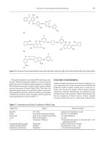
(see Fig. 13.1). The final two-step oxidation of hypoxanthine is
catalyzed by one and the same enzymes, xanthine oxidase (EC
1.1.3.22). The oxidation requires a reintroduction of oxygen in
the otherwise anaerobic pathway, which often limits the extent
of the xanthine oxidase reaction and causes inosine and hypoxanthine to accumulate. Hypoxanthine is an indicator of spoilage
and may contribute to bitter taste (Hughes and Jones 1966).
The hydrogen peroxide produced by the oxidation of hypoxanthine can be expected to cause oxidation, but the technological
Figure 13.1. The successive reactions of the main pathway for nucleotide degradation in fish. Substances listed below the dotted line are
generally considered to be indicators of spoilage. Pi is inorganic phosphate.
P1: SFK/UKS
BLBS102-c13
P2: SFK
BLBS102-Simpson
March 21, 2012
13:15
Trim: 276mm X 219mm
Printer Name: Yet to Come
251
13 Seafood Enzymes
implications of this are minor, since it is only after the quality of
the fish has declined beyond the level of acceptability that these
reactions take place on a large scale.
The relation between the accumulation of breakdown products
of ATP and the postmortem storage period of seafood products
has resulted in various freshness indicators being defined. Saito
et al. (1959) defined a freshness indicator K as a simplified way
of expressing the state of nucleotide degradation by a single
number:
K = 100 ×
[Inosine] + [Hypoxanthine]
[ATP] + [ADP] + [AMP] + [IMP]
+ [Inosine] + [Hypoxanthine]
Higher K values correspond to a lower quality of fish. The K
value often increases linearly during the first period of storage on
ice, but the K value at sensory rejection differs between species.
An alternative and simpler Ki value correlates well with the K
value of wild stock fish landed by traditional methods (Karube
et al. 1984):
Ki = 100 ×
[Inosine] + [Hypoxanthine]
[IMP] + [Inosine] + [Hypoxanthine]
Both K values reflect fish quality well, provided the numerical
values involved are not compared directly with the values from
other species. Still, K values necessarily remain less descriptive
than the concentrations of the degradation products themselves.
ATP is an important regulator of biochemical processes in
all animals and continues to be so during the postmortem processes. One of the most striking postmortem changes is that of
rigor mortis. When the concentration of ATP is low, the contractile filaments lock into each other and cause the otherwise
soft and elastic muscle tissue to stiffen. Rigor mortis is thus a
direct consequence of ATP depletion. The biochemical basis and
physical aspects of rigor mortis are reviewed by Hultin (1984)
and Foegeding et al. (1996).
ATP depletion and the onset of rigor mortis in fish are highly
correlated with the residual glycogen content at the time of death
and with the rate of ATP depletion. The onset of rigor mortis
may occur only a few minutes after death or may be postponed
for up to several days when rested harvest techniques, gentle
handling, and optimal storage conditions are employed (Azam
et al. 1990, Lowe et al. 1993, Skjervold et al. 2001). The strength
of rigor mortis is greater when it starts soon after death (Berg
et al. 1997). Although the recommended temperature for chilled
storage is generally close to 0◦ C, in some fish from tropical
and subtropical waters, the degradation of nucleotides has been
found to be slower, and the onset of rigor mortis to be further
delayed when the storage temperature is elevated to 5◦ C, 10◦ C,
and even 20◦ C (Saito et al. 1959, Iwamoto et al. 1987, 1991).
Rigor mortis impedes filleting and processing of fish. In the
traditional fishing industry, therefore, filleting is often postponed
until rigor mortis has been resolved. In aquaculture, however, it
has been shown that rested harvesting techniques can postpone
rigor mortis sufficiently to allow prerigor filleting to be carried
out (Skjervold et al. 2001). Fillets from prerigor filleted fish
shorten, in accordance with the rigor contraction of the muscle
fibers, by as much as 8%. Prerigor filleting has few problems with
gaping, since the rigor contraction of the fillet is not hindered by
the backbone (Skjervold et al. 2001). Gaping, which represents
the formation of fractures between segments of the fillets, is
described further in the Section Postmortem Proteolysis in Fresh
Fish.
All enzyme reactions are virtually stopped during freezing at
low temperatures. The ATP content of frozen prerigor fish can
thus be stabilized. During the subsequent thawing of prerigor
fish muscle, the leakage of Ca2+ from the organelles results in
a very high level of myosin Ca2+ –ATPase activity and a rapid
consumption of ATP. This leads to a strong form of rigor mortis
called thaw rigor. Thaw rigor results in an increase in drip loss, in
flavor changes, and in a dry and tough texture (Jones 1965) and
gaping (Jones 1969). Thaw rigor can be avoided by controlled
thawing, holding the frozen products at intermediate freezing
temperatures above −20◦ C for a period of time (Mcdonald and
Jones 1976, Cappeln et al. 1999). During this holding period,
ATP is degraded at moderate rates, allowing a slow onset of rigor
mortis in the partially frozen state.
Rigor mortis is a temporary condition, even though in the
absence of ATP, the actin-myosin complex is locked. The resolution of rigor mortis is due to structural decay elsewhere in the
muscular structure, as will be discussed later.
ENZYMATIC DEGRADATION OF
TRIMETHYLAMINE-N-OXIDE
The substance TMAO is found in all marine seafood species
but occurs in some freshwater fish as well (Anthoni et al. 1990,
Parab and Rao 1984, Niizeki et al. 2002). It contributes to cellular osmotic pressure, as previously described, but several other
physiological functions of TMAO have also been suggested.
TMAO itself is a harmless and nontoxic constituent, yet it forms
a precursor of undesirable breakdown products. In seafood products, TMAO can be degraded enzymatically by two alternative
pathways as described later.
The Trimethylamine-N-Oxide
Reductase Reaction
Many common spoilage bacteria reduce TMAO to trimethylamine (TMA) by means of the enzyme TMAO reductase (EC
1.6.6.9):
CH3
O=N CH3
CH3
CH3
NADH→N CH3
CH3
NAD
H2O
.
TMA has a strong fishy odor, and TMAO reductase activity is responsible for the typical off-odor of spoiled fish. Since
TMAO reductase is of microbial origin, the formation of TMA
occurs primarily under conditions such as cold storage that allow microbial growth to take place. TMA is thus an important
spoilage indicator of fresh seafood products. The formation of
P1: SFK/UKS
BLBS102-c13
P2: SFK
BLBS102-Simpson
March 21, 2012
13:15
Trim: 276mm X 219mm
252
Printer Name: Yet to Come
Part 2: Biotechnology and Enzymology
TMA is further discussed in the food microbiology literature
(e.g., Barrett and Kwan 1985, Dalgaard 2000).
The Trimethylamine-N-Oxide
Aldolase Reaction
TMAO is also the precursor of the formation of dimethylamine
(DMA) and formaldehyde:
O
=
HC
.
CH3
CH3
O = N CH3 → H N CH3
CH3
H
This reaction is catalyzed by trimethylamine-N-oxide aldolase
(TMAOase or TMAO demethylase, EC 4.1.2.32) but may also
to some extent be nonenzymatic, catalyzed by iron and various
reductants (Vaisey 1956, Spinelli and Koury 1981, Nitisewojo
and Hultin 1986, Kimura et al. 2002). In most cases in which
significant amounts of formaldehyde and DMA are accumulated, however, species possessing TMAOase enzyme activity
are involved. The reaction leads to cleavage of a C-N bond and
the elimination of an aldehyde, resulting in the classification of
TMAOase as a lyase (EC 4.1.2.32) and the International Union
of Biochemistry and Molecular Biology name aldolase.
DMA is a reactive secondary amine with a milder odor than
TMA. Formaldehyde is highly reactive and strongly affects the
texture of fish meat by making it tougher, harder, more fibrous,
and less juicy, as well as increasing the drip loss in the thawed
products. The quality changes are associated with a loss in protein solubility and in particular the solubility of the myofibrillar
proteins. For reviews, see Sikorski and Kostuch (1982), Hultin
(1992), Mackie (1993), Sikorski and Kolakowska (1994), and
Sotelo et al. (1995).
Formaldehyde can react with a number of chemical groups,
including some protein amino acid residues and terminal amino
groups, resulting in denaturation and possibly in the crosslinking of proteins, both of which are believed to be the cause
of the observed effects on seafood products. The formaldehyde
concentration in severely damaged seafood products may reach
240 µg/g (Nielsen and Jørgensen 2004). These concentrations
are generally considered nontoxic, but may still exceed various
national trade barrier limits.
Whereas TMAO is widespread, TMAOase is only found in a
limited number of animals, many of which belong to the order
of gadiform fish (pollock, cod, etc.). In gadiform species, the
highest levels of enzyme activity are found in the inner organs
(kidney, spleen, and intestine), while the enzyme activity in the
large white muscle is low. The formation of DMA and formaldehyde in various tissues of certain nongadiform fish, as well as of
crustaceans and mollusks, has also been reported, as reviewed
by Sotelo and Rehbein (2000), although the taxonomic distribution of the enzyme has not been investigated systematically.
The TMAOase content of gadiform fish exhibits large individual
variation, probably due to the influence of biological factors that
have not yet been adequately studied (Nielsen and Jørgensen
2004).
The enzyme is stable and tolerates both high salt concentrations and freezing. The accumulation of DMA and formaldehyde progresses only slowly, and most of the accumulation is
produced during prolonged frozen storage or cold storage of
the salted fish (e.g., salted cod and bacalao). During freezing,
the enzymatic reaction proceeds as long as liquid water and
substrate are available. In practice, the formation of DMA and
formaldehyde is found at temperatures down to approximately
−30◦ C (Sotelo and Rehbein 2000). At higher temperatures, the
rate of formation of formaldehyde is also higher; thus, freezing
of gadiform fish at insufficiently low temperatures may result in
dramatic changes in the solubility of myofibrillar proteins within
a few weeks (Nielsen and Jørgensen 2004).
Despite the TMAOase concentration of the white muscle being low, it is the enzyme activity of the white muscle that is
responsible for the accumulation of formaldehyde in whole fish
and in fillets (Nielsen and Jørgensen 2004). In the case of minced
products, even minor contamination by TMAOase-rich tissues
leads to a marked rise in the rate of accumulation (Dingle et al.
1977, Lundstrøm et al. 1982, Rehbein 1988, Rehbein et al. 1997).
Despite its dependency upon other factors, such as cofactors, the
rate of accumulation of formaldehyde can to a large extent be
predicted from the TMAOase enzyme activity of fish meat alone
(Nielsen and Jørgensen 2004).
The physiological function of TMAOase remains unknown.
The formation of formaldehyde could have a digestive function, yet this would not explain the high TMAOase content of
the kidney and spleen. Accordingly, it has been speculated that
TMAO may not even be its primary natural substrate (Sotelo
and Rehbein 2000).
The in vitro reaction rate is low without the presence of a number of redox-active cofactors. Although the overall TMAOase
reaction is not a redox reaction, its dependency on redox agents
shows that the reaction mechanism must include redox steps.
Little is known about the in vivo regulation of TMAOase,
but studies suggest the importance of nucleotide coenzymes
and iron (Hultin 1992). TMAOase appears to be membrane
bound and is only partly soluble in aqueous solutions. This has
complications for its purification and is one of the main reasons
why the complete characterization of the enzyme remains yet to
be done.
POSTMORTEM PROTEOLYSIS
IN FRESH FISH
Postmortem proteolysis is an important factor in many changes
in seafood quality. During cold storage, the postmortem proteolysis of myofibrillar and connective tissue proteins contributes
to deterioration in texture. Despite the natural tenderness of
seafood, texture is an important quality parameter, in fish
and shellfish alike. Deterioration in seafood quality through
proteolysis involves a softening of the muscle tissue. This reduces the cohesiveness of the muscle segments in fillets, which
promotes gaping, a formation of gaps and slits between muscle
segments. The negative character of this effect contrasts with
the effect of postmortem proteolysis on the meat of cattle and
P1: SFK/UKS
BLBS102-c13
P2: SFK
BLBS102-Simpson
March 21, 2012
13:15
Trim: 276mm X 219mm
Printer Name: Yet to Come
13 Seafood Enzymes
253
Figure 13.2. Gross anatomy of fish muscle. (Drawing courtesy of A. S. Matforsk, Norwegian Food Research Institute.)
pigs, in which the degradation of myofibrillar proteins produces
a highly desired tenderization in the conversion of muscle
to meat.
The proteolytic enzymes that cause a softening of seafood are
of basically the same classes as those found in terrestrial animals. The special effects of proteolysis on seafood result from
the combined action of the homologous proteolytic enzymes on
the characteristic muscle structures of seafood. The muscle of
fish differs on a macrostructural level from that of mammals, fish
muscle being segmented into muscle blocks called myotomes.
A myotome consists of a single layer of muscle fibers arranged
side by side and separated from the muscle fibers of the adjacent myotomes by collagenous sheets termed the myocommata
(Bremner and Hallett 1985). See Figure 13.2.
A myotome resembles a mammalian skeletal muscle in terms
of its intracellular structure (see Fig. 13.3) and the composition
of the extracellular matrix. Each muscle fiber is surrounded
by fine collagen fibers, the endomysium, joined with a larger
network of collagen fibers, the perimysium, which is contiguous
with the myocommata (Bremner and Hallet 1985). Although the
endomysium, perimysium, and myocommata are considered to
be discrete areas of the extracellular matrix, they join to form a
single weave.
Such phenomena in seafood as softening, gaping, and the
resolution of rigor are believed to be caused by hydrolysis of the
myofibrillar and the extracellular matrix proteins. Initially, the
disintegration of the attachment between the myocommata and
muscle fibers leads to the resolution of rigor (Taylor et al. 2002).
Endogenous proteolytic enzymes that cleave muscle proteins
under physiological conditions and at neutral pH can be a factor
in the resolution of rigor mortis. The continuation of this process
is regarded as one of the main causes of gaping (Taylor et al.
2002, Fletcher et al. 1997).
The softening of fish muscle appears to be the result of multiple changes in muscle structure. Histological studies have shown
that the attachment between muscle fibers in fish muscle is broken during ice storage and has been associated with a loss of
“hardness” as measured by instrumental texture analysis (Taylor
et al. 2002). During cold storage, the junction between the myofibrils and connective tissue of the myocommata is hydrolyzed
(Bremner 1999, Fletcher et al. 1997, Taylor et al. 2002), and the
collagen fibers of the perimysium surrounding the bundles of
muscle fibers are degraded (Ando et al. 1995, Sato et al. 2002).
It appears that cleavage of the costameric proteins that link
the myofibrils (the sarcomere) with the sarcolemma (the cell
membrane), and of the basement membrane, which is attached
Figure 13.3. A longitudinal view of the intracellular structure of a skeletal muscle fiber showing the contractile elements (actin and myosin),
the cytoskeleton, and the attachment to the extracellular matrix (endomysium and perimysium). (Adapted from Lødemel 2004.)
P1: SFK/UKS
BLBS102-c13
P2: SFK
BLBS102-Simpson
March 21, 2012
13:15
254
Trim: 276mm X 219mm
Printer Name: Yet to Come
Part 2: Biotechnology and Enzymology
to the fine collagen fibers of the endomysium (see Fig. 13.3),
leads to muscle fibers being detached, which results in softening.
Studies have shown that the costameric proteins found in fish
muscle are already degraded 24 hours postmortem (Papa et al.
1997). This emphasizes the importance of early postmortem
changes.
It has been shown that changes in the collagen fraction of
the extracellular matrix and the softening of fish are related.
Collagen V, a minor constituent of the pericellular connective
tissue of fish muscle, becomes soluble when rapid softening
takes place, whereas no solubilization is observed in fish that
do not soften (Sato et al. 2002). Recent studies also indicate
a degradation of the most abundant collagen in fish muscle,
collagen I, during 24 hours of cold storage (Shigemura et al.
2004). This indicates that the degradation of collagen and the
resulting weakening of the intramuscular pericellular connective
tissue also play a role in the textural changes in fish muscle that
occur early postmortem.
A number of studies have revealed structural changes in the
myofibrillar proteins in fish muscle during cold storage. Analysis of myofibrillar proteins from the muscle of salmon stored at
0◦ C for as long as 23 days has shown that several new protein
fragments form (Lund and Nielsen 2001). Other studies suggest
that predominantly proteins of the cytoskeletal network, such
as the high molecular weight proteins titin and nebulin, are degraded (Busconi et al. 1989, Astier et al. 1991). The extent of
degradation of the muscle myofibrillar proteins varies among
species. It has been found, for example, that the intermediate filament protein desmin is clearly degraded during the cold storage
of turbot and sardines but that no degradation occurs during the
cold storage of sea bass and brown trout (Verrez-Bagnis et al.
1999).
As research shows, both myofibrillar and extracellular matrix
proteins in the muscle of many fish are degraded during storage,
and textural changes can be expected to result from degradation
of proteins from both structures.
Proteases in Fish Muscle
The structural and biochemical changes just described can be
considered to largely represent the concerted action of different endogenous proteolytic enzymes. Proteolytic enzymes of all
major classes have been documented in the muscle of various
fish species. An overview of the different proteases believed to
play a role in the postmortem proteolysis of seafood is presented
later. Further information on the proteases found in fish and marine invertebrates can be found in reviews by Kolodziejska and
Sikorski (1995, 1996).
Matrix Metalloproteinases
Matrix metalloproteinases are extracellular enzymes involved in
the in vivo catabolism (degradation) of the extracellular matrix
(degradation of the helical regions of the collagens). They have
been isolated from rainbow trout (Saito et al. 2000), Japanese
flounder (Kinoshita et al. 2002) and Pacific rockfish (Bracho and
Haard 1995). Collagenolytic and gelatinolytic activities have
also been detected in the muscle of winter flounder (Teruel and
Simpson 1995), yellowtail (Kubota et al. 1998), ayu (Kubota
et al. 2000), salmon, and cod (Lødemel and Olsen 2003). Type
I collagen is solubilized and degraded by matrix metalloproteinases in rainbow trout (Saito et al. 2000), Japanese flounder
(Kubota et al. 2003), and Pacific rockfish (Bracho and Haard
1995), suggesting that these proteases can participate in postmortem textural changes.
Cathepsins
Lysosomal proteinases such as cathepsins B, D, and L have
been isolated from a number of fish species, including herring
(Nielsen and Nielsen 2001), mackerel (Aoki and Ueno 1997),
and tilapia (Jiang et al. 1991). In vitro studies show that certain
cathepsins are capable of cleaving myofibrillar proteins (Ogata
et al. 1998, Nielsen and Nielsen 2001) and may also participate in
degradation of the extracellular matrix since they can cleave both
nonhelical regions of the collagen (Yamashita and Konagaya
1991) and collagen that has already been partly degraded by
matrix metalloproteinases. Since cathepsins in the muscle of
living fish are located in the lysosomes, they are not originally
in direct contact with either the myofibrils or the extracellular
matrix, although it has been shown that the enzymes leak from
the lysosomes in fish muscle postmortem, and from lysosomes in
bovine muscles postmortem as well (Geromel and Montgomery
1980, Ertbjerg et al. 1999).
Calpains
Calpains have been reported in the muscle of various fish species
(Wang and Jiang 1991, Watson et al. 1992). They are only active
in the neutral pH range, although research shows that they also
remain active at the slightly acidic postmortem pH (Wang and
Jiang 1991) and are capable of degrading myofibrillar proteins
in vitro (Verrez-Bagnis et al. 2002, Geesink et al. 2000). This
suggests a participation in the postmortem hydrolysis of fish
muscle. Watson et al. (1992) found evidence for calpains being
involved in the degradation of myofibrillar proteins in tuna, leading to the muscle becoming pale and grainy, which is referred to
as “burnt tuna.”
20S Proteasome
The 20S proteasome enzyme is a 700 kDa multicatalytic proteinase with three major catalytic sites. The term 20S refers to
its sedimentation coefficient. In the eukaryotic cells, the enzyme
exists in the cytoplasm, either in a free state or associated with
large regulatory complexes. In vivo, it is involved in nonlysosomal proteolysis and apoptosis. Research strongly indicates
that this proteasome is involved in meat tenderization in cattle
(Sentandreu et al. 2002). Although proteasomes have been
detected in the muscle of such fish species as carp (Kinoshita
et al. 1990a), white croaker (Busconi et al. 1992), and salmon
(Stoknes and Rustad 1995), their postmortem activity in fish
muscle has not been clarified.