Fundamental & Advanced Nursing Skills_2 pptx
Bạn đang xem bản rút gọn của tài liệu. Xem và tải ngay bản đầy đủ của tài liệu tại đây (22.35 MB, 772 trang )
Skill 6-7 Applying a Condom Catheter
Skill 6-8 Inserting an Indwelling
Catheter: Male
Skill 6-9 Inserting an Indwelling
Catheter: Female
Skill 6-10 Routine Catheter Care
Skill 6-11 Obtaining a Residual Urine
Specimen from an
Indwelling Catheter
Skill 6-12 Irrigating a Urinary Catheter
Skill 6-13 Irrigating the Bladder Using
a Closed-System Catheter
Skill 6-14 Removing an Indwelling
Catheter
Skill 6-15 Catheterizing a Noncontinent
Urinary Diversion
Skill 6-16 Maintaining a Continent
Urinary Diversion
Skill 6-17 Pouching a Noncontinent
Urinary Diversion
Skill 6-18 Administering Peritoneal
Dialysis
Skill 6-19 Administering an Enema
Skill 6-20 Digital Removal of Fecal
Impaction
Skill 6-21 Inserting a Rectal Tube
Skill 6-22 Irrigating and Cleaning a
Stoma
Skill 6-23 Changing a Bowel Diversion
Ostomy Appliance: Pouching
a Stoma
Skill 6-1 Inserting and Maintaining a
Nasogastric Tube
Skill 6-2 Assessing Placement of a
Large-Bore Feeding Tube
Skill 6-3 Assessing Placement of a
Small-Bore Feeding Tube
Skill 6-4 Removing a Nasogastric
Tube
Skill 6-5 Feeding and Medicating Via
a Gastrostomy Tube
Skill 6-6 Maintaining Gastrointestinal
Suction Devices
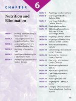
Nutrition and
Elimination
CHAPTER 6
6
645
646
> ASSESSMENT
1. Assess client’s consciousness level to determine
the ability of the client to cooperate during the
procedure.
2. Check the client’s chart for any previous medical
history of nostril surgery or injury or unusual nos-
tril bleeding. Reduces risk of injury from the tube.
3. Use a penlight to assess nostrils for a deviated sep-
tum. Facilitates choice of nostril and size of tube.
4. Ask the client to breathe through each nostril oc-
cluding the other with a finger. Facilitates choice
of nostril and decreases chance that tube will in-
terfere with respirations.
> DIAGNOSIS
1.1.2.2 Altered Nutrition: Less Than Body
Requirement
6.5.1.1 Swallowing Impairment
1.6.1.4 Risk for Aspiration
1.3.1.2 Risk for Diarrhea
1.6.2.1.1 Altered Oral Mucous Membranes
1.4.1.2.2.1 Risk for Fluid Volume Deficit
9.1.1 Pain
1.6.2.1.2.1 Impaired Skin Integrity
> PLANNING
Expected Outcomes:
1. Client’s nutritional status will improve, as indi-
cated by increased body weight, physical strength,
and mental status.
2. Client’s nutritional needs will be met with the as-
sistance of tube feeding.
Inserting and
Maintaining a
Nasogastric Tube
Hsin-Yi Tang, RN, MS, and Jung-Chen Chang, RN, MN
SKILL 6-1
SKILL 6-1
Decompression
Double lumen
Gastric content
Gastrointestinal
surgery
Levin’s tube
Nasogastric tube
Peptic ulcer
Salem sump tube
Single lumen
Tube feeding
KEY TERMS
> OVERVIEW OF THE SKILL
Nasogastric (NG) tubes are used for several purposes,
including feeding for nutrition when the client is co-
matose, semiconscious, or unable to consume sufficient
nutrition orally. Nasogastric suction tubes are used for
decompression of gastric content after gastrointestinal
surgery, and to obtain gastric specimens for diagnosis of
peptic ulcer. Tubes are used for irrigation to clean and
flush the stomach after oral ingestion of poisonous sub-
stances. Finally, NG tubes are used to document the
presence of blood in the stomach, monitor the amount
of bleeding from the stomach, and identify the recur-
rence of bleeding in the stomach.
The two most commonly used NG tubes are the
single lumen Levin’s tube, and the double lumen
Salem sump tube.
The gastrointestinal tract is considered to be a
clean area rather than a sterile one. The procedure to
place an NG tube is performed using clean technique
unless it is performed in conjunction with gastroin-
testinal surgery.
3. Client will maintain a patent airway, as evidenced
by absence of coughing, no shortness of breath,
and no aspiration.
4. Client will not have diarrhea due to nasogastric
feeding.
5. Mouth mucous membranes will remain moist and
intact.
6. Client will maintain a normal fluid volume, as evi-
denced by good skin texture, muscle tone, and
blood volume.
7. Client’s comfort level will increase.
8. Skin around the tube will remain intact, with no
redness or blisters.
Equipment Needed:
• Nasogastric tube: adult, 14 to 18 French; child/
infant, 5 to 10 French; single lumen (Levin’s sump):
feeding; double lumen (Salem sump tube): feeding,
suction, irrigation (see Figure 6-1-2)
• Water-soluble lubricant
• Syringe with catheter tip or adapter, 20-50 ml
• Glass of tap water with straw, or ice
• Towel or tissue
• Emesis basin with ice chips
• Tongue blade
• pH chemstrip
• Stethoscope
• Disposable gloves (nonsterile), goggles, gown
• Hypoallergenic tape, rubber band, and safety pin
• Penlight or flashlight
• Disposable irrigation set (if needed)
• Wall mount or portable suction equipment (if needed)
• Administration set with pump or controller for
feeding tube
> CLIENT EDUCATION NEEDED:
1. Inform the client of the purpose of the NG tube.
2. Explain the procedure of insertion and any ex-
pected discomfort.
3. Establish and clarify a “hand signal” to indicate the
need to temporarily stop the NG insertion.
4. Explain how the client can cooperate during tube
insertion, especially by swallowing water when
asked to do so.
5. Explain potential complications, such as diarrhea,
mouth dryness, and nostril irritation.
6. Review the skills and procedures of maintaining
tube.
7. Instruct to chew on ice chips to satisfy the basic
need to eat (if there is no fluid intake restriction).
8. Encourage physical activity to enhance gastroin-
testinal mobility (if there is no activity restriction).
9. If a client with dentures is conscious, encourage to
wear the dentures to maintain the normal shape of
oral cavity.
SKILL 6-1 Inserting and Maintaining a Nasogastric Tube 647
Estimated time to complete the skill:
15–20 minutes
Figure 6-1-2 Double-lumen nasogastric tube
IMPLEMENTATION—ACTION/RATIONALE
ACTION RATIONALE
1. To assess for any nostril surgery and abnormal
bleeding.
2. Decreases anxiety and promotes cooperation.
1. Review client’s medical history.
2. Assess client’s consciousness and ability to un-
derstand. Explain the procedure and develop a
hand signal (see Figure 6-1-3).
continues
648 CHAPTER 6 Nutrition and Elimination
3. Facilitates an efficient procedure.
4. Facilitates insertion and prevents back strain.
5. Practices clean technique.
6. Choosing the more patent nostril for insertion
decreases discomfort and unnecessary trauma.
3. Prepare the equipment, putting tissues, a cup
of water, and an emesis basin nearby (see Fig-
ure 6-1-4).
4. Prepare the environment; raise the bed and
place it in a high Fowler’s position (45 to
60 degrees). Cover the chest with a towel.
5. Wash hands and then put on gloves.
6. Use a penlight to view the client’s nostrils. As-
sess client’s nostrils with penlight and have the
client blow her nose one nostril at a time (see
Figure 6-1-5).
7. Using the NG tube, measure the distance from
the bridge of the nose to the earlobe and then
to the xiphoid process of the sternum and
mark this distance on the tube with a piece of
tape (see Figure 6-1-6).
8. Lubricate first 4 inches of the tube with water-
soluble lubricant.
9. Ask the client to slightly flex the neck backward.
7. Determines the approximate amount of tube
needed to reach the stomach.
8. Facilitates passage into the naris.
9. Makes insertion easier.
Figure 6-1-4 Put an emesis basin, cup with straw, and
tissues nearby.
Figure 6-1-3 Explain the procedure; demonstrate head
position and tube insertion.
Figure 6-1-5 Assess the client’s nostrils before intro-
ducing the nasogastric tube.
Figure 6-1-6 Measure the distance from nose to ear-
lobe to the xiphoid process to determine how much
tube will need to be inserted to reach the stomach.
SKILL 6-1 Inserting and Maintaining a Nasogastric Tube 649
10. Promotes passage of tube with minimal
trauma to mucosa.
10. Gently insert the tube into a naris (see Fig-
ure 6-1-7)
11. Ask the client to tip the head forward once the
tube reaches the nasopharynx. If the client
continues to gag, stop a moment.
12. Advance the tube several inches at a time as
the client swallows water or ice chips (see Fig-
ure 6-1-8).
13. Withdraw the tube immediately if there are
signs of respiratory distress.
14. Advance the tube until the taped mark is
reached (see Figure 6-1-9).
15. Split a 4-inch strip of tape lengthwise 2 inches.
Secure the tube with the tape by placing the
wide portion of the tape on the bridge of
the nose and wrapping the split ends around
the tube (see Figure 6-1-10).Tape to cheek as
well if desired (see Figure 6-1-11).
15. Prevents tube displacement.
11. Tipping the head forward facilitates passage of
the tube into the esophagus instead of the tra-
chea.Tube may stimulate gag reflex. Allows the
client to rest, reduces anxiety,and prevents
vomiting.
12. Assists in pushing the tube past the oropharynx.
13. Prevents trauma to bronchus or lung.
14. Enables the tube to reach the stomach.
continues
Figure 6-1-7 Gently insert the tube into the naris. Figure 6-1-8 Advance the tube slowly.The client swal-
lows small sips of water to assist in pushing the tube past
the oropharnyx.
Figure 6-1-9 Advance the tube until the taped mark is
at the opening of the naris.
Figure 6-1-10 Secure the tube to the nose.
650 CHAPTER 6 Nutrition and Elimination
16. Ensures correct placement. (A pH below 4 indi-
cates the tube is in the stomach; a pH range of
6–7 indicates intestinal sites.)
16. Check the placement of the tube:
• Attach the syringe to the end of the tube for
injecting 10 cc of air and auscultate over the
epigastric area (upper left quadrant); see
Figure 6-1-12.
• Aspirate sample gastric content and mea-
sure with chemstrip pH (see Figure 6-1-13).
• Prepare the client for x-ray check-up, if
prescribed.
17. Connect the distal end of the tube to suction,
draining bag, or adapter (see Figure 6-1-14).
18. Secure the tube with rubber band and safety
pin to client’s gown or bed sheet.
19. Remove gloves, dispose of contaminated ma-
terials in proper container, and wash hands.
20. Position client comfortably and place the call
light in easy reach.
21. Document procedure.
17. Establishes an appropriate pathway for
intervention.
18. Enhances the level of comfort and secures the
tubing system.
19. Implements the principles of infection control.
20. Decreases client’s anxiety and provides access
to help if needed.
21. Records implementation of intervention and
promotes continuity of care.
Figure 6-1-11 Tape the tube to the cheek as well, if de-
sired, to provide extra support.
Figure 6-1-12 Auscultate over the epigastric area.
Figure 6-1-13 Aspirate a sample of gastric content to
check for pH.
Figure 6-1-14 Connect the distal end of the tube to
suction or drainage to complete the procedure.
SKILL 6-1 Inserting and Maintaining a Nasogastric Tube 651
22. Reduces the transmission of microorganisms.
23. Prevents complications from dislocation of the
tube.
24. Prevents complications from the loss of benefi-
cial effects from the tube.
25. Rotation or irrigation may disturb incisions.
26. Enhances client’s comfort and the integrity of
skin and nose mucosa.
27. Reduces the transmission of microorganisms.
Maintaining a Nasogastric Tube
22. Wash hands and apply gloves.
23. Follow the steps in Action 16 to check the
proper tubing position before instilling any-
thing per NG tube or at least every 8 hours.
24. Assess for signs that the tube has become
blocked, including epigastric pain and vomit-
ing, and/or the inability to pass medications or
feedings through the tube.
25. Remember never to irrigate or rotate a tube
that has been placed by the physician or quali-
fied practitioner during gastric or esophageal
surgery.
26. Provide oral hygiene and assist client to clean
nares daily.
27. Remove gloves, dispose of contaminated ma-
terials in proper container, and wash hands.
▼ REAL WORLD ANECDOTES
Mr. Klotz had just been admitted to the hospital with severe abdominal distention. NG tube
placement was ordered for abdominal decompression. Mr. Klotz was not to have any fluids by mouth
but he could have ice chips. The nurse provided Mr. Klotz with ice chips and instructed him to suck
on a few chips and swallow as she inserted the NG tube. The nurse inserted the NG tube into Mr.
Klotz’s right naris but was unable to advance the tube any further than an inch. After several
> EVALUATION
• Client’s nutritional status improves, as indicated by
increased body weight, physical strength, and men-
tal status.
• Client’s nutritional needs are met with the assis-
tance of tube feeding.
• Client maintains a patent airway, as evidenced by
absence of coughing, no shortness of breath, and no
aspiration.
• Client does not have diarrhea due to nasogastric
feeding.
• Mouth mucous membranes remain moist and
intact.
• Client maintains a normal fluid volume, as evi-
denced by good skin texture, muscle tone, and
blood volume.
• Client’s comfort level increases.
• Skin around the tube remains intact, with no red-
ness or blisters.
> DOCUMENTATION
Nurses’ Notes
• Document the type of NG tube inserted, the naris
used, how the client tolerated the procedure, and
the methods used to verify placement.
• Document care provided to the client to increase
comfort of the NG insertion naris.
• Note any unusual findings.
Intake and Output Record
• Note the amount of fluid the client drank to aid in-
sertion of the NG tube.
• Note the amount of gastric contents removed for
testing.
continues
> CRITICAL THINKING SKILL
Introduction
Nurses must be able to evaluate the effectiveness of NG
tube insertion, maintenance, or removal.
Possible Scenario
The family of your home care client has been assisting
in her care, including the care of her feeding tube. You
have educated them on the tube and its placement. Al-
though they state they secured the tube in a proper
place and the end of the tube is currently positioned
higher than the stomach, you observe the tube is filled
with gastric content.
Possible Outcome
Client has a continuous risk for infection, electrolyte
imbalance, and potential aspiration.
Prevention
Assess that the caregiver is properly securing the end of
the tube at a level higher than the stomach. Assess the
client’s vital signs and respiratory pattern for infection,
electrolyte imbalance, or aspiration. Reeducate the care-
givers on assessing for correct tube placement, and re-
view with them common situations where the tube
might move.
652 CHAPTER 6 Nutrition and Elimination
▼ REAL WORLD ANECDOTES continued
attempts to advance the tube, the nurse tried Mr. Klotz’s left naris. It required several gentle attempts
and lots of lubricant to pass the tube into the nasopharynx, the nurse was finally able to advance the
tube into Mr. Klotz’s stomach. After Mr. Klotz had received some relief from his distention, he did
mention to the nurse that he had broken his nose many years earlier.
▼ VARIATIONS
Geriatric Variations:
• For elderly clients who wear dentures, oral hygiene and denture care should not be overlooked
simply because an NG tube is in place.
Pediatric Variations:
• Dispose of, or securely tape any small parts, such as plastic connectors or plugs, to prevent small
children from accidentally aspirating or swallowing them.
Home Care Variations:
• Periodically assess the family member’s ability to check the placement of the tube, check residual
gastric contents, administer tube feedings, or connect the tube properly with suction.
Long-Term Care Variations:
• Teach family members or caregivers to assess client’s nutritional status and assess for any sign of
complications related to the NG tube.
▼COMMON ERRORS—ASK YOURSELF
Possible Error:
The nurse is unable to auscultate air bubbles but assumes the NG tube is in place anyway.
Ask Yourself:
How do I prevent this error?
> NURSING TIPS
• Adjust the height of the bed to eliminate back strain.
• Prepare the split tape before putting on gloves.
• This can be an anxiety-provoking procedure. Good
communication skills decrease anxiety and promote
the client’s cooperation.
• The size of the NG tube used depends on client size,
client history of damage to the structure of the
nose, and the purpose of the procedure.
• Tincture of Benzoine may be used to prep the skin.
This acts as an adhesive as well as a skin prep.
• Carefully observe client’s verbal and nonverbal re-
sponses during the entire procedure.
• When feasible, engage family members or caregivers
to assist in NG tube insertion.
• Sump tubes should whistle continuously on low
suction.
SKILL 6-1 Inserting and Maintaining a Nasogastric Tube 653
▼COMMON ERRORS—ASK YOURSELF continued
Prevention:
If you are unable to verify NG tube position by auscultating air, use another method of verification. Attempt
to aspirate gastric contents. Place the end of the NG tube in a glass of water and check for air bubbles that
correspond to the client’s exhalations. If you are unable to verify NG tube placement, do not instill anything
through the tube. Notify the client’s qualified practitioner. Send the client for an x-ray to verify placement if
this is within institutional guidelines.
654
> ASSESSMENT
1. Check the physician’s or qualified practitioner’s
order for the type and size of feeding tube to en-
sure accurate placement of the correct tube.
2. Review the client’s medical record for a history of
prior tube use or displacement since recurring
tube displacement may increase the risk of pul-
monary placement.
3. Assess the client for signs and symptoms of inad-
vertent respiratory placement since coughing,
choking, and cyanosis may indicate placement of
the tube in an airway.
4. Assess the client for signs and symptoms that in-
crease the client’s risk of tube dislocation. Cough-
ing, retching, and nasotracheal suctioning may
cause the tube to become dislodged.
Assessing Placement of a
Large-Bore Feeding Tube
Kathy Lilleby, RN
SKILL 6-2
SKILL 6-2
Aspiration
Large-bore feeding
tube
Nasoduodenal tube
Nasogastric tube
PEG
PEJ
pH
KEY TERMS
> OVERVIEW OF THE SKILL
Clients who cannot take food or fluids orally may
require the placement of a feeding tube for enteral
nutrition. These clients may be unconscious, unable
to respond to the thirst reflex, unable to swallow,
or receiving a hyperosmotic enteral preparation.
The large-bore nasogastric feeding tube requires
a physician’s or qualified practitioner’s order to be
placed. The tube can be a firm, polyvinyl large-bore
tube or a soft, flexible polyurethane or silicone tube.
After insertion, the placement should be checked
by x-ray to determine that it is in the stomach or
in the intestine as ordered and not in an airway.
After the initial x-ray for placement, it is the
nurse’s responsibility to verify the tube’s position
before each intermittent feeding or medication or
once a shift if the client is receiving continuous
feedings.
There are several types of large-bore feeding tubes.
A nasogastric tube is for short-term use; the major
complication of its use is aspiration pneumonia. The
nasoduodenal tube is also used short term. There is less
risk of aspiration with this tube, since the tip is
weighted and rests in the duodenum. But it is also more
difficult to place, and some institutions require that a
physician or qualified practitioner insert this type of
tube. The gastrostomy tube (GT) is placed surgically
by laparoscopy for long-term use. The more common
percutaneous endoscopic gastrostomy (PEG) tube is
placed at the bedside under local anesthesia and con-
scious sedation. A PEG tube is used for long-term feed-
ings. The percutaneous endoscopic jejunostomy (PEJ)
tube may also be placed at the bedside by the physician
or qualified practitioner. It is more comfortable for the
client and carries minimal risk of aspiration.
> DIAGNOSIS
1.1.2.1 Altered Nutrition: More Than Body
Requirements
1.1.2.2 Altered Nutrition: Less Than Body
Requirements
1.4.1.2.2.2 Risk for Fluid Volume Deficit
1.6.1.4 Risk for Aspiration
6.5.1.1 Impaired Swallowing
> PLANNING
Expected Outcomes:
1. The tube will remain in place and intact.
2. The tube feeding or medication will infuse into
the client’s gastrointestinal (GI) tract.
3. The client will not experience any respiratory
distress.
4. The client will not experience any pain.
5. The client will be able to describe the reason for
checking the tube’s placement.
Equipment Needed (see Figure 6-2-2):
• Catheter tip syringe, 60 ml
• Stethoscope
• Gloves
• pH indicator strip
• Emesis basin
•Towel
> CLIENT EDUCATION NEEDED:
1. Tell the client the rationale for checking the place-
ment of the feeding tube.
2. Ask the client to tell you if they are having respira-
tory difficulties.
3. Instruct the caregiver how to check the tube
for correct placement before each feeding or
medication.
4. Provide written and oral instructions about how
to check for correct tube placement.
5. Tell the client and caregiver not to proceed with a
feeding if there is any doubt about the tube’s
proper placement.
SKILL 6-2 Assessing Placement of a Large-Bore Feeding Tube 655
Estimated time to complete the skill:
5 minutes
Figure 6-2-2 Stethoscope, syringe, and pH strips are used to
assess placement of the tube.
IMPLEMENTATION—ACTION/RATIONALE
ACTION RATIONALE
1. Ensures accurate placement of the tube.
2. Reduces the transmission of microorganisms.
3. This method is less reliable than checking for
gastric contents, but it is the simplest way to
assess for placement of the feeding tubes.
• Allows nurse to hear sound of air.
1. Check physician’s or qualified practitioner’s
order for the feeding tube.
2. Wash hands (see Figure 6-2-3). Apply gloves.
3. Assess placement of the tube by auscultation:
• Place stethoscope over left upper quadrant
of the abdomen.
• Quickly inject 10–20 ml air with the 60-ml
syringe (see Figure 6-2-4).
continues
656 CHAPTER 6 Nutrition and Elimination
• If resistance is felt, tells nurse to attempt to
aspirate GI contents.
• A whooshing or gurgling sound can be
heard as air enters the stomach.
4. Gastric contents have pH of 1–4. Intestinal con-
tents have pH of 6–7.
• Measure the pH of the gastric aspirate before
instilling anything through the feeding tube.
• The tube opening may be lying against the
gastric wall.
• To obtain accurate results.
• Assess for resistance.
• Listen for sound.
4. Measure pH of GI contents:
• Aspirate 10 cc of GI contents with 60-cc
syringe (see Figure 6-2-5).
• If unable to aspirate, reposition client on
side and try again.
• Measure pH of GI contents with pH indica-
tor strip.
5. Proceed with feeding and medication (see Fig-
ures 6-2-6 and 6-2-7). Continue to monitor the
client for discomfort.
6. Recheck tube placement following the tube
feeding.
• Flush tube with 30 cc warm water after med-
ication or tube feeding (see Figure 6-2-8).
• Wait 1 hour before testing pH.
5. To provide the client with nutrition and treat-
ment. Continuing to assess for signs of tube
displacement ensures client safety.
6. Continuing to assess for signs of tube displace-
ment ensures client safety.
• Flushes out residual formula or medication.
• Allows for digestion of the formula or assim-
ilation of the medication.
Figure 6-2-4 Inject 10-20 ml of air.Figure 6-2-3 Wash hands prior to beginning procedure.
Figure 6-2-5 Aspirate 10 cc of gastric contents to
check pH.
Figure 6-2-6 After verifying placement of the tube, pro-
ceed with feeding.
SKILL 6-2 Assessing Placement of a Large-Bore Feeding Tube 657
• Assesses placement of the feeding tube.
• Assesses placement of the feeding tube.
7. Reduces transmission of microorganisms.
• Flush tube with 30 cc air and auscultate for
sound as in Action 3.
• Aspirate 10 cc of GI contents and check for
pH as in Action 4.
7. Remove gloves and wash hands.
Figure 6-2-8 After feeding or administering medica-
tions, flush the tube with 30 cc of warm water to rinse
out residue.
Figure 6-2-7 Proceed with administering medications.
> EVALUATION
• The tube remains in place and intact.
• The tube feeding or medication is infusing into the
client’s GI tract.
• The client has not experienced any respiratory
distress.
• The client is not experiencing any pain.
• The client is able to describe the reason for checking
the tube’s placement.
> DOCUMENTATION
Nurses’ Notes
• Document the type of tube placed.
• Note the character of GI contents.
• Record the pH measurement.
• Document the assessment of air injected into stomach.
• Note any client complaints or unusual findings.
Intake and Output Record
• Record the amount of any fluid infused or removed
in the appropriate category.
▼ REAL WORLD ANECDOTES
Claudia, 18 years old, was comatose after a motor vehicle accident. She had a head injury and
broken clavicle, radius, and pelvis. After the accident she had been in the intensive care unit on a ven-
tilator for 5 days but later was transferred to a medical floor. She was breathing on her own but had
not regained consciousness. A large-bore nasogastric feeding tube had been placed for enteral feed-
ings and medication administration. The feedings were intermittent so the nurse began her assess-
ment of its placement before starting the feeding. When the nurse injected air, she heard the charac-
teristic gurgle, but when she started the feeding, Claudia started coughing. The nurse immediately
stopped the feeding and assessed her for respiratory distress. She notified the physician, who ordered
an x-ray. The tube was found to be looped with the end near Claudia’s bronchus. The physician repo-
sitioned the tube and obtained another x-ray, which showed it was in good position and the feeding
was restarted.
658 CHAPTER 6 Nutrition and Elimination
▼ VARIATIONS
Geriatric Variations:
• Elderly clients may have more fragile tissue that could be damaged with a large-bore feeding
tube.
• Older clients with other respiratory conditions are at increased risk for respiratory complications
if the feeding tube migrates to the pulmonary tree.
Pediatric Variations:
• Inject only 0.5 to 1.0 ml of air into a pediatric feeding tube.
• Be sure the child is quiet and calm while checking for placement so you can hear the air being
injected.
Home Care Variations:
• Assess the sanitation of the home to determine the client’s risk for infection.
• The caregiver should be taught the normal range of pH for GI contents.
• The caregiver should be taught the signs and symptoms of feeding tube displacement and what to
do if displacement is suspected.
Long-Term Care Variations:
• The staff should be taught the normal range of pH for GI contents.
• Equipment for verifying tube placement should be at the client’s bedside at all times.
• Staff members should be taught the signs and symptoms of tube displacement and to whom to re-
port the symptoms.
• Staff members should be taught how to discontinue a feeding if they suspect tube displacement.
> CRITICAL THINKING SKILL
Introduction
Displacement of a large-bore feeding tube may occur
when a client coughs, gags, or vomits. The nurse needs
to assess the client for any symptoms that may cause a
tube displacement.
Possible Scenario
Brian was 16 years old when he needed to have a tube
placed for feeding. He hated the feeling of the tube in
his throat and could not get used to it. He gagged and
coughed, sometimes so vigorously that he vomited.
When the nurse came to assess the tube placement, she
noted that the end of the tube was visible in the back of
Brian’s throat.
Possible Outcome
The nurse could see the tube was not in its proper posi-
tion and knew that it could not be repositioned. So she
gently removed the entire tube and notified the physi-
cian. He reevaluated Brian’s need for the tube, consid-
ered the risk of aspiration and Brian’s difficulty with the
tube, and decided to stop the tube feedings and to let
Brian try to eat on his own.
Prevention
Client teaching and reassurance are an important part
of maintaining a large-bore feeding tube in place.
Antiemetic or antianxiety medications may be needed
to help clients tolerate the tube if it is placed orally or
nasally.
▼COMMON ERRORS—ASK YOURSELF
Possible Error:
You do not wait 1 hour after the last tube feeding has finished to check for tube placement so the pH of the
aspirated GI contents is inaccurate.
> NURSING TIPS
• A muffled or faint sound of injected air may signal
that the tube is in the lungs.
• It may be necessary to inject air two or three times
in obese clients since the sound of injected air may
be faint.
• Do not withdraw or advance the tube into clients
who have had gastric resections or other abdominal
surgery as it could damage the suture lines and
cause hemorrhage.
SKILL 6-2 Assessing Placement of a Large-Bore Feeding Tube 659
▼COMMON ERRORS—ASK YOURSELF continued
Ask Yourself:
How do I prevent this error?
Prevention:
Plan a schedule for feeding, medications, and checking for tube placement so that an accurate measurement
will be obtained. Write this plan into the client’s plan of care so all staff can comply. If this error does occur,
flush the tube with 30 ml warm water. Wait 1 hour. Begin tube placement assessment again.
660
> ASSESSMENT
1. Assess client for any signs of respiratory distress
such as choking, coughing, shallow breathing, or
decreasing oxygen saturations. These symptoms
could be indicative of aspiration of the feeding
tube.
2. Check for a tape marker on the tube, near the
nose, which indicates the length of tube inserted.
If tube has become displaced, marker will be far-
ther away from nose.
3. Assess sputum for distinguishing features that
would indicate aspiration, such as blue color
(tube feeding formula is mixed with blue food
coloring to distinguish feeding from normal
white sputum). Blue sputum could signify
aspiration of feeding, which could lead to
pneumonia.
> DIAGNOSIS
1.1.2.2 Altered Nutrition: Less Than Body
Requirements
1.6.1.4 Risk for Aspiration
> PLANNING
Expected Outcomes:
1. The client’s feeding tube will be intact in the
ordered area of the GI tract.
2. The client will not experience aspiration sec-
ondary to tube feedings.
Equipment Needed:
• Syringe: 20 or 60 ml for adults, 5 or 10 ml for
pediatrics.
• Stethoscope
Assessing Placement of a
Small-Bore Feeding Tube
Nancy E. Chambers, RN, BSN
SKILL 6-3
SKILL 6-3
Enteral nutrition
Feeding tube
Gastric contents
Nasogastric tube
Nasointestinal tube
pH
KEY TERMS
> OVERVIEW OF THE SKILL
Clients with a small-bore feeding tube must have
placement of the tube verified at time of insertion
and every shift to prevent insertion/migration of the
tube into the esophagus, trachea, or lungs and aspira-
tion of feeding. Placement of a feeding tube is easy to
disrupt because the tubes are small, flexible, and se-
cured only with tape on the nose. There are three ef-
fective methods of verifying placement. The first
method is to inject air through the feeding tube and
simultaneously auscultate the air bubble over the
stomach. The second is to aspirate a sample of gastric
contents and check pH levels. Finally, the most pre-
cise way to verify placement is to obtain an abdomi-
nal x-ray.
• pH testing equipment (see Figure 6-3-2)
• Progress notes/flow sheets
> CLIENT EDUCATION NEEDED:
1. Explain reason for verifying placement.
2. Explain steps of procedure.
3. Answer questions from client/family.
4. Instruct client to notify staff immediately if
experiencing respiratory distress or blue
sputum.
5. Explain purpose of x-rays, if needed.
SKILL 6-3 Assessing Placement of a Small-Bore Feeding Tube 661
Estimated time to complete the skill:
5–30 minutes
Figure 6-3-2 Equipment used to test pH
IMPLEMENTATION—ACTION/RATIONALE
ACTION RATIONALE
1. Practices clean technique.
2. Promotes efficiency and speed.
3. Prevents spilling of feeding.
1. Wash hands and apply clean gloves.
2. Prepare equipment, put pH testing equipment
nearby.
3. Clamp the tube feeding infusion if it has al-
ready been running (see Figure 6-3-3).
4. Locate the connection between the feeding
tube and feeding bag tubing (see Figure 6-3-4).
5. Disconnect infusion tubing from feeding tube
and attach a cap to tubing and feeding tube
(see Figure 6-3-5).
6. Draw 10–20 ml of air into syringe.
7. Attach syringe to proximal end of feeding tube.
4. To disconnect the tubing.
5. Prevents contamination of tubing.
6. Provides enough air to hear an air bubble as it
is inserted.
7. Allows for insertion of air.
continues
Figure 6-3-4 Find the connection between the feeding
tube and feeding bag tubing.
Figure 6-3-3 Clamp the tube feeding infusion.
662 CHAPTER 6 Nutrition and Elimination
8. Facilitates accurate auscultation.
9. Facilitates auscultation of air rush.
10. Air bubbles may be difficult to hear due to
client position or gastric contents.
11. To provide gastric contents for visual inspec-
tion and pH testing.
12. The pH of the fluid aspirate can help to verify
tube placement.
• The pH reading can be altered by the pres-
ence of medication or formula, so pH should
be tested after the client’s stomach has
been empty for approximately 1 hour.
8. Place diaphragm of stethoscope in epigastric,
area over stomach: upper left quadrant near
midline.
9. Inject air quickly into feeding tube and listen
for air rush.
10. If unsuccessful in hearing rush of air, repeat Ac-
tions 6 to 9. It may be necessary to reposition
stethoscope over stomach, use more air,or in-
ject more slowly.
11. While syringe is connected to feeding tube, as-
pirate approximately 20 ml of gastric contents
(see Figure 6-3-6).
12. Check the contents and obtain pH level (see
Figures 6-3-7 and 6-3-8).
• pH below 4 means tube is in stomach.
• pH range of 6–7 means tube is in intestine.
13. Gastric contents may be green, tan, off-white,
bloody,or brown. Intestinal contents may be
clear yellow or bile-colored. Pleural contents
may be tan, off-white, or pale yellow.
13. Assess the color of aspirate (see Figure 6-3-9).
Figure 6-3-6 While the syringe is connected to the
feeding tube, aspirate approximately 20 ml of gastric
contents.
Figure 6-3-5 Disconnect and attach a cap to both the
tubing and feeding tube.
Figure 6-3-8 Read and record the results of the gastric
pH test.
Figure 6-3-7 Check the pH of gastric contents.
SKILL 6-3 Assessing Placement of a Small-Bore Feeding Tube 663
14. X-ray is most precise method of verifying
placement of tube in stomach. Keep physician
or qualified practitioner informed of progress.
15. Provides continuity for other staff and legal
documentation.
16. Ensures adequate nutrition and consistent pre-
vention of aspiration.
17. Reduces transmission of microorganisms.
14. If unable to aspirate contents or unsure of results
of visualization, call physician or qualified practi-
tioner and consider confirmation with x-ray.
15. Record method of verification and results in
flow sheets/progress notes.
16. If placement in stomach is verified, reattach
feeding tubing and resume tube feedings (see
Figure 6-3-10). Recheck placement in 4 hours if
feeding is continuous.
17. Wash hands.
Figure 6-3-10 Once placement is verified, reattach the
feeding tube and resume the feeding.
Figure 6-3-9 Assess the color of the gastric aspirate.
> EVALUATION
• The client’s feeding tube continues to be intact in
the ordered area of the GI tract.
• The client has not experienced aspiration secondary
to the tube feedings.
> DOCUMENTATION
Nurses’ Notes
• Document the time and method of verification of
tube placement.
• Note the color of any aspirate and the pH if it was
tested.
• Note any unusual findings or suspicion of
migration.
• If migration is suspected or placement cannot be
verified, note the interventions implemented.
• Record the client’s condition and response to any
possible aspiration.
▼ REAL WORLD ANECDOTES
After inserting a small-bore feeding tube in a comatose client, the nurse attempted to verify the tube
placement by aspirating gastric contents. She was unable to aspirate any fluid through the tube and
thought that perhaps the tube was collapsing under the vacuum of the aspiration. She then attempted
to verify the tube position by instilling air through the tube and listening for air bubbles. This too was
unsuccessful. The policy in this institution was to verify all new tube placements with abdominal x-rays
as well as the traditional methods and a portable flat abdominal x-ray was performed. When the x-ray
was read, there was no sign of the feeding tube in the abdomen or the lungs. The nurse had inserted
nearly 2 feet of tubing, and she was concerned about where that tubing might have gone. Finally it oc-
curred to her to check the back of the client’s mouth. There, curled up tightly was the entire length of the
feeding tube. The nurse removed the tube and successfully reinserted a new small-bore feeding tube.
> CRITICAL THINKING SKILL
Introduction
Feeding tubes are generally secured only by tape to the
nose and face. It is easy to disconnect or completely
remove a tube.
Possible Scenario
Clara is an 80-year-old woman who is now disoriented
and restless at midnight. Upon arrival, her nurse discovers
Clara with a respiratory rate of 35, productive cough of
blue-tinged sputum, and the tape marker on her feeding
tube pulling a fair distance away from her nose. The tape,
which secured the tube to her nose, has been pulled off.
Possible Outcome
When the nurse tries to verify placement, she is unable
to hear the air rush. The nurse removes the feeding
tube and pages the doctor to the room immediately.
She assesses for additional signs and symptoms of
aspiration.
Prevention
Secure the tube well with tape to the nose, a transparent
dressing over the tube on the cheek or forehead, and
tape around the tubing secured to the gown. Observe
confused clients very closely, and restrain as needed to
prevent injury and aspiration.
664 CHAPTER 6 Nutrition and Elimination
▼ VARIATIONS
Geriatric Variations:
• Older clients may have problems with confusion. Secure the tubing well and monitor the client closely.
Pediatric Variations:
• Infants will require less air for the injection into stomach. Use a pediatric stethoscope and a
smaller syringe.
• Due to the much smaller anatomy of a child, a feeding tube has a much shorter distance to
migrate before it is in the trachea or lungs. Be sure to assess the tube feeding placement prior to
instilling anything into the feeding tube or at least every 4 hours during a continuous feeding.
Home Care Variations:
• Teach family members to verify tube placement when administering tube feedings.
• Teach the client or caregivers what to do if tube migration is suspected.
Long-Term Care Variations:
• Clients with long-term respiratory conditions may cough intensely enough to dislodge a feeding
tube. Be sure to assess tube placement regularly.
• Be sure the staff members caring for a tube feeding client are aware of the signs and symptoms of
aspiration and tube migration.
• Teach the staff what to do and who to notify if they believe a feeding tube has migrated into the
pulmonary tree.
▼COMMON ERRORS—ASK YOURSELF
Common Error:
The nurse doesn’t place the stethoscope correctly to hear the air bubbles while assessing tube placement.
Ask Yourself:
How do I prevent this error?
Prevention:
Keep the stethoscope firmly in place over the epigastric region. If unable to hear air rush, always reassess, or
ask a coworker to assist. Use one hand for syringe and one hand to hold diaphragm of stethoscope.
> NURSING TIPS
• Elevate the bed to a good height for you.
• A 60-ml syringe works best if you expect a lot of
aspirate.
• Involve the client; ask them to hold the tubing if
you need help.
• Remove tube and replace if unable to verify place-
ment in stomach or small intestine.
• Reevaluate placement before starting a new feeding,
giving boluses, every 4 hours while continuous feed
or every shift when the tube is not in use.
• Keep the client’s head elevated at 30° while receiving
feeding to prevent aspiration.
• Small, thin feeding tubes may collapse with at-
tempted aspiration. The inability to aspirate any-
thing via the feeding tube is not necessarily an indi-
cation of a misplaced tube. Use a second method to
verify placement.
SKILL 6-3 Assessing Placement of a Small-Bore Feeding Tube 665
666
> ASSESSMENT
1. Assess client’s consciousness level to determine
the ability of the client to cooperate during the
NG tube removal.
2. Check the client’s chart for orders to remove the
tube. Reduces the risk for a nursing error and the
need to reinsert the tube.
3. Use a penlight to assess nostrils for irritation and
dryness. Establishes a baseline and identifies the
risk for nasal irritation and bleeding.
> DIAGNOSIS
1.6.1.4 Risk for Aspiration
1.6.2.1.1 Altered Mucous Membranes
Removing a
Nasogastric Tube
Hsin-Yi Tang, RN, MS, and Jung-Chen Chang, RN, MN
SKILL 6-4
SKILL 6-4
Nasogastric tube
Tube feeding
Tube irrigation
Tube removing
Tube suction
(decompression)
KEY TERMS
> OVERVIEW OF THE SKILL
Once the reason for the nasogastric tube (NG) has
been resolved, the physician or qualified practi-
tioner will order the tube removed. Prior to removal,
the nurse should check the orders and assess the
client.
If the tube was placed to keep the stomach
empty during and after surgery, auscultate all four
quadrants of the abdomen to verify that peristalsis is
present. Ask the client if he or she is passing gas, or
flatus. If the tube was in place to measure and mon-
itor gastric bleeding, make sure that little or no
blood is being produced. Make sure the tube is not
draining large amounts of gastric secretions, which
could indicate poor gastric emptying, obstruction,
or ileus.
If any problems are noted, report these findings
and verify the order to remove the tube before pro-
ceeding. After the removal, the nurse should monitor
the client’s condition, watching especially for signs
that the tube may need to be reinserted. Nausea, vom-
iting, abdominal distention, vomiting blood, and
complaints of pain or gastric distress are all signs that
should be reported.
If the tube has been in place for more than a
few days, the potential for complications from the
tube arises. Gastric ulceration occurs when the suc-
tion from the tube erodes the gastric wall. Sinusitis
and esophagitis can occur from irritation from the
NG tube.
9.1.1 Pain
1.6.2.1.2.1 Impaired Skin Integrity
> PLANNING
Expected Outcomes:
1. Client will be able to tolerate the removal of the tube
without undue anxiety, nausea, pain, or distress.
2. Client will understand the reasons for tube removal.
3. Skin around the tube will remain intact, with no
redness or blisters.
4. Client will understand signs and symptoms to re-
port of potential complications.
Equipment Needed (see Figure 6-4-2):
• Syringe with catheter tip or adapter, 20–50 ml
• Towel and tissue, or disposable waterproof pad
• Emesis basin
• Tongue blade
• Stethoscope
• Disposable gloves (nonsterile), gargle, gown
• Penlight or flashlight
> CLIENT EDUCATION NEEDED:
1. Inform the client of the reason the NG tube is be-
ing removed.
2. Explain the procedure and any expected discom-
fort. Tell the client removing the tube will not be
nearly as uncomfortable or lengthy a procedure as
the NG tube insertion was.
3. Establish and clarify a “hand signal” to indicate the
need to temporarily stop the NG tube removal.
4. Explain how the client can cooperate during tube
removal.
5. Explain potential complications, such as gastric
distention or vomiting, if there is a possibility that
the tube might need to be reinserted.
SKILL 6-4 Removing a Nasogastric Tube 667
Estimated time to complete the skill:
15–20 minutes
Figure 6-4-2 Stethoscope, syringe, and penlight are used to
assess placement of the tube.
IMPLEMENTATION—ACTION/RATIONALE
ACTION RATIONALE
1. Reduces the transmission of microorganisms.
2. Reduces the risk of removing the tube
prematurely.
3. Decreases client’s anxiety level and promotes
cooperation.
4. Facilitates an efficient procedure.
5. Elevated position helps removal of the tube
and prevents the chance of aspiration if the cli-
ent vomits. Prevents strain on the nurse’s back.
Removing a Nasogastric Tube
1. Wash hands.
2. Check the qualified health care provider’s
order for tube removal.
3. Assess client’s consciousness and ability to un-
derstand and explain the procedure.
4. Prepare the equipment: gloves, gown, goggles,
tissue, 20-cc syringe, 20 cc normal saline, eme-
sis basin.
5. Prepare the environment; privacy curtain, and
place the client in high Fowler’s position (see
Figure 6-4-3).
continues
668 CHAPTER 6 Nutrition and Elimination
6. Practices clean technique.
7. Enhances cleanliness and the comfort of the
client.
8. Keeps these items handy for the client in case
of gagging when the tube is removed.
9. Prevents the spillage of gastric secretions or
tube feeding solution. Protects the esophageal
tissue from suction pressure damage.
10. Ensures correct placement before flushing.
11. Clears the tube of gastric drainage, which
could irritate the esophagus and nasal mucosa
or be aspirated into the lungs during removal.
6. Put on gloves.
7. Place a clean towel over client’s chest.
8. Have the client hold emesis basin and a towel
or tissue while the tube is removed.
9. Disconnect suction or feeding pump, if any.Re-
move the tape and safety pin.
10. Check placement of the tube.
11. Flush tube with 10–20 cc normal saline and
follow by injecting 10 cc air into the tube (see
Figure 6-4-4).
12. Ask the client to take a deep breath and hold
still while you are pulling the tube out (coiling
the tube around your hand as you are pulling).
Remove the tube slowly but evenly over the
course of 3–6 seconds (see Figure 6-4-5).
12. Facilitates removal of the tube. Coiling the
tube prevents spillage of gastric contents.
Removing a Nasogastric Tube continued
Figure 6-4-4 Flush tube and inject with 10 cc of air.
Figure 6-4-3 Position the client in high Fowler’s posi-
tion to help facilitate removal of the tube.
Figure 6-4-5 Remove the tube slowly but evenly.
SKILL 6-4 Removing a Nasogastric Tube 669
13. Seeing the tube can cause nausea or distress.
Removing it quickly will minimize this risk.
14. Promotes the client’s comfort.
15. Reduces the transmission of microorganisms.
16. Records implementation of intervention and
promotes continuity of care.
17. Reduce transmission of microorganisms.
18. Allows the nurse to provide the qualified prac-
titioner with feedback regarding the client’s
tolerance of the tube removal.
13. Cover or wrap the tube in a towel and remove
from the client’s bedside.
14. Provide oral hygiene and assist the client to
clean the nares.
15. Remove gloves, dispose of contaminated ma-
terials in proper container, and wash hands.
16. Document the NG tube removal and client’s
responses.
17. Wash hands.
18. Review the original purpose of the tube. As-
sess for signs that the tube may need to be
reinserted.
> EVALUATION
• The client was able to tolerate the removal of the
tube without undue anxiety, nausea, pain, or
distress.
• The client understands the reasons for tube
removal.
• Skin around the tube remained intact, with no red-
ness or blisters.
• Client understands signs or symptoms to report of
complications.
> DOCUMENTATION
Nurses’ Notes
• Document NG tube removal and the client’s
responses.
• Document any signs of irritation around the nares
or complaints of nose or throat pain.
Intake and Output Record
• If the NG tube was attached to suction or a feeding
pump, record the amount of intake or drainage.
> CRITICAL THINKING SKILL
Introduction
The nurse must continuously reassess the client’s con-
dition and symptoms.
Possible Scenario
Mrs. Marino is a very demanding client. Everything the
nurses do seems to cause her pain,and nothing is ever quite
right.The NG tube that has been in place for approximately
1 week has been a major source of complaint, and the
nurses are finding it difficult to listen and respond with
much compassion.As predicted, removing the tube causes
screams of anguish. The nurse quickly wipes Mrs.Marino’s
nose,offers a tissue, and leaves the room with the tube.
Possible Outcome
Upon discarding the tube, the nurse notices it has
blood on the outside. Reassessing the client, she
▼ REAL WORLD ANECDOTES
A nurse addressed the client’s anxiety by giving her a hand signal to use when she wanted the
removal procedure paused. The nurse did not address the client’s anxiety directly with support and
education about the procedure. The client was so frightened that she used the hand signal every time
she felt the tube moving. It took a long time to get the tube out, and the procedure was made more
complicated and traumatic for an already upset client. The nurse should have taken the time to care-
fully address the client’s fears by explaining the procedure and what the client would feel during the
process.