Small animal ophthalmology a problem oriented approach WWW VETBOOKSTORE COM
Bạn đang xem bản rút gọn của tài liệu. Xem và tải ngay bản đầy đủ của tài liệu tại đây (32.37 MB, 327 trang )
We dedicate this book to our families, our pets, and our patients.
For Elsevier:
Commissioning Editor Joyce Rodenhuis
Development Editor Louisa Welch
Project Manager Morven Dean/Jane Dingwall
Designer Erik Bigland
Illustration Manager Kirsteen Wright
© 2009, Elsevier Limited. All rights
reserved.
No part of this publication may be
reproduced, stored in a retrieval system,
or transmitted in any form or by any
means, electronic, mechanical,
photocopying, recording or otherwise,
without either the prior permission of the
publishers or a licence permitting
restricted copying in the United Kingdom
issued by the Copyright Licensing Agency,
90 Tottenham Court Road, London W1T
4LP. Permissions may be sought directly
from Elsevier’s Health Sciences Rights
Department in Philadelphia, USA: phone:
(+1) 215 238 7869, fax: (+1) 215 238 2239,
e-mail:
You may also complete your request
on-line via the Elsevier homepage
(), by selecting
‘Customer Support’ and then ‘Obtaining
Permissions’.
First published 1989
Second edition 1996
Third edition 2001
Fourth edition 2009
ISBN: 978-0-7020-2861-8
British Library Cataloguing in
Publication Data
A catalogue record for this book is
available from the British Library
Library of Congress Cataloging in
Publication Data
A catalog record for this book is available
from the Library of Congress
Knowledge and best practice in this field
are constantly changing. As new research
and experience broaden our knowledge,
changes in practice, treatment and drug
therapy may become necessary or
appropriate. Readers are advised to check
the most current information provided (i)
on procedures featured or (ii) by the
manufacturer of each product to be
administered, to verify the recommended
dose or formula, the method and duration
of administration, and contraindications. It
is the responsibility of the practitioner,
relying on their own experience and
knowledge of the patient, to make
diagnoses, to determine dosages and the
best treatment for each individual patient,
and to take all appropriate safety
precautions. To the fullest extent of the law,
neither the publisher nor the author
assumes any liability for any injury and/or
damage.
The Publisher
Working together to grow
libraries in developing countries
www.elsevier.com | www.bookaid.org | www.sabre.org
Printed in China by RDC
Group Limited
The
publisher’s
policy is to use
paper manufactured
from sustainable forests
www.pdfgrip.com
Contributors
Peter GC Bedford BVetMed PhD DVOphthal DipECVO FRCVS
GBDA Professor of Canine Medicine and Surgery
Royal Veterinary College
Hatfield, UK
Ellen Bjerkås DVM PhD DipECVO
Professor
Department of Companion Animal Clinical Sciences
Norwegian School of Veterinary Sciences
Oslo, Norway
Cynthia S Cook DVM PhD DipACVO
Veterinary Vision
San Carlos, CA, USA
Björn Ekesten DVM PhD
Professor of Clinical Neurophysiology
Department of Clinical Sciences
Swedish University of Agricultural Sciences
Uppsala, Sweden
Bruce H Grahn DVM Diplomate ABVP ACVO
Professor of Veterinary Ophthalmology
Department of Small Animal Clinical Sciences
Western College of Veterinary Medicine
University of Saskatchewan
Saskatoon, Saskatchewan, Canada
R Gareth Jones BVSc CertVOphthal MRCVS
The Park Veterinary Group
Leicester, UK
Olivier Jongh DMV
Clinique Vétérinaire du Val de Saône
Neuville sur Saône, France
www.pdfgrip.com
vii
CONTRIBUTORS
Mary L Landis MS VMD
Resident in Ophthalmology
Bucks County Animal Ophthalmology
Doylestown, PA, USA
Sebastien Monclin DVM
Resident of Ophthalmology
University of Liège
Belgium
Domenico Multari DVM SCMPA PhD
Centro Veterinario Oculisto ‘Fontane’
Treviso, Italy
Kristina Narfström DVM PhD DipECVO
Professor of Veterinary Ophthalmology
Department of Veterinary Medicine & Surgery
University of Missouri
Columbia, MO, USA
Robert L Peiffer Jr DVM PhD DipACVO
Bucks County Animal Ophthalmology
Doylestown, PA, USA
Simon M Petersen-Jones DVetMed PhD DVOphthal DipECVO MRCVS
Assistant Professor of Comparative Ophthalmology
Department of Small Animal Clinical Sciences
Veterinary Medical Center
Michigan State University
East Lansing, MI, USA
Peter W Renwick MA VetMB DVOphthal MRCVS
Willows Referral Service
Shirley, Solihull, UK
Serge G Rosolen DVM PhD
Eye Veterinary Clinic
Asnières, France
Robin Stanley BVSc(Hons) FACVSc
Animal Eye Care
East Malvern, Victoria, Australia
viii
Wendy M Townsend DVM MS DipACVO
Assistant Professor of Comparative Ophthalmology
Small Animal Clinical Sciences
Veterinary Teaching Hospital
Michigan State University
East Lansing, MI, USA
www.pdfgrip.com
Mike Woods MVB CertVOphthal MRCVS
Practice Principal & Ophthalmologist
Primrose Hill Veterinary Hospital
Dun Laoghaire, Co Dublin, Ireland
CONTRIBUTORS
Joe Wolfer DVM DipACVO
Veterinary Ophthalmologist
Animal Eye Clinic
Toronto, Ontario, Canada
ix
www.pdfgrip.com
Preface to First Edition
Ophthalmology has blossomed and matured as a recognized, valued specialty
of veterinary medicine and surgery; ophthalmic exposure is generally emphasized in the professional curriculum; the competency and sophistication of the
general practitioner is continually improving; and several excellent contemporary comprehensive textbooks are available on the subject.
Then why this text? We have recognized a need by the general practitioner
for an informative source that he or she can turn to as a guide to the management of a particular problem. Appropriate management implies two inseparable principles – accurate diagnosis and adequate therapy. We have attempted
to address each with equal emphasis. We perceive a need by the student for a
text that condenses a large amount of information into a ‘friendly’ manual that
emphasizes problem solving rather than memorization and that provides more
usable information than lecture notes without the depth of a reference text. We
hope this manual meets these needs.
Why these authors? The profession and the specialty are evolving and changing. Although I am somewhat reluctant to classify myself as ‘mature’ as a clinical ophthalmologist, I cannot help but be impressed by the energy, enthusiasm,
and ideas of a younger generation of amazingly well-trained ophthalmologists.
All of the contributors fit this mold, and I hope that they and their colleagues
who follow will continue to probingly question the established as well as
addressing unsolved problems. Experience is almost always tainted by dogmatism, which in turn can cloud truth; I have encouraged Drs Cook, Leon,
Cottrell, and Petersen-Jones to express their ideas and philosophies without
unwarranted respect for sacred cows. The product is exciting.
We have attempted not to reproduce a comprehensive text but to produce
a clinical manual; references are not included. As conditions may present
with more than one presenting sign, there is some repetition; conditions are
discussed in detail under their most obvious or significant sign. We have
discussed in detail only those surgical procedures that are likely to be routinely
performed by the practitioner, and details of these procedures are described
with their pictorial presentation rather than in the text. Emphasis is placed
on techniques that have proven to be most valuable and effective for the
authors, and readers should recognize that there may indeed be quite acceptable alternative approaches to clinical problems. We do hope that this
www.pdfgrip.com
xi
PREFACE TO FIRST EDITION
handbook will prove a ready and valuable reference to the general practitioner
presenting with a challenging ophthalmic case and when reviewed in its entirety
will provide a practical overall approach to small animal ophthalmology.
Bob Peiffer
Chapel Hill
1989
xii
www.pdfgrip.com
Preface to Fourth Edition
When the first edition of Small Animal Ophthalmology: a Problem-Oriented
Approach was published in 1989 I would not have foreseen struggling with
the Preface to the Fourth Edition almost two decades later. The children
have grown and moved away, and a German Shorthair and Redbone Coon
Hound have been replaced by a pair of Labrador Retrievers. The cat, I suspect,
is reincarnate of his predecessors, and the Pennsylvania winters are a bit
longer and colder than those in the South. I have been fortunate to have
Simon Petersen-Jones to share the labor from the second edition onward
and both myself and the text have benefited from his diligence and insight.
While the world has changed, the scope and intent of the text remain constant
– to provide the student or general practitioner with a practical reference
that condenses an ever-expanding base of knowledge in small animal ophthalmology into an affordable user-friendly clinical manual that emphasizes
problem-solving in dealing with patients that present with ophthalmic signs.
This was a novel approach at the time, and the fact that the book has been
translated into Japanese, Spanish, and French, and oft mimicked since, speaks
to its utility.
We have maintained the theme of recruiting accomplished contributors who
provide broad, contemporary, and international perspectives. All share a commitment to excellence in the management of their patients that is reflected in
the quality of their work.
As I compare their contributions to those in the first edition I realize that
progress is made in small steps; successful management of canine glaucoma is
still largely an exercise in frustration in spite of new potent drugs and the contemporary technologies of laser and implants. Treatment of tear film deficiencies still requires long-term management and a motivated and educated pet
owner, although the lacrimostimulants have obviated the necessity of parotid
duct transposition in many. Technologies and methodologies in imaging, cataract surgery, and retinal detachment repair have remarkably enhanced outcomes for many of our patients. The potential of molecular medicine beckons
from a seemingly distant horizon. Practicing ophthalmology during these times
has been an adventure and a privilege indeed.
We are grateful for the competence and professionalism of the Elsevier staff
who have provided encouragement, guidance, and the occasional nudge that
www.pdfgrip.com
xiii
PREFACE
VERSO
TO FOURTH
RUNNING
EDITION
HEAD
these projects seem to require. The opportunity to include a CD-Rom allows
us to expand the visual impact of observation to formulate differential diagnoses. We will be content with our labors if readers emerge from their study more
proficient in the management of their ophthalmic cases.
Bob Peiffer
Doylestown, Pennsylvania. 2008
I am delighted to join with Bob again to help edit another edition of Small
Animal Ophthalmology: a Problem-Oriented Approach. I well remember over
20 years ago writing a chapter for the first edition. I was an ophthalmology
resident visiting Dr Peiffer (as have many aspiring young ophthalmologists
before and after me) when he asked if I would be interested to write a chapter
for the book he was developing. I jumped at the opportunity, never suspecting
that I would join Bob to edit the subsequent editions.
Veterinary ophthalmology has a rapidly expanding knowledge base but the
problem-oriented approach still works well. Our patients present to us with
certain clinical signs that fall into the broad categories of the chapters in the
book, rather than with a diagnosis of, for example, retinal detachment or distichiasis. It is our job to identify the clinical signs and through a systematic and
thorough eye examination reach a diagnosis. The aim of the book is to help
practitioners achieve this goal.
In this latest edition we have added a CD-Rom that allows for case presentations – we hope that this will be useful and educational for our readers.
Simon Petersen-Jones
East Lansing, Michigan. 2008
xiv
www.pdfgrip.com
Clinical basic science
Cynthia S. Cook, Robert L. Peiffer, Jr
and Mary L. Landis
OCULAR EMBRYOLOGY
1
The ocular primordia appear during the first weeks of gestation as bilateral
evaginations of the neural ectoderm of the forebrain. These optic sulci gradually enlarge and approach the surface ectoderm as optic vesicles connected to
the forebrain by the optic stalks. Thickening of the overlying surface ectoderm
to form the lens placode (Fig. 1.1A,B) occurs as a result of inductive influences
by the optic vesicle. Invagination of the lens placode occurs concurrently with
that of the optic vesicle to form a hollow lens vesicle within a bilayered optic
cup (Fig. 1.1C,D), the inner layer of which will form the stratified layers of
the neural retina and the inner epithelial layer of the iris and ciliary body; the
outer layer becomes the cuboidal monolayered retinal pigment epithelium, the
outer pigmented epithelial layer of the iris and ciliary body, and, in the dog
and cat, the pupillary sphincter and dilator muscles (the only muscles in the
body of neural ectodermal origin). The potential space between the two
apposed layers becomes formed and fluid-filled in retinal detachment and uveal
cysts. The stalk attaching the lens vesicle to the surface ectoderm atrophies
through a combination of cell death and active migration of cells out of the
stalk (Fig. 1.1E,F).
Invagination to form the optic cup occurs eccentrically, with formation of a
slit-like opening called the optic (choroid) fissure located inferiorly (Fig. 1.1F).
The vascular supply to the embryonic eye, the hyaloid artery (or primary vitreous), enters the optic cup through this opening and arborizes extensively around
the lens to form the tunica vasculosa lentis. Embryonic remnants of this vascular structure may persist as insignificant posterior capsular opacities (including Mittendorf’s dot, located inferior to the suture junction), persistent tunica
vasculosa lentis, or, more significant clinically, persistent hyperplastic primary
vitreous (PHPV). The term persistent embryonic vasculature, or PEV, encompasses the entire spectrum. Failure of the optic fissure to close normally may
result in congenital defects anteriorly (iridial coloboma) or posteriorly (chorioretinal or optic nerve coloboma). Microphthalmos or anophthalmos may occur
as a result of deficiencies in the early formation of the optic sulcus or vesicle,
or from incomplete closure of the optic fissure with failure to establish early
intraocular pressure (Fig. 1.2).
www.pdfgrip.com
1
SMALL ANIMAL OPHTHALMOLOGY
2
A
B
C
D
E
F
The posterior lens epithelial cells elongate, forming primary lens fibers that
obliterate the space within the lens vesicle. Secondary lens fibers are formed by
elongation of cells at the equator (lens bow); these fibers pass circumferentially
around the embryonal lens nucleus. Note that the sutures are associated only
with the fetal and adult lens fibers. This marvellous differentiaton of the young
posterior epithelial cells accounts for the unchanging 3–6 μm thick posterior
capsule (the bane of the cataract surgeon) compared to the more robust anterior capsule, which progressively thickens with age as basement membrane
produced by the lens epithelial cells accumulates.
Thickening of the future neural retina occurs with segregation into inner and
outer neuroblastic layers. Cellular proliferation takes place in the outer neuroblastic layer, with migration to form the inner layer. The ganglion cells are the
first to achieve final differentiation, extending axons that form the nerve fiber
layer and collectively form the optic nerve. The horizontal, amacrine, and
www.pdfgrip.com
Fig. 1.2 Microphthalmia in a merle Australian Shepherd pup. This genetic syndrome
(merle ocular dysgenesis) occurs in dogs with a predominantly white coat color.
Microphthalmia occurs through multiple mechanisms including hypoplasia of the optic
vesicle.
www.pdfgrip.com
CLINICAL BASIC SCIENCE
Fig. 1.1 Sequential development of ocular structures. These scanning electron
micrographs are of mouse embryos on days 10 and 11 of gestation, corresponding to days
17–24 of gestation in the dog. The sequence in most mammals is quite similar. (A) On
external examination the invaginating lens placode can be seen (arrow). Note its position
relative to the maxillary (Mx) and mandibular (Mn) prominences of the first visceral arch.
(B) Embryo of the same age as that in (A). Frontal fracture through the lens placode
(arrow) illustrates the associated thickening of the surface ectoderm (E). Mesenchyme
(M) of neural crest origin is present adjacent to the lens placode. The distal portion of the
optic vesicle concurrently thickens as the precursor of the neural retina (NR), while the
proximal optic vesicle becomes a shorter, cuboidal layer which is the anlage of the retinal
pigment epithelium (PE). The cavity of the optic vesicle (V) becomes progressively
smaller. (C) The epithelium of the lens placode continues to invaginate (L). There is an
abrupt transition between the thicker epithelium of the placode and the adjacent surface
ectoderm, which is not unlike the transition between the future neural retina (NR) and the
future pigmented epithelium (PE) (periodic acid–Schiff). (D) As the lens vesicle enlarges,
the external opening, or lens pore (arrow), becomes progressively smaller. The lens
epithelial cells at the posterior pole of the lens elongate to form the primary lens fibers
(L). NR = anlage of the neural retina; PE = anlage of the pigmented epithelium (now a
very short cuboidal layer) (magnification ×221). (E) External view of the lens pore
(arrowhead) and its relationship to the maxillary prominence (Mx). (F) Frontal fracture
reveals the optic fissure (*) where the two sides of the invaginating optic cup meet. This
forms an opening in the cup allowing access to the hyaloid artery (H), which ramifies
around the invaginating lens vesicle (L). The former cavity of the optic vesicle is
obliterated except in the marginal sinus (S), at the transition between the neural retina
(NR) and the pigmented epithelium. E = surface ectoderm. Arrowhead = stalk of
separating lens vesicle. (Reprinted with permission from Vet. Comp. Ophthalmol. (1995) 5:
109–123.)
3
SMALL ANIMAL OPHTHALMOLOGY
Müller cells also differentiate in the inner neuroblastic layer. The bipolar cells
and photoreceptors develop in the outer neuroblastic layer and form the inner
and outer nuclear layers in the adult. Retinal dysplasia may result from disorganized development of the neural retina, with formation of rosettes. The
retinal pigment epithelium is the determining factor for the differentiation of
the layers on each side, namely the retina and the choroid and sclera.
Following detachment of the lens vesicle from the surface ectoderm, development of the anterior chamber structures progresses. A specialized population
of the neural ectoderm called the neural crest cells migrate between the surface
ectoderm and lens vesicle to form the corneal endothelium, which secretes its
basement membrane, Descemet’s membrane. Additional neural crest cells form
the corneal stroma between the surface epithelium and endothelium. The pupillary membrane and anterior iris stroma develop from neural crest cells migrating onto the anterior surface of the optic cup; persistence or dysplasia of the
pupillary membrane results in uveal attachments between the iris and lens
and/or cornea (Figs 1.3 & 1.4). Neural crest cells also form the outer two coats
of the posterior globe, the choroid (including the tapetum) and sclera.
OCULAR ANATOMY, PHYSIOLOGY, AND BIOCHEMISTRY
Orbit
The orbit in the cat and dog is formed by contributions of the frontal, palatine, lacrimal, maxillary, zygomatic, and presphenoid bones. The bony orbit
is incomplete superotemporally, where it is bridged by the dense orbital ligament spanning the frontal process of the zygomatic bone and the zygomatic
process of the frontal bone. The lacrimal gland lies superiorly, under this
orbital ligament. The orbital contents are covered by a connective tissue layer,
the periorbita, which is firmly attached to the orbital margins rostrally. Seven
extraocular muscles innervated by the third, fourth, and sixth cranial nerves
Fig. 1.3 Peter’s anomaly
in a cat. Note the persistent
pupillary membranes
attached to the anterior lens
capsule with associated
anterior subcapsular
opacity.
4
www.pdfgrip.com
A
C
D
CLINICAL BASIC SCIENCE
B
Fig. 1.4 Schematic of components of Peter’s anomaly (anterior segment dysgenesis)
which result from incomplete or delayed separation of the lens vesicle from the surface
ectoderm. (A) Persistent pupillary membranes; (B) corneal opacity with absence of
endothelium and Descemet’s membrane; (C) iris hypoplasia; (D) anterior lenticonus and
anterior polar cataract associated with anterior capsular defects. (Courtesy of Farid
Mogannam.)
control movement of the globe. There is a variable amount of fat between
the periorbita and the bony wall and surrounding the extraocular muscles.
The zygomatic salivary gland is located inferotemporally, deep to the zygomatic arch, and may be a site of infection or mucocele formation.
The wall of the bony orbital wall is thinner medially and may allow extension
of infectious or neoplastic processes originating in the nasal cavity or periorbital sinuses. Infectious processes involving the roots of the molar teeth may
also extend to involve the orbit.
Space-occupying orbital lesions include both inflammatory and neoplastic
etiologies. Due to the incomplete nature of the bony orbit, both inferiorly and
superotemporally, a space-occupying process may become quite advanced
before exophthalmos and/or deviation of the globe is noted. Diagnosis and
management of such conditions are discussed in subsequent chapters.
Eyelids
The eyelids form the initial barrier to mechanical damage to the eye. They also
serve to distribute the tear film and, through the meibomian glands, provide
an oily secretion to slow tear evaporation. The eyelids consist of:
1. An outer layer of thin, pliable skin
2. A small amount of loose connective tissue containing modified sweat
glands and the circumferential fibers of the orbicularis oculi muscle
(innervated by branches of the facial nerve)
3. The more rigid fibrous connective tissue of the tarsal plate
4. The radial fibers of the levator palpebrae superioris (innervated by the
oculomotor nerve) and Müller’s (sympathetic innervation via branches of
the trigeminal nerve) muscles
5. The palpebral conjunctiva containing goblet cells.
www.pdfgrip.com
5
SMALL ANIMAL OPHTHALMOLOGY
Cilia are found on the margin of the upper lid; posterior to these follicles
are the openings of the sebaceous (meibomian) glands; these gland orifices
are found along the eyelid margin (Figs 1.5 & 1.6). Dysplasia or metaplasia
of these glands results in formation of aberrant hair follicles (distichia or
ectopic cilia), which may contact the cornea and result in epiphora and,
rarely, keratitis.
Surgical manipulations of the eyelids require delicate handling to minimize
swelling and careful apposition of surgical or traumatic wound margins. Particular attention should be paid to maintenance of a smooth eyelid margin.
Closure of full-thickness defects should utilize a two-layer pattern; the tarsal
plate has the greatest strength and should be included in the subcutaneous
layer.
Lacrimal system
The precorneal tear film consists of three distinct layers:
1. A mucous layer located closest to the cornea and produced by the
conjunctival goblet cells
2. A thick aqueous layer
3. An outer oily layer produced by the meibomian glands of the eyelids.
The aqueous portion of the tear film is the combined product of the orbital
lacrimal gland and a gland located at the base of the third eyelid. The major
lacrimal gland is located in the superotemporal area of the orbit beneath the
orbital ligament and supraorbital process of the frontal bone; its secretions gain
access to the conjunctival sac from numerous small ducts in the superior fornix.
The tears are distributed over the surface of the cornea through the action of
the eyelids and exit through the nasolacrimal puncta. These two openings are
located nasally, superior, and inferior to the medial canthus, just inside the
eyelid margin (see Fig. 1.5). The puncta open into two canaliculi joining to
form the nasolacrimal duct, which passes through a bony canal in the maxilla
to open ventrolaterally in the nasal cavity.
Pupil
Cilium
Limbus
Dorsal (superior)
punctum
Lateral
(temporal)
canthus
Medial (nasal)
canthus
Ventral (inferior)
punctum
Conjunctiva
Third eyelid
6
Iris
Fig. 1.5 External appearance of the canine eye depicting the adnexal structures. With
the exception of the pupillary shape, the feline eye is identical.
www.pdfgrip.com
CLINICAL BASIC SCIENCE
Orbicularis oculi m.
Levator palpebrae
superioris m.
Palpebral conjunctiva
Müller’s m.
Fornix
Bulbar conjunctiva
Tarsal plate
Gland of Zeis
and Moll
Zonules
Cilium
A
Retinal
vessels
Meibomian
gland
Tapetum
Lens
Pupil
Optic
nerve
Anterior
chamber
Iris
Myelinated
fibers
Iridocorneal
angle
Ciliary body
Optic
disk
B
A
Stroma
Endothelium
Epithelium
Descemet’s
membrane
B
Inner limiting membrane
Nerve fiber layer
Ganglion cell layer
Inner plexiform layer
Inner nuclei layer
Outer plexiform layer
Outer nuclei layer
Rods and cones
Pigment epithelium
Choroid
Sclera
Fig. 1.6
Ganglion cell
Ganglion cell axons
forming optic nerve
Bipolar cell
Outer limiting membrane
Nuclei of photoreceptors
Schematic anatomy of the canine and feline eye.
7
www.pdfgrip.com
SMALL ANIMAL OPHTHALMOLOGY
Conjunctiva and third eyelid
The conjunctiva is a mucous membrane that covers the globe between the fornix
and the cornea, the third eyelid, and the inner surface of the eyelids (see Fig.
1.6). Over the surface of the globe, the conjunctiva blends with Tenon’s capsule,
which attaches firmly to the limbus. The conjunctiva is a highly vascular, delicate tissue containing many mucus-secreting goblet cells. The vascularity and
mobility of the conjunctiva can be used to the surgeon’s advantage to act as a
graft for corneal defects. The stroma is rich in lymphatics and the conjunctiva
is a site of localization of lymphocytes, and provides a reservoir of immunocompetent cells for the globe, playing an important role in the inflammatory
responses of the avascular cornea.
The third eyelid is a mobile, semi-rigid structure located inferonasal to the
globe (see Fig. 1.5). It is covered on both palpebral and bulbar surfaces by
conjunctiva. The third eyelid owes its rigidity to a T-shaped piece of hyaline
cartilage located within its substantia propria. At the base of the cartilage is a
seromucoid lacrimal gland that produces approximately one third of the precorneal tear film. Poorly defined connective tissue attaches the gland and base
of the cartilage to the sclera and periorbita inferiorly. Inadequacy of these
attachments with prolapse of the gland occurs not uncommonly, particularly
in the American Cocker Spaniel and English Bulldog breeds. Removal of the
gland in such cases is contraindicated as it may predispose to future development of keratoconjunctivitis sicca; the gland should be repositioned and fixated
as described in Chapter 4 (pp. 88–90).
Cornea
8
The cornea is the transparent, avascular, anterior portion of the outer fibrous
coat of the eye (see Fig. 1.6A). The cornea consists of surface epithelium, collagenous stroma, and Descemet’s membrane, which is the basement membrane
produced by the inner endothelial monolayer. As the cornea is avascular, its
oxygen and nutritional needs are met by diffusion externally from the precorneal tear film and internally from the aqueous humor; the peripheral cornea is
also oxygenated by the limbal capillary plexus. Corneal transparency is a
product of several factors unique to corneal physiology. Relative dehydration
of the cornea is maintained by an active Na+-K+ ATPase-associated pump
mechanism within the endothelial monolayer. The regular arrangement of the
collagen fibrils in the corneal stroma minimizes scattered light and thus enhances
transparency. The normal absence of pigment and blood vessels in the stroma
is also a requirement for optical transparency.
The cornea has remarkable healing capabilities. Simple epithelial defects
are covered by a combination of sliding of adjacent cells and mitosis to restore
normal architecture. Wounds that extend into the stroma heal first by reepithelialization, with a longer period of time required to fill the stromal
defect. Corneal scarring is a result of the irregular pattern created by replacement collagen fibrils. Vascularization is expected to accompany any corneal
injury or inflammatory condition that persists longer than 7–10 days and
contributes to the granulation tissue that initially fills a deep corneal wound.
Descemet’s membrane is elastic and tends to resist tearing during an injury.
Wounds extending to Descemet’s membrane (descemetocele) and full-thickness
lacerations are indications for immediate surgical management. Some regen-
www.pdfgrip.com
Iris and ciliary body
The iris and ciliary body comprise the anterior portion of the middle, vascular coat of the eye, called the uvea (see Fig. 1.6). The iris creates a pupillary opening of variable diameter to adjust the quantity of light that is able
to pass through the lens to reach the photosensitive retina. This variable
aperture is maintained by the sympathetically supplied radial dilator muscle
and the parasympathetically supplied circumferential sphincter muscle. Both
muscles are located on the posterior side of the iris, adjacent to the pigmented epithelial layer. The iris anterior to these muscles consists of a loose,
vascular connective tissue that is variably pigmented. Full-thickness corneal
wounds often seal with prolapsed iris tissue, which must be replaced into
the anterior chamber (if viable) or excised. Surgical manipulations of the
iris are frequently accompanied by hemorrhage that may complicate postoperative healing.
The ciliary body is the posterior continuation of the iris and consists of an
anterior portion called the pars plicata (with the ciliary processes) and a posterior portion called the pars plana. The ciliary body is lined by a bilayered epithelium of which only the inner layer is pigmented. Aqueous humor is produced
by the ciliary epithelium through a combination of passive ultrafiltration and
active secretion involving carbonic anhydrase. The passive production of
aqueous humor is influenced by mean arterial blood pressure. Inflammation of
the anterior uvea will result in reduced active aqueous secretion and thus
lowered intraocular pressure. The stroma of the ciliary body contains the
smooth fibers of the parasympathetically innervated ciliary muscle, which is
important in accommodation of the lens for near vision.
Aqueous humor circulates from the ciliary processes into the posterior
chamber of the eye, through the pupil, to exit via the trabecular meshwork
within the iridocorneal angle. During this process, metabolites are exchanged
with the avascular lens and cornea. Morphologic or physiologic barriers to
aqueous circulation and outflow are responsible for elevations in intraocular
pressure (glaucoma).
CLINICAL BASIC SCIENCE
erative properties are attributed to the canine endothelium, fewer to the
feline.
Lens
The lens is a transparent, biconvex structure anchored equatorially to the
ciliary body by collagenous zonular fibers (see Fig. 1.6). Contraction of the
ciliary muscle alters the degree of curvature of the lens, thereby changing its
optical power. The lens is surrounded by an outer capsule; deep to the anterior
portion of the capsule is a monolayer of cuboidal epithelium. These epithelial
cells are metabolically active and undergo mitosis throughout life. As the cells
multiply they migrate to the equator of the lens where they elongate and gradually lose their nucleus and other organelles to form the lens fibers. These fibers
are added in a circumferential arrangement so that older fibers are within the
deeper portion of the lens. The fiber ends meet anteriorly at the upright Y
suture and posteriorly at the inverted Y suture.
The anterior epithelial cells utilize glucose, which diffuses into the lens from
the circulating aqueous humor and is broken down anaerobically to lactic acid.
www.pdfgrip.com
9
SMALL ANIMAL OPHTHALMOLOGY
Saturation of the normal pathways for glucose metabolism occurs in diabetes
mellitus and results in accumulation of sorbitol within the lens. Sorbitol accumulation causes the lens to imbibe water by osmosis, which leads to the formation of a clinically observable cataract that usually progresses rapidly.
Retina
The retina (see Fig. 1.6) is a complex photosensory structure consisting of ten
layers:
1.
2.
3.
4.
5.
6.
7.
8.
9.
10.
Pigment epithelium
Photoreceptors (rod and cone outer segments)
External limiting membrane (Müller cell processes)
Outer nuclear layer (photoreceptor nuclei)
Outer plexiform layer
Inner nuclear layer (nuclei of Müller; amacrine, horizontal, and bipolar
cells)
Inner plexiform layer
Ganglion cell layer
Nerve fiber layer (axons of ganglion cells)
Inner limiting membrane (Müller cell processes).
The principal neuronal connections of the retina involve the photoreceptors,
which synapse with the bipolar cells that then synapse with the ganglion cells
in the inner plexiform layer. The axons of the ganglion cells form the nerve
fiber layer and join to make up the optic nerve at the posterior pole. The amacrine and horizontal cells form internal connections between bipolar cells and
may thus exert a regulatory influence. Müller cells are a non-neuronal constituent that forms a supporting matrix and the barriers of the inner and outer
limiting membranes.
Inherited retinal degenerative processes and sudden acquired retinal degeneration (SARD) initially involve the photoreceptors, either rods or cones, or
both. With time the condition usually progresses to involve the other retinal
layers, and diffuse thinning and blindness results.
Tapetum
The tapetum is a modification of the choroid located deep to the pigment epithelium and choriocapillaris. It is composed of a highly organized arrangement
of cells containing zinc and riboflavin, which results in a reflective appearance.
The color of the tapetum ranges from green to blue to yellow and varies with
the species, breed, and age. Thinning of the overlying retina (as occurs in retinal
degeneration) results in a hyper-reflective appearance of the tapetum.
Optic nerve and central visual pathways
10
The optic nerve consists of combined axons of the ganglion cells and is surrounded by all three meningeal layers of the central nervous system. The optic
disk is the origin of the optic nerve within the globe; its irregular triangular
appearance in the dog is a result of the variable quantity of myelin surrounding
the nerve fibers of the optic disk (see Fig. 1.6). The optic nerve exits the orbit
at the optic foramen. The right and left optic nerves meet at the optic chiasm,
located rostral to the pituitary gland. In cats and dogs, the majority (65–75%)
www.pdfgrip.com
Visual fields:
R
L
L
CLINICAL BASIC SCIENCE
of nerve fibers cross in the chiasm to travel as the optic tracts to the contralateral lateral geniculate nucleus. This decussation is responsible for coordinated
bilateral vision as well as the occurrence of a consensual pupillary light reflex
(Fig. 1.7).
The majority of axons in the optic tracts terminate in the lateral geniculate
nucleus, synapsing on neurons whose axons form the optic radiations and terminate in the occipital cortex. This pathway is responsible for conscious visual
perception.
The remaining optic tract axons bypass the lateral geniculate nucleus and
terminate in the rostral colliculus of the pretectal area. Parasympathetic axons
originating here synapse in the oculomotor nucleus of the midbrain, origin of
the oculomotor nerves, whose axons synapse in the ciliary ganglion prior to
entering the globe as the short ciliary nerves to the pupillary sphincter muscles.
This pathway is responsible for the direct and consensual pupillary light
responses. The cat has two short ciliary nerves whereas the dog has several.
R
Constrictor
Dilator
Retina
Optic nn.
Ciliary ganglion
Optic tract
Chiasm
Oculomotor
nerve
Lateral
geniculate
nucleus
Optic tract
Forebrain
Midbrain
Oculomotor
nucleus
Middle
ear
Cranial
cervical
ganglion
Cervical spinal cord
Cervical
sympathetic
trunk
Thoracic spinal cord T1–T3
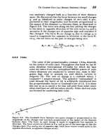
Fig. 1.7 Pupillary reflex pathways.
www.pdfgrip.com
11
SMALL ANIMAL OPHTHALMOLOGY
Sympathetic control of the pupillary dilator muscle originates in the hypothalamus, the axons from which synapse with preganglionic neurons in the first
three or four segments of the thoracic spinal cord. These axons join the sympathetic trunk terminating in the cranial cervical ganglion. Postganglionic fibers
travel to the eye after crossing the roof of the middle ear cavity and are distributed to the ciliary muscle, pupillary dilator, third eyelid, and the Müller’s
muscle of the upper lid. Compromise of sympathetic innervation to the globe
and adnexa results in the classic signs of Horner’s syndrome: ptosis (drooping
of the upper lid), miosis (pupillary constriction), and protrusion of the third
eyelid.
OCULAR PATHOLOGY
The systematic examination of surgical and necropsy-obtained ocular tissue
is essential for optimal patient management, the career-long educational
process, and enhancing understanding of ocular disease in animals. Maximal
benefit is obtained from optimally fixed tissues; in almost all cases, immersion
fixation in 10% formalin is adequate. Fixation should be expedient as the
retina, especially, undergoes rapid autolysis; trimming of periocular tissues
enhances penetration of fixatives, and injection of 0.5 ml of the fixative into
the vitreous cavity with a 27-gauge needle at the equator will minimize neurosensory retinal separation artifact. Otherwise, submit globes intact so that
the pathologist can appreciate the intertissue relationships. Use adequate
volumes of fixative (at least 100 ml for dog and cat eyes), and allow 72 h for
fixation to occur.
Ocular response to disease
A detailed discussion of ocular pathology would fill a text of its own; principles
and concepts of importance to clinicians are discussed with particular disease
processes throughout the following chapters. Three related features warrant
note:
1. The propensity of the ocular tissues (especially the epithelium of lens,
uvea, and retina, but also the corneal endothelium and uveal vasculature)
to undergo reactive changes of hypertrophy, hyperplasia, and metaplasia
(in the case of feline ocular sarcomas, perhaps neoplasia as well)
2. In contrast to the above, the fact that many of the specialized ocular
tissues are post-mitotic, with limited regenerative potential
3. Because of the dependence of the ocular tissues on tissue transparency
and intertissue relationships for normal function, the devastating effect
that these changes can have on vision. A focus of hepatitis may resolve
with scarring and minimal, if any, functional significance, while a
comparable process in the eye may lead to blindness.
12
Fibroplasia in the cornea, for example, will result in scarring and opacification. In the anterior chamber, peripheral anterior and posterior synechia and
membranes are associated with secondary glaucoma. Iris neovascularization,
also known as rubeosis irides or pre-iridal fibrovascular membrane, is a common
cause of intraocular hemorrhage and secondary angle closure glaucoma.
www.pdfgrip.com
CLINICAL BASIC SCIENCE
Hypertrophy, hyperplasia, and metaplasia of lens epithelium are an integral
part of cataractogenesis, and the bane of the cataract surgeon who has to deal
with postoperative capsular fibrosis. Vitreous detachment, fibrosis, and neovascularization lead to cyclitic membranes and their dire consequences of
retinal detachment and phthisis bulbi. The clinical ophthalmologist wages
a relentless pharmacologic battle against these processes with anti-inflammatories and antimetabolites, and new approaches will likely play an important
role in the future management of ocular disease.
13
www.pdfgrip.com
SMALL ANIMAL OPHTHALMOLOGY
Diagnostics
Serge G. Rosolen, Domenico Multari,
Mike Woods and Olivier Jongh
INTRODUCTION
2
The ophthalmic examination, combined with history and signalment, provides
the foundation for obtaining an accurate diagnosis. Ophthalmic diagnosis is
achieved by a combination of basic knowledge, the mastering of simple instrumentation, and critical observation. The former includes an understanding of
anatomy, physiology, and disease mechanisms. Instrumentation facilitates
critical observation. Basic equipment and simple techniques, including a magnifying loupe, bright focal illumination, Schirmer tear test strips, diagnostic
dyes, cytology, direct ophthalmoscopy, and Schiøtz tonometry should be
readily available in any practice, and in experienced hands will be adequate to
manage the great majority of ophthalmic cases. More expensive and sophisticated instrumentation and technologies, including the slit-lamp biomicroscope,
indirect ophthalmoscope, applanation tonometry, electrophysiology, gonioscopy, ultrasonography, and other imaging modalities, fluorescein angiography,
keratoscopy, and retinoscopy represent the next level of diagnostics and are
available to specialists or to those with a particular interest in the field. A systematic approach to examination should be followed and modified for each
individual case based upon the history and signs. Technical competency in
diagnostics is achieved simply by practice; making an ophthalmic examination
a part of every routine physical examination will hone skills for the occasion
upon which they are more urgently required.
INSTRUMENTS AND BASIC DIAGNOSTIC TECHNIQUES
Magnifying loupe
A binocular magnifying loupe of ×2 to ×4 magnification and a focal length of
15–25 cm is useful not only for diagnostics but also for surgery; it allows
freedom of both hands for manipulation and a loupe-mounted diffuse illuminator facilitates observation.
Focal illumination
14
A transilluminator provides an excellent light source for external eye examination and to evaluate the pupillary light reflexes (PLRs). For the latter, it is
www.pdfgrip.com
Schirmer tear test (STT)
DIAGNOSTICS
important to use a narrow beam of bright light with a constant source of energy
(such as a rechargeable handle) directed toward the posterior pole. One of the
most common causes of abnormal PLRs is a dim light source.
This test is used quantitatively to evaluate the aqueous component of the tear
film and thus aid in the diagnosis of keratoconjunctivitis sicca (KCS). The STT
is indicated in all patients with external ocular disease. Individually wrapped
sterile filter paper test strips may be dye impregnated to facilitate reading; these
strips are typically 5 mm wide and 50 mm in length. If performing a STT, it
should be undertaken before any other procedures or tests; if there is discharge
in or around the eye, dry cotton swabs should be used gently to clean the area,
avoiding irritation and reflex lacrimation. The strips have a notch near one end
where they are folded prior to use; fold the strip without touching it with fingers
while it is still in the overwrap. Then open the package and, grasping the strip
from the end opposite the notch with fingers or forceps, place it into the lower
conjunctival sac approximately midway between the medial and lateral canthus
with the short folded end in the fornix and the notch on the eyelid margin (Fig.
2.1). The lower lid can be rolled outward with the thumb to facilitate insertion,
but care should be applied not to compress the eye, which may likewise elicit
reflex lacrimation. The lids may be maintained in an open position, or closed
by gentle pressure on the upper lid if blinking and retention of the strip becomes
a problem. After 1 min, the moistened distance from the notch in the longer
part is measured. Normal values in the dog are 15–25 mm/min; values lower
than 10 mm/min are suggestive of a deficit in aqueous tear production. Most
clinical cases of KCS have a wetting of less than 5 mm; cats have slightly lower
and more variable normal values. There is a wide range of normal readings,
and results should be interpreted in association with clinical signs. Increased
aqueous tear production may occur if conditions causing ocular irritation are
present.
Fig. 2.1
Schirmer tear test being performed in a feline patient.
www.pdfgrip.com
15
SMALL ANIMAL OPHTHALMOLOGY
Diagnostic stains
Fluorescein stain
Fluorescein is a water-soluble dye; owing to its lipid insolubility, it does not
penetrate intact corneal epithelium. Epithelial erosions or ulcers, which expose
the hydrophilic stroma, allow penetration and retention of the dye. The barrier
to penetration in the healthy eye resides in the outermost cells of the corneal
epithelium. As Descemet’s membrane does not retain fluorescein, descemetoceles will not stain. Fluorescein is available as impregnated paper strips or as
a solution; the solution may become contaminated with multiple usage, and
individually wrapped strips are preferred.
Fluorescein staining is indicated in all patients with ocular pain or observable corneal lesions. The tip of the fluorescein-impregnated strip is moistened
with a drop of sterile saline and gently applied to the superior bulbar conjunctiva. If the patient exhibits severe blepharospasm, local anesthetic can be
instilled but may result in a mild diffuse positivity that is usually readily
discernible from significant retention. Blinking will distribute the dye over
the corneal surface. The excess dye is immediately flushed with a sterile saline
rinse and the eye is then examined with a focal light and magnification (Fig.
2.2). A cobalt blue filter will facilitate detection of subtle lesions. To evaluate
nasolacrimal patency, apply the fluorescein as described above, but do not
rinse the eye. If the ipsilateral nostril shows dye within 5 to 10 min, the
nasolacrimal drainage system on that side is patent; the absence of dye passage,
however, does not necessarily mean the contrary, and negative passage is
followed by cannulation and irrigation. Dye may be seen in the nasopharynx
related to alternative duct openings.
Biomicroscopic observation of the fluorescein-stained tear film while holding
the lids open enables evaluation of the tear break-up time (BUT) as an indirect
method of evaluating the non-aqueous components of the tear film; mucus
deficiency will result in shortening of the BUT from the 20–30 s normally
encountered.
Rose bengal and lissamine green
These dyes stain cells of the cornea and conjunctiva that are not covered
by mucin; usually these are degenerating cells. The stains are taken up by neoplastic cells as well and may be useful in defining the extent of epithelial neo-
Fig. 2.2 Fluorescein
uptake by the corneal
stroma associated with a
boxer ulcer.
16
www.pdfgrip.com