Progress in the chemistry of organic nat
Bạn đang xem bản rút gọn của tài liệu. Xem và tải ngay bản đầy đủ của tài liệu tại đây (10.64 MB, 301 trang )
Progress in the Chemistry of Organic Natural Products
A. Douglas Kinghorn · Heinz Falk
Simon Gibbons · Jun’ichi Kobayashi
Yoshinori Asakawa · Ji-Kai Liu Editors
108
Progress in the
Chemistry of
Organic Natural
Products
www.pdfgrip.com
Progress in the Chemistry of Organic Natural
Products
Founded by László Zechmeister
Series Editors
A. Douglas Kinghorn, Columbus, OH, USA
Heinz Falk, Linz, Austria
Simon Gibbons, London, UK
Jun’ichi Kobayashi, Sapporo, Japan
Yoshinori Asakawa, Tokushima, Japan
Ji-Kai Liu, Wuhan, China
Editorial Board
Giovanni Appendino, Novara, Italy
Verena Dirsch, Wien, Austria
Nicholas H. Oberlies, Greensboro, USA
Yang Ye, Shanghai, PR China
www.pdfgrip.com
The volumes of this classic series, now referred to simply as “Zechmeister” after its
founder, Laszlo Zechmeister, have appeared under the Springer Imprint ever since
the series’ inauguration in 1938. It is therefore not really surprising to find out that
the list of contributing authors, who were awarded a Nobel Prize, is quite long: Kurt
Alder, Derek H.R. Barton, George Wells Beadle, Dorothy Crowfoot-Hodgkin, Otto
Diels, Hans von Euler-Chelpin, Paul Karrer, Luis Federico Leloir, Linus Pauling,
Vladimir Prelog, with Walter Norman Haworth and Adolf F.J. Butenandt serving as
members of the editorial board. The volumes contain contributions on various topics
related to the origin, distribution, chemistry, synthesis, biochemistry, function or use
of various classes of naturally occurring substances ranging from small molecules to
biopolymers. Each contribution is written by a recognized authority in the field and
provides a comprehensive and up-to-date review of the topic in question. Addressed
to biologists, technologists, and chemists alike, the series can be used by the expert
as a source of information and literature citations and by the non-expert as a means of
orientation in a rapidly developing discipline. Listed in Medline. All contributions
are listed in PubMed.
More information about this series at />
www.pdfgrip.com
A. Douglas Kinghorn • Heinz Falk •
Simon Gibbons • Jun’ichi Kobayashi •
Yoshinori Asakawa • Ji-Kai Liu
Editors
Progress in the Chemistry of
Organic Natural Products
Volume 108
With contributions by
R. Mata Á M. Figueroa Á A. Navarrete Á I. Rivero-Cruz
S. Fiorito Á F. Epifano Á F. Preziuso Á V. A. Taddeo Á S. Genovese
D. I. Bernardi Á F. O. das Chagas Á A. F. Monteiro Á G. F. dos Santos Á
R. G. de Souza Berlinck
www.pdfgrip.com
Editors
A. Douglas Kinghorn
College of Pharmacy
The Ohio State University
Columbus, OH, USA
Heinz Falk
Institute of Organic Chemistry
Johannes Kepler University
Linz, Austria
Simon Gibbons
UCL School of Pharmacy
University College London, Research
London, UK
Jun’ichi Kobayashi
Grad. School of Pharmaceutical Science
Hokkaido University
Fukuoka, Japan
Yoshinori Asakawa
Faculty of Pharmaceutical Sciences
Tokushima Bunri University
Tokushima, Japan
Ji-Kai Liu
School of Pharmaceutical Sciences
South-Central Univ. for Nationalities
Wuhan, China
ISSN 2191-7043
ISSN 2192-4309 (electronic)
Progress in the Chemistry of Organic Natural Products
ISBN 978-3-030-01098-0
ISBN 978-3-030-01099-7 (eBook)
/>Library of Congress Control Number: 2018965903
© Springer Nature Switzerland AG 2019
This work is subject to copyright. All rights are reserved by the Publisher, whether the whole or part of the
material is concerned, specifically the rights of translation, reprinting, reuse of illustrations, recitation,
broadcasting, reproduction on microfilms or in any other physical way, and transmission or information
storage and retrieval, electronic adaptation, computer software, or by similar or dissimilar methodology
now known or hereafter developed.
The use of general descriptive names, registered names, trademarks, service marks, etc. in this publication
does not imply, even in the absence of a specific statement, that such names are exempt from the relevant
protective laws and regulations and therefore free for general use.
The publisher, the authors, and the editors are safe to assume that the advice and information in this
book are believed to be true and accurate at the date of publication. Neither the publisher nor the authors or
the editors give a warranty, express or implied, with respect to the material contained herein or for any
errors or omissions that may have been made. The publisher remains neutral with regard to jurisdictional
claims in published maps and institutional affiliations.
This Springer imprint is published by the registered company Springer Nature Switzerland AG
The registered company address is: Gewerbestrasse 11, 6330 Cham, Switzerland
www.pdfgrip.com
Contents
Chemistry and Biology of Selected Mexican Medicinal Plants . . . . . . .
Rachel Mata, Mario Figueroa, Andrés Navarrete, and Isabel Rivero-Cruz
Biomolecular Targets of Oxyprenylated Phenylpropanoids and
Polyketides . . . . . . . . . . . . . . . . . . . . . . . . . . . . . . . . . . . . . . . . . . . . .
Serena Fiorito, Francesco Epifano, Francesca Preziuso,
Vito Alessandro Taddeo, and Salvatore Genovese
Secondary Metabolites of Endophytic Actinomycetes: Isolation,
Synthesis, Biosynthesis, and Biological Activities . . . . . . . . . . . . . . . . .
Darlon Irineu Bernardi, Fernanda Oliveira das Chagas, Afif Felix Monteiro,
Gabriel Franco dos Santos, and Roberto Gomes de Souza Berlinck
1
143
207
v
www.pdfgrip.com
Chemistry and Biology of Selected Mexican
Medicinal Plants
Rachel Mata, Mario Figueroa, Andrés Navarrete, and Isabel Rivero-Cruz
Contents
1 Introduction . . . . . . . . . . . . . . . . . . . . . . . . . . . . . . . . . . . . . . . . . . . . . . . . . . . . . . . . . . . . . . . . . . . . . . . . . . . . . . . . . . .
2 Mexican Medicinal Plants Employed for Treating Major National Health Problems . . . . . .
2.1 Diabetes . . . . . . . . . . . . . . . . . . . . . . . . . . . . . . . . . . . . . . . . . . . . . . . . . . . . . . . . . . . . . . . . . . . . . . . . . . . . . . . . .
2.1.1 Swietenia humilis . . . . . . . . . . . . . . . . . . . . . . . . . . . . . . . . . . . . . . . . . . . . . . . . . . . . . . . . . . . . . . .
2.1.2 Mexican “Copalchis”: Hintonia latiflora, Hintonia standleyana,
and Exostema caribaeum . . . . . . . . . . . . . . . . . . . . . . . . . . . . . . . . . . . . . . . . . . . . . . . . . . . . . . .
2.1.3 Salvia circinata . . . . . . . . . . . . . . . . . . . . . . . . . . . . . . . . . . . . . . . . . . . . . . . . . . . . . . . . . . . . . . . . .
2.2 Smooth Muscle-Relaxant Agents for Gastrointestinal and Cardiovascular Illnesses .
2.2.1 Scaphyglottis livida, Maxillaria densa, and Nidema boothii . . . . . . . . . . . . . . . . . .
2.3 Antiulcer Agents . . . . . . . . . . . . . . . . . . . . . . . . . . . . . . . . . . . . . . . . . . . . . . . . . . . . . . . . . . . . . . . . . . . . . . . .
2.3.1 Amphipterygium adstringens . . . . . . . . . . . . . . . . . . . . . . . . . . . . . . . . . . . . . . . . . . . . . . . . . . .
2.3.2 Ligusticum porteri . . . . . . . . . . . . . . . . . . . . . . . . . . . . . . . . . . . . . . . . . . . . . . . . . . . . . . . . . . . . . .
2.3.3 Hippocratea excelsa . . . . . . . . . . . . . . . . . . . . . . . . . . . . . . . . . . . . . . . . . . . . . . . . . . . . . . . . . . . .
2.4 Analgesic and Anti-inflammatory Agents . . . . . . . . . . . . . . . . . . . . . . . . . . . . . . . . . . . . . . . . . . . . . .
2.4.1 Hofmeisteria schaffneri . . . . . . . . . . . . . . . . . . . . . . . . . . . . . . . . . . . . . . . . . . . . . . . . . . . . . . . . .
2.4.2 Artemisia ludoviciana . . . . . . . . . . . . . . . . . . . . . . . . . . . . . . . . . . . . . . . . . . . . . . . . . . . . . . . . . .
2.5 Antiparasitics . . . . . . . . . . . . . . . . . . . . . . . . . . . . . . . . . . . . . . . . . . . . . . . . . . . . . . . . . . . . . . . . . . . . . . . . . . .
2.5.1 Dysphania graveolens . . . . . . . . . . . . . . . . . . . . . . . . . . . . . . . . . . . . . . . . . . . . . . . . . . . . . . . . . .
2.5.2 Geranium niveum . . . . . . . . . . . . . . . . . . . . . . . . . . . . . . . . . . . . . . . . . . . . . . . . . . . . . . . . . . . . . . .
2.6 Cytotoxic Activity . . . . . . . . . . . . . . . . . . . . . . . . . . . . . . . . . . . . . . . . . . . . . . . . . . . . . . . . . . . . . . . . . . . . . .
2.6.1 Annona mucosa . . . . . . . . . . . . . . . . . . . . . . . . . . . . . . . . . . . . . . . . . . . . . . . . . . . . . . . . . . . . . . . . .
2.6.2 Annona purpurea . . . . . . . . . . . . . . . . . . . . . . . . . . . . . . . . . . . . . . . . . . . . . . . . . . . . . . . . . . . . . . .
2.7 Anxiolytic and Sleep-Aid Agents . . . . . . . . . . . . . . . . . . . . . . . . . . . . . . . . . . . . . . . . . . . . . . . . . . . . . .
2.7.1 Valeriana procera . . . . . . . . . . . . . . . . . . . . . . . . . . . . . . . . . . . . . . . . . . . . . . . . . . . . . . . . . . . . . .
2
3
3
4
9
18
22
22
31
31
35
40
46
46
51
64
65
69
72
73
77
81
82
R. Mata (*) · M. Figueroa (*) · A. Navarrete · I. Rivero-Cruz
Departamento de Farmacia, Facultad de Qmica, Universidad Nacional Autónoma de México,
Ciudad de México, México
e-mail: ; mafi; ;
© Springer Nature Switzerland AG 2019
A. D. Kinghorn, H. Falk, S. Gibbons, J. Kobayashi, Y. Asakawa, J.-K. Liu (eds.),
Progress in the Chemistry of Organic Natural Products, Vol. 108,
/>
1
www.pdfgrip.com
2
R. Mata et al.
2.8 Antiasthmatic Agents . . . . . . . . . . . . . . . . . . . . . . . . . . . . . . . . . . . . . . . . . . . . . . . . . . . . . . . . . . . . . . . . . . 87
2.8.1 Pseudognaphalium liebmannii . . . . . . . . . . . . . . . . . . . . . . . . . . . . . . . . . . . . . . . . . . . . . . . . 88
3 Concluding Remarks . . . . . . . . . . . . . . . . . . . . . . . . . . . . . . . . . . . . . . . . . . . . . . . . . . . . . . . . . . . . . . . . . . . . . . . . 92
Appendix . . . . . . . . . . . . . . . . . . . . . . . . . . . . . . . . . . . . . . . . . . . . . . . . . . . . . . . . . . . . . . . . . . . . . . . . . . . . . . . . . . . . . . . . . 93
References . . . . . . . . . . . . . . . . . . . . . . . . . . . . . . . . . . . . . . . . . . . . . . . . . . . . . . . . . . . . . . . . . . . . . . . . . . . . . . . . . . . . . . . 118
1 Introduction
Mexico is a multifaceted and heterogeneous country with high cultural richness and
10–12% of the world’s biodiversity. This country ranks 4th in the variety of vascular
plants with about 31,000 different species; of this stock more than 3350 form part of
the medicinal flora. When the Spanish conquerors arrived to ancient Mexico, they
found existing civilizations with a holistic view of illnesses and healing. These early
Mesoamericans inhabitants used religious, magic rituals and a variety of plant-based
remedies to improve health. The abundance and variety of Mexican medicinal flora
can be traced from published work written from the sixteenth century to modern
times. Crucial and most important sources of information about traditional Mexican
medicine were recently reviewed [1].
The use of herbal medicines survives to this day in modern Mexico; the original
Aztec beliefs and practices are interlaced with strands of the European medicine
introduced by the Spaniards in the sixteenth century. They are an integral element of
alternative medical care and the best testimony of their efficacy and cultural value is
the persistence of medicinal plants in present-day Mexican markets, where the
highest percentage of medicinal and aromatic plants is sold.
For more than 100 years, researchers have explored Mexican medicinal flora from
the ethnobotanical, anthropological, chemical, pharmacological, and biotechnological points of view; in a few cases some clinical investigations have been pursued.
The most important investigations have been carried out at the Instituto Nacional de
Antropología, Instituto Mexicano del Seguro Social, Universidad Autónoma de
Nuevo León, Universidad Autónoma del Estado de Morelos, Instituto Tecnológico
y de Estudios Superiores de Monterrey, Universidad Autónoma Metropolitana,
Centro de Investigación y Estudios Avanzados del Instituto Politécnico Nacional,
and Universidad Nacional Autónoma de México. The no-longer existing Instituto
Médico Nacional and Instituto Mexicano de Plantas Medicinales deserve special
mention since they were devoted to the study of Mexican medicinal plants in
different periods of the twentieth century. Both are good examples of important
institutions dedicated to the comprehensive analysis of the national Materia Medica,
and were pioneering institutions in bioprospecting matters.
In the twenty-first century, the commerce of medicinal plants in Mexico has grown
due to a global resurgence of herbal-based remedies. Furthermore, according to a
recent survey, 54% of health professionals and 49% of physicians have used medicinal
plants as an alternative therapy for several diseases. Twenty-eight percent of health
professionals and 26% of physicians, have recommended or prescribed medicinal
plants to their patients, in particular for digestive and respiratory ailments; finally, 73%
www.pdfgrip.com
Chemistry and Biology of Selected Mexican Medicinal Plants
3
of health professionals would agree to receiving academic information regarding the
use and prescribing of medicinal plants [2].
Concomitantly, a loss of biodiversity, over-exploitation, biopiracy, and weak
regulations on the use of medicinal plants are the major impediments to the growth
of herbal medicine as an important national industry [3]. Therefore, current research
on medicinal plants should also involve conservation issues and the sustainable
search for bioactive natural products based on traditional knowledge, regulation, and
quality control of the most important species; these are essential issues for the
growth of a rational herbal medicine usage.
In the following sections, some work from the authors’ laboratories will be
highlighted. The most relevant phytochemical and pharmacological profiles of a
selected group of plants widely used for treating major national health problems will
be discussed.
2 Mexican Medicinal Plants Employed for Treating Major
National Health Problems
2.1
Diabetes
The global prevalence of diabetes in adults has been increasing over recent decades,
making this disease a major public health threat in countries all over the world. The
International Diabetes Federation estimated the global prevalence to be 425 million in
2017, which implied a health expenditure of 673 billion USD [4]. The prevalence of
diabetes in adults aged 20–79 years is predicted to rise to 10.4% in 2040. Of the total
diabetics, about 95% have type 2 diabetes mellitus (T2DM). Mexico is one of the
countries most affected by this metabolic disease, in particular indigenous people
owing to changes in their traditional lifestyle and the effects of industrialization on
both environmental and sociocultural norms. In 2017, there were more than 12 million
people affected by diabetes, representing a prime cause of mortality. In Mexico as in
other regions of the world, people use plants to treat the symptoms of diabetes. More
than 300 different plants have been described as reputedly beneficial for the diabetic
patient [5–7], but most claims are subjective and few have received any suitable
scientific evaluation. So far, about 200 plants have been investigated scientifically in
Mexico in order to establish their antidiabetic potential. Most studies have been
limited to the preclinical evaluation of extracts prepared with selected solvents using
different pharmacological models [6]; the depth of their analysis is variable since some
authors have reported in detail the mode of action of the extracts while others just
measured their hypoglycemic activity. Other studies have determined both the active
principles and the preclinical efficacy of the traditional preparations. Finally, only a
very few studies have pursued in-depth clinical observations. Most of the work of the
present author group falls into the second category, involving detailed phytochemical
work coupled with substantial preclinical biological observations.
Some examples of our work on antidiabetic plants are described in the following
sections. In addition, other investigations, from other authors and ourselves, carried out
www.pdfgrip.com
4
R. Mata et al.
after a survey on diabetic plants was published in 2005 [6], are summarized in the
Appendix Table.
2.1.1
Swietenia humilis
Swietenia humilis Zuccarini (Meliaceae), locally known as “zopilote”, “cobano”,
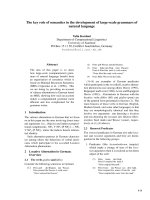
“flor de venadillo” and “caoba”, is a medium-sized deciduous tree (Fig. 1). The
species is regarded as one of the three true American mahogany species. It grows in a
very wide ecological range within its native Pacific watershed of Central America
and Mexico. The seeds are wind dispersed and highly valued for medicinal purposes.
The plant is also a much appreciated hardwood species in the neotropics and is
seriously threatened owing to overexploitation and habitat destruction. Therefore, a
multilateral treaty called the Convention on International Trade in Endangered
Species of Wild Fauna and Flora lists S. humilis in Appendix II (all parts and
derivatives except the seeds) [8]. Also, it is categorized in the International Union
for Conservation of Nature Red List of Threatened Species as “vulnerable” [9].
The medicinal use of the seeds of S. humilis can be traced to the sixteenth century;
the Spanish royal physician Francisco Hernández, in his magnificent manuscript
“Four Books on the Nature and Virtues of Plants and Animals for Medicinal
Purposes in New Spain”, described the antiulcer, astringent, antitussive, and emollient properties of these seeds. In the middle of the twentieth century, their astringent
effects were also described [10]. In the present day, decoctions of the seeds of
S. humilis (SHD), alone or in combination with other plants, are valued for treating
indigestion, stomachache, amebic dysentery, and diarrhea. The ground raw seeds or
their decoctions are also ingested as a blood depurative and antidiabetic agent [5, 6].
In general, for conducting our studies focused on the determination of any
pharmacological properties of traditional extracts, first acute preclinical toxicity
using the Lorke procedure is assessed [11]. This method measures acute toxicity
for 14 days in mice using a range of doses between 10 and 5000 mg/kg, in two
phases. The dried seeds and SHD (10–5000 mg/kg) showed no acute toxic effects
when assessed by the Lorke procedure. The calculated LD50 values of the preparation and crude drug were higher than 5000 mg/kg.
Fig. 1 Leaves, stems (A), and seeds (A and B) of Swietenia humilis
www.pdfgrip.com
Chemistry and Biology of Selected Mexican Medicinal Plants
5
Since the plant preparation lacked acute toxic effects, it was next tested for
antidiabetic action in vivo by means of animal models using a standard protocol.
By means of this protocol, initially the acute hypoglycemic activity in normoglycemic
and hyperglycemic animals (ICR mice or Wistar rats) is assessed. If feasible,
subchronic (14 days) or chronic (30 days) experiments are also performed. Then,
the antihyperglycemic action of the extracts or purified compounds after a glucose
(1 g/kg; oral glucose tolerance test, OGTT), sucrose (2 g/kg; oral sucrose tolerance
test, OSTT) or starch (2 g/kg; oral starch tolerance test, OStTT) challenge is assessed
using normal and hyperglycemic animals. These tests provide relevant information
regarding peripheral utilization or absorption of glucose. In all tests, the animals are
made hyperglycemic with streptozotocin (STZ, 130 mg/kg for mice; and 50 mg/kg for
rats), after previous protection with nicotinamide (NAA, 40 mg/kg for mice; and
65 mg/kg for rats). After 7 days of NAA-STZ administration, the animals are
generally hyperglycemic and can be included in the studies conducted subsequently.
The NAA-STZ model affords a similar biochemical blood profile and pathogenesis to
T2DM in humans. Glibenclamide (15 mg/kg), metformin (200 mg/kg) or acarbose
(5 mg/kg) are used as positive controls, depending of the type of experiment. The
percentage variation of glycemia for each group of animals is calculated with respect
to the initial values at different periods of time. The results are plotted indicating
blood glucose values or percentage of variation versus time at several doses [12].
In a series of experiments conducted in NAA-STZ hyperglycemic mice, SHD
(100–316 mg/kg) caused a significant reduction in blood glucose levels and inhibited
the postprandial peak provoked by a glucose load during an OGTT. On the other
hand, SHD (100–316 mg/kg) did not inhibit the postprandial peak at any of the doses
tested during an OSTT in normoglycemic mice, ruling out an inhibition of
α-glucosidases at the intestinal level [13].
The antihyperglycemic, hypoglycemic, and hypolipidemic effects of S. humilis
seeds were corroborated in rats with fructose-fed metabolic syndrome. SHD
(100 and 316 mg/kg) caused a significant inhibition of the postprandial peak during
an OGTT when compared with a vehicle-treated group. Moreover, daily administration of SHD (100 mg/kg) for a week provoked a significant hypoglycemic effect,
and reductions in both serum triglycerides and uric acid, without any significant
changes in fasting insulin levels or body weight. In addition, a reduction in the
abdominal fat of the test animals, and an increment in hepatic glycogen, were
observed. Altogether, the results suggested that the traditional preparation of
S. humilis induced modifications in peripheral glucose uptake, rather than by inhibition of the intestinal α-glucosidases. The reduction of the postprandial peak
observed during the OGTT, and the increment of hepatic glycogen in rats with
fructose-fed metabolic syndrome indicated that the hypoglycemic effect of SHD
involves an insulin-sensitizing mechanism. The reduction in blood triglycerides is
compatible with an increment in glucose uptake in adipose tissue, where energy is
stored as triglycerides. These effects are also consistent with the use of this species as
blood depurative (purifying) agent [13].
In order to identify the compounds responsible for these pharmacological effects,
both the active aqueous and an organic extracts of S. humulis seeds were fractionated
extensively by chromatographic procedures. These processes led to the isolation of
www.pdfgrip.com
6
R. Mata et al.
eight new limonoids of the mexicanolide type, namely, humilinolides A–H (1–8)
along with humulin B (9), methyl-2-hydroxy-3β-isobutyroxy-1-oxomeliac-8(30)enate (10), methyl-2-hydroxy-3β-tigloyloxy-1-oxomeliac-8(30)-enate (11), swietenin
C (12), swietemahonin C (13) and 2-hydroxy-destigloyl-6-deoxyswietenine acetate
(14) (Fig. 2) [13]. These mexicanolides can be categorized into two structural subclasses by considering the degree of oxidation at C-8/C-30 of the basic methyl-1oxomeliacate nucleus. The first one comprises limonoids with an 8,30 double bond,
while the second includes those with an 8,30 epoxide function. The compounds in
each group differ in the number and position of oxygenated substituents. The acid
residues esterifying the hydroxy group at C-3 could be either isobutyric, tiglic or acetic
acid. All structures were elucidated using one- and two-dimensional NMR spectroscopic techniques, and with that of humulinolide G (5) confirmed by X-ray diffraction
analysis [13].
Chromatographic analysis of SHD revealed that compounds 9, 11, and 14 are its
major components, although the remaining limonoids isolated were also identified.
These limonoids were isolated in adequate amounts to perform in vivo assays. As
expected, the three major compounds (3.16–31.6 mg/kg) showed hypoglycemic and
antihyperglycemic actions when tested in the NAA-STZ mice model using the acute
hypoglycemic assay and the OGTT, respectively (Fig. 3). Although limonoids 9, 11,
and 14 were found as the major hypoglycemic and antihyperglycemic limonoids of
the decoction, the remaining compounds could also contribute to the pharmacological action displayed by SHD. Furthermore, they could be acting synergistically on
different molecular targets to produce antidiabetic and hypolipidemic effects. Likewise, the mixture of components in SHD might enhance the bioavailability of one or
several compounds of the extract, thus improving their pharmacological actions. It is
worth mentioning that none of the isolates inhibited α-glucosidases.
The antihyperalgesic effects of SHD and compound 14 were assessed in
NAA-STZ hyperglycemic mice using the formalin method. The formalin test in
mice is a valid and reliable model of nociception and is sensitive to various classes of
analgesic drugs. The noxious stimulus is an injection of dilute formalin (1% in
O
O
O
R3
O
O
O
O
O
O
R3
O
O
O
A
O
O
R1
R1
=
O
B
OR2
OR2
O
=
1 (humilinolide A) R1 = OH, R2 = A, R3 = OH
2 (humilinolide B) R1 = OH, R2 = A, R3 = B
6 (humilinolide F) R1 = B, R2 = C, R3 = B
8 (humilinolide H) R1 = B, R2 = A, R3 = H
9 (humulin B) R1 = OH, R2 = A, R3 = H
13 (swietemahonin C) R1 = H, R2 = A, R3 = B
3 (humilinolide C) R1 = B, R2 = C, R3 = H
4 (humilinolide D) R1 = OH, R2 = D, R3 = B
5 (humilinolide E) R1 = OH, R2 = C, R3 = B
7 (humilinolide G) R1 = B, R2 = A, R3 = H
10 R1 = OH, R2 = A, R3 = H
11 R1 = OH, R2 = C, R3 = H
12 (swietenin C) R1 = H, R2 = A, R3 = OH
14 R1 = OH, R2 = D, R3 = H
Fig. 2 Limonoids isolated from Swietenia humilis
O
C
=
O
D
=
www.pdfgrip.com
Chemistry and Biology of Selected Mexican Medicinal Plants
7
Fig. 3 Effect of SHD (A), mexicanolide 14 (B), humulin B (9) (C), and methyl-2-hydroxy3β-tigloyloxy-1-oxomeliac-8(30)-enate (11) (D) on blood glucose levels in NAA-STZ-hyperglycemic mice during an OGTT. VEH: vehicle; MTF: metformin. Values are expressed as the means
from six data points ỈSEM. *p < 0.05, **p < 0.01 and ***p < 0.001. Adapted from [13]
saline), placed under the skin of the dorsal surface of the right hind paw. The
response observed is the amount of time the animals spend licking the injected
paw. Two distinct periods of high licking activity can be identified, an early phase
lasting for the first 5 min and a late phase lasting from 20 to 30 min after the injection
of formalin. The two phases in the formalin test may have different nociceptive
mechanisms. The early phase seems to be caused predominantly by C-fiber activation due to the peripheral stimulus, while the late phase appears to be dependent on
the combination of an inflammatory reaction in the peripheral tissue and functional
changes in the dorsal horn of the spinal cord; this pain can be inhibited by antiinflammatory drugs [14]. Thus, local injection of SHD (10–177 μg) and
mexicanolide 14 (0.5–3.5 μg) provoked a concentration-dependent antihyperalgesic
action in NAA-STZ hyperglycemic mice (Fig. 4). Ketanserin (6 μg), a 5-HT2A/C
receptor antagonist, and flumazenil (6 μg), a GABAA receptor antagonist, abolished
the antihyperalgesic effect of mexicanolide 14 (3 μg) (Fig. 5). On the other hand,
naloxone (3 μg), L-arginine (50 μg), and Nω-nitro-L-arginine methyl ester hydrochloride (L-NAME; 150 μg) diminished the antihyperalgesic effect of mexicanolide
14 (Fig. 6). The aqueous extract of the seeds possesses significant antihyperalgesic
action [15]. Thus, S. humilis seeds have shown also promising results for managing
secondary complications (neuropathic pain) of diabetes.
www.pdfgrip.com
8
R. Mata et al.
Fig. 4 Antihyperalgesic effect of mexicanolide 14 in NAA-STZ hyperglycemic mice during
phases 1 (A), 2 (B), and the total area under the curve (C) in the formalin test. VEH: vehicle;
GBP: gabapentin (30 μg per paw) was used as positive control. Each bar represents the mean area
under the curve (AUC, time of licking against time, sec  min) from six data points ỈSEM.
*p < 0.01 and **p < 0.001. Adapted from [15]
Fig. 5 Possible antihyperalgesic mechanism of mexicanolide 14 (3 μg per paw) in NAA-STZ
hyperglycemic mice during the formalin test: serotoninergic, GABAergic (A), and opioid modulation (B). VEH: vehicle, ketanserin (KET, 6 μg per paw), flumazenil (FLU, 6 μg per paw), and
naloxone (NLX, 3 μg per paw). (A) Each bar represents the mean area under the curve (AUC, time
of licking against time, sec  min) from six data points ỈSEM. *p < 0.05, **p < 0.01 and
***p < 0.001. (B) Each point represents the mean of the time of licking (sec) from six data points
ỈSEM. *p < 0.05, **p < 0.01 and ***p < 0.001. Adapted from [15]
Swietenia humilis and/or its limonoids represent promising alternatives for development as safer and cheaper phytotherapeutic agents. The overall results described
in the paragraphs above support the use of seeds of this tropical species for treating
diabetes in contemporary Mexico. Finally, it is worth mentioning that the potential
antiamebiasis effects of the limonoids and extracts from this plant were tested, with
negative results being obtained. Moreover, most of the limonoid constituents and an
organic extract from S. humilis were active against the European corn borer, Ostrinia
nubilalis, affecting important life cycle parameters such as reduction of the %
pupation and the % of adult emergence, in a similar way to the positive control
toosendanin [16, 17]. They also inhibited radical growth of a several weed species
www.pdfgrip.com
Chemistry and Biology of Selected Mexican Medicinal Plants
9
Fig. 6 Possible antihyperalgesic mechanism of mexicanolide 14 (3 μg per paw) in NAA-STZ
hyperglycemic mice during phases 1 (A) and 2 (B) on the formalin test: nitrergic modulation.
VEH: vehicle, L-NAME (150 μg per paw), L-arginine (ARG, 50 μg per paw), and
3-morpholinosydnonimine hydrochloride (SIN-1, 200 μg per paw). Each bar represents the mean
area under the curve (AUC, time of licking against time, sec  min) from six data points ỈSEM.
*p < 0.05, **p < 0.01 and ***p < 0.001. Adapted from [15]
when tested in vitro [18]. In consequence, this species seems valuable not only as a
medicinal agent but also as a pesticide.
2.1.2
Mexican “Copalchis”: Hintonia latiflora, Hintonia standleyana,
and Exostema caribaeum
Hintonia latiflora (Sessé & Moc. ex DC.) Bullock (Rubiaceae) is a species endemic
to Mexico, while H. standleyana Bullock has a wider distribution area up to
Northern Central America. Hintonia standleyana was considered to be synonym
of H. latiflora, which is still widely accepted by some authors, however, recent
molecular evidence has revealed that these two species are significantly different
[19–21]. Both species are known commonly as “copalquin” and “copalchi”, among
other colloquial names. The plants are shrubs or trees up to 8 m tall, with gray stems;
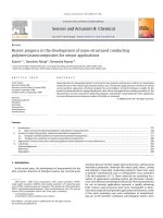
the leaves are bright green and covered with hairs on the back (Fig. 7). The main area
Fig. 7 Mexican “Copalchis”: Hintonia latiflora (A), Hintonia standleyana (B) and Exostema
caribaeum (C)
www.pdfgrip.com
10
R. Mata et al.
supplying the commercial “copalchi” is the northern state of Guerrero, Mexico. Teas
from the bark of these species are used in modern Mexico for a variety of health
problems, including malaria, stomach ulcers, diabetes, obesity, infections and fevers.
In addition, the Tarahumaras have used H. latiflora on body sores [22].
Exostema caribaeum (Jacq.) Schult. (Rubiaceae), the Caribbean prince wood, is
an evergreen slender shrub or small tree up to 12 m height (Fig. 7). The plant occurs
on all islands within the Bahamian Archipelago, as well as the rest of the Caribbean
region, Florida, Mexico, and Central America. In Mexico, the plant is gathered from
the wild for local use as a medicine to treat fevers, especially those related to malaria,
and also a source of lighting and timber. This species is also regarded as “copalchi”,
and in some local markets its stem bark is mixed with those of H. latiflora or
H. standleyana [23].
The hypoglycemic and diuretic properties of H. latiflora were discovered clinically by researchers at the Instituto Médico Nacional in Mexico City at the beginning
of twentieth century (Fig. 8). They also discovered some chemical compounds
present that were later on rediscovered by German, French, and Mexican
researchers. It is notable that in 1913, when the Instituto Médico Nacional closed,
“copalchi” was reintroduced in Europe for the treatment of diabetes. Later on,
researchers in Germany and France corroborated the earlier work of the Mexican
scientists. Recently, the most relevant historical aspects about this species as well as
the research carried out by other scientists were reviewed [12]. Perhaps the most
relevant aspect of these historical events was that, after the Royal Botanical Expedition to New Spain (1787–1803), led by Martín Sessé and José Mociño, H. latiflora,
Fig. 8 Hypoglycemic and diuretic effects exerted by a Hintonia latiflora hydroalcoholic extract in
a clinical trial conducted at IMN, Mexico City. Urine volume during a 24-h period (black line);
amount of glucose in urine during a 24-h period (green line); proportion of glucose per liter (red
line). Adapted from [12]
www.pdfgrip.com
Chemistry and Biology of Selected Mexican Medicinal Plants
11
under its synonym Coutarea latiflora Sessé & Moc. ex DC., and E. caribaeum
appeared in the list of the most important “Medicinal Plants of New Spain”. They
were also included in the well-known “Torner Collection” of Sessé and Mociño
biological illustrations. Thus, in the following paragraphs, we will review mostly the
work carried out by our group.
Phytochemical analysis of the stem bark of these three plants allowed the
discovery of cucurbitacins in the Rubiaceae family, as well as the characterization
of several 4-phenylcoumarins, with most being new chemical entities, and the indole
alkaloid desoxycordifolinic acid (15) [23–31]. The basic core of the cucurbitacins
16–19 is dihydrocucurbitacin F (16) (Fig. 9). The 4-phenylcoumarins 20–36 of the
three species are 5,7,30 ,40 - or 5,7,40 -substituted with oxygenated functionalities, with
the former having the most common pattern; the sugar unit is usually a monosaccharide (β-D-galactose, β-D-glucose, 600 -acetyl-β-D-glucose or 600 -acetyl-β-D-galactose), although some disaccharides have been found (β-D-apiofuranosyl-(1!6)-β-Dglucopyranose or β-D-xylopyranosyl-(1!6)-β-D-glucopyranose) (Fig. 9). In all
cases, the saccharide unit is attached to the hydroxy group at C-5. During the course
of our investigations it was demonstrated that 4-phenylcoumarins undergo oxidative
cyclization under aerobic alkaline conditions to give oxido-4-phenylcoumarins.
CO2H
O
HO
OR
N
OR2
O
O
N
H
OH
HO
HO2C
R1O
15 (desoxycordifolinic acid) R = b -D-glucopyranosyl
R2
O
O
16
17
18
19
(dihydrocucurbitacin F) R1 = H, R2 = H
R1 = H, R2 = Ac
R1 = b -D-glucopyranosyl, R2 = H
R1 = b -D-glucopyranosyl, R2 = Ac
R1
R3
R4
R1
O
O
20 R1 = R2 = R4 = OCH3, R3 = H
O
21 R1 = R2 = R4 = OCH3, R3 = OH
4
1
2
3
22 R = O-b -D-galactopyranosyl, R = OCH3, R = R = OH
R3
23 R1 = R3 = R4 = OH, R2 = OCH3
R2
24 R1 = O-b -D-glucopyranosyl, R2 = OCH3, R3 = R4 = OH
35 R1 = OCH3, R2 = R3 = OH
25 R1 = O-b -D-glucopyranosyl, R2 = R3 = R4 = OH
36 R1 = R2 = OCH3, R3 = OH
26 R1 = R2 = R3 = R4 = OH
27 R1 = 6''-O-acetyl-O-b -D-glucopyranosyl, R2 = R3 = R4 = OH
28 R1 = O-b -D-galactopyranosyl, R2 = OCH3, R3 = H, R4 = OH
29 R1 = O-b -D-glucopyranosyl, R2 = R4 = OCH3, R3 = OH
30 R1 = 6''-O-acetyl-O-b -D-galactopyranosyl, R2 = R4 = OH, R3 = H
31 R1 = 6''-O-acetyl-O-b -D-galactopyranosyl, R2 = R3 = R4 = OH
32 R1 = O-b -D-apiofuranosyl-(1 6)-b -D-glucopyranosyl, R2 = OCH3, R3 = R4 = OH
33 R1 = O-b -D-xylopyranosyl-(1 6)-b -D-glucopyranosyl, R2 = OCH3, R3 = R4 = OH
34 R1 = O-b -D-xylopyranosyl-(1 6)-b -D-glucopyranosyl, R2 = R4 = OCH3, R3 = H
Fig. 9 Compounds isolated from Mexican “Copalchis”
www.pdfgrip.com
12
R. Mata et al.
Thus, 7-methoxy-5,30 ,40 -trihydroxy-4-phenylcoumarin (23) was converted to
7-methoxy-40 ,50 -dihydroxy-4-phenyl-5,20 -oxido-coumarin (35) by treatment with
potassium hydroxide in methanol. Since the reaction took place only in basic
conditions and in the presence of air, it might proceed via an oxidative phenol
coupling process.
Preclinical toxicity studies have revealed that none of the aqueous (traditional
preparations) extracts from the stem bark of the two above-mentioned Hintonia
species and E. caribaeum were toxic to mice (LD50 > 5 g/kg). These results thus
suggest the preclinical safety of the traditional preparations of these three plants
[32, 33]. The organic extracts of H. latiflora and E. caribaeum, however, showed
LD50 values of 2900 and 700 mg/kg, respectively; the extract of E. caribaeum
generated tremors, respiratory distress as well as decreases in motor activity and in
body weight, by 27.1% with respect to the vehicle-treated animals. The organic
extract of H. standleyana had an LD50 of > 5 g/kg. Moreover, none of the extracts
induced mutagenic effects when assayed by the Ames test [33]. Rivera et al. [34]
reported that a methanol extract from H. latiflora induced genotoxic effects,
piloerection, excitability, dyspnea, anoxia, mydriasis, tachycardia, overcrowding,
decreased muscle tone, burying behavior, and ambulatory movements in a dosedependent manner in mice. These effects were only observed during the first 24 h of
the experiment. At the end of the study (15 days after treatment), all surviving mice
showed a normal behavior [34]. Our group has worked extensively with Hintonia
species and never observed such effects, even in long-term experiments. Unfortunately, the authors did not provide chromatographic profiles of their extract nor a
voucher number to compare the plant material they analyzed [34].
The long-term hypoglycemic effect of the organic extracts (CH2Cl2:MeOH ¼
1:1) of the three species (H. latiflora, H. standleyana, and E. caribaeum) and a
commercial mixture of “copalchi” (composed by H. standleyana and E. caribaeum),
and compounds 18, 22, 24, 25, and 32 (15 mg/kg each time) was established
(Fig. 10) [31]. The extract of H. latiflora and compound 25 restored blood glucose
levels to normal values, with the effect being comparable to that of glibenclamide.
Compounds 22 and 24 also restored blood glucose levels to near normal values by
the end of the experiment. During this study, it was also demonstrated that the extract
of H. latiflora regulated both hepatic glycogen and plasma insulin levels ( p < 0.05)
(Fig. 11). These data suggested that its hypoglycemic effect is due in part to
stimulation of insulin secretion and regulation of hepatic glycogen metabolism.
Comparison of the hypoglycemic activity of the 4-phenylcoumarins tested
established that the most active compounds possess a free hydroxy group at C-7 in
the 4-phenylcoumarin core. On the other hand, comparison of the activity of all
glycosides tested indicated that the nature of the sugar moiety (glucose, galactose, βD-apiofuranosyl-(1!6)-β-D-glucopyranose or β-D-xylopyranosyl-(1!6)-β-D-glucopyranose) had little or no influence on the resultant biological activity [31]. The
hypoglycemic activity of such cucurbitacin-type compounds was demonstrated for
the first time in these studies.
Infusions of the stem bark of both Hintonia species and E. caribaeum also
showed hypoglycemic and antihyperglycemic effects. The later was demonstrated
www.pdfgrip.com
Chemistry and Biology of Selected Mexican Medicinal Plants
13
Fig. 10 Long-term effect of extracts of Hintonia latiflora, Hintonia standleyana, Exostema
caribaeum, and CM (commercial mixture of “copalchi”) on blood glucose levels in NAA-STZdiabetic rats. Each value is the mean from six data points ỈSEM. *p < 0.05. Adapted from [31]
during an OSTT in mice suggesting that the plants contained inhibitors of
α-glucosidases [12]. In addition, the major components of the infusions were
4-phenylcoumarin glycosides: in the case of H. latiflora, the most abundant compound was 33, for H. standleyana, the most relevant was 32, while for E. caribaeum
this was 22. The α-glucosidase inhibitory activities of several glycosides present in
the infusions and the aglycones 23 and 26 were demonstrated in vitro using different
enzymes. This assay was carried out using a well-known spectrophotocolorimetric
procedure that measures the ability of any α-glucosidases (baker’s yeast,
Ruminococcus obeum or mammalian) to hydrolyze a suitable substrate ( pnitrophenyl-α-D-glucopyranoside) in the presence of the potential inhibitor; acarbose
was also used as positive control. Docking studies, using AutoDock software,
predicted that the aglycone 23 (IC50 ¼ 3.0 μM vs. 0.41 mM for acarbose), which
turned out to be the most active inhibitor, binds to the yeast α-glucosidases in the
same pocket as acarbose. Coumarin 23 and glycoside 32 were also very active in an
OStTT, thus indicating that both compounds possess also α-amylase inhibitory
activity [12].
For the crude drug (stem bark) of the three species, reliable, reproducible, and
accurate high-performance liquid chromatography (HPLC)-UV methods were
developed for the quantitative determination of the active compounds (Fig. 12).
These methods were included in the second edition of the “Mexican Herbal Pharmacopoeia” [23, 35]. The development of the composition and identity tests for the
three “copalchi” species have been very useful to detect the adulteration of
www.pdfgrip.com
14
R. Mata et al.
Fig. 11 Effect of extracts of Hintonia latiflora, Hintonia standleyana, Exostema caribaeum, and
CM (commercial mixture of “copalchi”), cucurbitacin 18, and 4-phenylcoumarins 22, 24, 25, and
32 on plasma insulin levels in STZ-diabetic rats. Each value is the mean from six data points
ỈSEM. *p < 0.05. Adapted from [31]
H. latiflora with E. caribaeum. Indeed, the analysis of the commercial crude drugs or
preparations made up with “copalchi” samples, Mexican or from abroad, have
revealed clearly that most preparations contain a mixture of the two “copalchi”
components, with E. caribeum almost always the more abundant in the mixture.
The organic extracts [CH2Cl2-MeOH (1:1)] and infusions from the leaves of
H. standleyana and H. latiflora were also hypoglycemic and antihyperglycemic, in
both normal and hyperglycemic rats. These extracts did not provoke death or
damage, behavioral alterations, lesions or bleeding of the internal tissues to the
animals, throughout the experiments conducted [32]. Therefore, the leaves of
Hintonia species represent a therapeutic alternative to the stem bark in terms of
conservation, since these species have been extensively exploited and commercialized locally and outside of Mexico from harvesting wild plants. In consequence, the
populations of the plants are now scarce and in danger of extinction. From the active
leaf extracts, three additional 4-phenylcoumarins (37–39) were obtained (Fig. 13)
[32]. In addition, HPLC profiles of the leaf extracts of both plants revealed the
presence of several hypoglycemic 4-phenylcoumarins isolated from the stem bark as
well as ursolic (40) and chlorogenic (41) acids (Fig. 13). The overall results indicated
www.pdfgrip.com
Chemistry and Biology of Selected Mexican Medicinal Plants
15
Fig. 12 UHPLC-PDA chromatogram of Exostema caribaeum stem bark aqueous extract under
optimized conditions; detection wavelength 327 nm. Adapted from [23]
R2
O
O
R1
R3
R4
37 R1 = 6''-O-acetyl-5-O-b -D-galactopyranosyl, R2 = R4 = OH, R3 =H
38 R1 = 6''-O-acetyl-5-O-b -D-galactopyranosyl, R2 = R3 = R4 = OH
39 R1 = O-b -D-xylopyranosyl-(1
6)-b -D-glucopyranosyl, R2 = R4 = OCH3, R3 = H
HO CO2H
O
CO2H
HO
O
OH
HO
OH
OH
40 (ursolic acid)
41 (chlorogenic acid)
Fig. 13 Structures of 4-phenylcoumarins 37–39, and ursolic (40) and chlorogenic (41) acids
that the leaves of both species possess similar antidiabetic actions to their stem bark.
Phenological and geographical analysis of the leaves from two different regions of
Mexico (Chihuahua and Michoacán), using a validated UPLC method, revealed that
the best harvesting season for the leaves of H. latiflora is between the leaf renovation
and senescence stages, avoiding the flowering period [32]. In addition, no significant
differences were found among the two different geographical populations analyzed.
www.pdfgrip.com
16
R. Mata et al.
Recently, a dry concentrated extract from the stem bark of H. latiflora in capsule
form was tested in an open prospective clinical study in 41 dietetically stabilized
subjects with T2DM for 6 months [36]. The results revealed that fasting and
postprandial glucose levels and HbA1c values all declined significantly. Moreover,
the tolerance was excellent, and liver and lipid values tended to be positively
affected. In particular, no side effects and no hypoglycemic episodes or worsening
of diabetic symptoms occurred. These results were in agreement with earlier studies
[36]. Thus, the use of Hintonia dry extract for treating mild to moderate T2DM can
be regarded as safe and useful in cases where dietary measures alone cannot provide
adequate control of the disease.
Vierling and collaborators showed also that an extract of H. latiflora exerted a
vasodilatory effect in vitro in aortic rings of the guinea pig and in vivo in rabbits
[37]. Aortic rings pre-contracted with noradrenaline (NA) could be relaxed
completely by the extract (EC50 ¼ 51.98 mg/dm3). The aglycone also inhibited
NA-induced contractions of aortic rings (EC50 ¼ 32.55 mg/dm3) and in aortic
cells suppressed calcium transients evoked by vasopressin at a concentration of
60 mg/dm3, suggesting a possible inhibition of G-protein-induced intracellular
calcium release. Ultrasound measurements in conscious rabbits showed that the
extract induced vasodilation and lowering of blood flow velocity in the abdominal
aorta and the carotid artery. The combination of a blood glucose-lowering with a
vasodilating effect may be helpful for reducing vascular long-term complications in
patients with T2DM.
During our earlier work on H. latiflora, the in vitro anti-Plasmodium falciparum
activity of the extracts or isolates was not demonstrated. The same type of results
were obtained by Noster and Kraus in Germany [38], who found only a moderate
effect in vitro with E. caribaeum extracts. However, in a paper by Argotte-Ramos
et al. [39], it was reported that an ethyl acetate extract of the stem bark of H. latiflora
provoked suppression of induced parasitemia with P. berghei in mice. Bioassaydirected fractionation of the active extract using in vitro and in vivo assays indicated
that 23 is the active principle. Also, Rivera and coworkers [34] found that a
methanolic extract of H. latiflora had an excellent chemosuppression of P. yoelii
total parasitemia and schizonts number in CD1 male mice in a 4-day test protocol. As
indicated above, in this work no voucher nor chemical composition of the plant was
provided. Hence, there is an uncertainty about the precise identity of the plant
material that was actually analyzed.
The organic extract of H. standleyana showed a potent antinociceptive effect
when tested in hot-plate and writhing tests [29]. The hot-plate test is a suitable
method to evaluate central antinociceptive activity. In addition, this model measures
animal behavior and has good sensitivity and specificity. It is based on the principle
that when rodents are placed onto a hot surface they will initially demonstrate
aversive behaviors to the thermal stimulus by licking their paws and jumping.
Compounds that alter the nociceptive threshold increase the latency period to the
observed licking/jumping. On the other hand, the acetic acid-induced writhing test,
is a classical visceral pain model useful to detect painful symptoms associated with
inflammatory disorders of the internal organs such as the stomach or intestines.
www.pdfgrip.com
Chemistry and Biology of Selected Mexican Medicinal Plants
17
The metabolite responsible for this antinociceptive activity of the extract was
found to be compound 18, which significantly reduced the acetic acid-induced
abdominal constrictions in mice. In addition, this compound produced a significant
increase in thermal latency in the hot-plate test (Fig. 14) [29]. The effect of 18 was
Fig. 14 Antinociceptive effects of desoxycordifolinic acid (15), cucurbitacin 18 and
4-phenylcoumarin 32 in mice submitted to the writhing (A) and hot-plate (B) tests. Metamizol
(MET) and morphine (MOR) were used as positive controls in the writhing and hot-plate tests. In
both cases, bars are the means from six data points ÆSEM. *p < 0.05 and **p < 0.01. Adapted
from [29]
www.pdfgrip.com
18
R. Mata et al.
mediated by the nitric oxide-cyclic GMP pathway at the peripheral and/or central
levels, opening of ATP-sensitive K+ channels, and activation of the opioid receptors
or increasing the levels of endogenous opioids. Altogether, these results suggested
that the extract of H. standleyana investigated is able to reduce inflammatory and
central pain in mice [29].
The aqueous extracts from the stem bark and leaves of H. latiflora and
H. standleyana (80.5%, 80.2%, 75.1%, and 76.9% of gastroprotection, respectively),
as well as compounds 32 (ED50 ¼ 15 mg/kg) and 38 (ED50 ¼ 26 mg/kg), were able
to inhibit ethanol-induced gastric lesions in rats [40]. Intragastric application of
absolute ethanol to produce gastric lesions in experimental animals is a wellknown and reproducible method to investigate cytoprotective agents. The mode of
action of 32 and 38 involved non-protein sulfhydryl endogenous compounds,
because when the rats were pretreated with N-ethylmaleimide (NEM), a sulfhydryl
alkylator, their gastroprotective action was inhibited [40].
Both Hintonia species contain more than one active constituents that act on
different targets. Accordingly, these plants exhibit a wide range of biological
properties, which collectively could be useful for treating a multifactorial disease
such as diabetes.
2.1.3
Salvia circinata
Salvia circinata Cav. (syn. S. amarissima Ortega) (Lamiaceae) is a perennial shrub
native to Mexico (Fig. 15). It grows up to 1.5 m tall. The whitish green oval leaves
are rough or wrinkled and usually downy. The flowers, usually pale blue, feature
tubular two-lipped corollas and produce nutlet fruits [41]. Salvia circinata was also
listed as a medicinal in the catalog of plants from the Royal Botanical Expeditions to
New Spain. A tea brewed from the aerial parts of the plant is used in Mexican folk
medicine for treating ulcers, helminthiasis, and diabetes. According to the Lorke
criterion, this tea did not provoke behavior alterations, macroscopic tissue injury, or
weight loss, when tested during 14-day in mice; the estimated LD50 was higher than
5 g/kg [41].
Single oral administration of the traditional preparation (100–570 mg/kg) to
normal and NAA-STZ hyperglycemic mice induced a perceptible decrease of
blood glucose level. During an OSTT, the infusion (31.6, 100, and 316 mg/kg)
also significantly reduced the postprandial peak when compared with a vehicletreated group; the effect observed was comparable to that of the positive control,
acarbose. However, the preparation (100–570 mg/kg) did not induce a significant
drop in the postprandial peak after a glucose challenge in normal and hyperglycemic
mice. These results strongly suggested that the antihyperglycemic effect of
S. circinata infusion could be due to the presence of α-glucosidase inhibitors,
which may be able to prevent postprandial hypersecretion of insulin and reactive
hypoglycemia [41]. The active compounds included a few new glucosides of
www.pdfgrip.com
Chemistry and Biology of Selected Mexican Medicinal Plants
19
Fig. 15 Salvia circinata
tricyclic neo-clerodane type diterpenoids with the six-membered rings trans-fused,
and the side chain fragment forming ethylbutenolides, ethylfuran or linear substructures. These terpenoids were given the trivial names amarisolides A–E (42–46)
(Fig. 16). Several flavonoids (47–50) were also among the active principles
(Fig. 16). All compounds were characterized on the basis of their spectroscopic
properties. The absolute configurations at the stereocenters of diterpenoids 42–46
were established by comparison of the experimentally obtained and calculated
electronic circular dichroism spectra [41].
Compounds 42–50 were tested in vitro against rat small intestine α-glucosidase.
The more active compounds were the flavonoids, in particular, compound 47, which
showed an IC50 value of 39 μM and was 2.5 times more active than acarbose
(IC50 ¼ 100 μM). Flavonoids 48–50 exhibited IC50 values of 810, 200, and
1800 μM, respectively. Regarding the diterpenoids, the most active compound was
42, with an IC50 value of 500 μM. Compounds 42 and 48 were also tested against a
recombinant α-glucosidase with maltase-glucoamylase activity from Ruminococcus
obeum, a bacterium found in the human intestine that is involved in carbohydrate
metabolism. This enzyme is phylogenetically closer to human N-maltaseglucoamylase than those of rats. The results of the assays showed that 42 and 48
inhibited the activity of the pure enzyme with IC50 values of 400 and 60 μM,