Excited states in organic chemistry and biochemistry proceedings of the tenth jerusalem syposium on quantum chemistry and biochemistry held in jerusa
Bạn đang xem bản rút gọn của tài liệu. Xem và tải ngay bản đầy đủ của tài liệu tại đây (33.43 MB, 444 trang )
www.pdfgrip.com
EXCITED STATES IN ORGANIC CHEMISTRY AND BIOCHEMISTRY
www.pdfgrip.com
THE JERUSALEM SYMPOSIA ON
QU ANTUM CHEMISTRY AND BIOCHEMISTRY
Published by the Israel Academy of Sciences and Humanities,
distributed by Academic Press (N. Y.)
1st JERUSALEM SYMPOSIUM:
2nd
JERUSALEM SYMPOSIUM:
3rd JERUSALEM SYMPOSIUM:
4th JERUSALEM
5th
SYMPOSIUM:
JERUSALEM SYMPOSIUM:
The Physicochemical Aspects of Carcinogenesis
(October 1968)
Quantum Aspects of Heterocyclic Compounds in
Chemistry and Biochemistry (April 1969)
Aromaticity, Pseudo-Aromaticity, Antiaromaticity
(April 1970)
The Purines: Theory and Experiment
(April 1971)
The Conformation of Biological Molecules and
Polymers (April 1972)
Published by the Israel Academy of Sciences and Humanities,
distributed by D. Reidel Publishing Company (Dordrecht and Boston)
6th
Jl'ltUSALEM SYMPOSIUM:
Chemical and Biochemical Reactivity
(April 1973)
Published and distributed by D. Reidel Publishing Company
(Dordrecht and Boston)
7th JERUSALEM SYMPOSIUM:
8th JERUSALEM SYMPOSIUM:
9th JERUSALEM SYMPOSIUM:
Molecular and Quantum Pharmacology
(Marchi April 1974)
Environmental Effects on Molecular Structure and
Properties (April 1975)
Metal-Ligand Interactions in Organic Chemistry
and Biochemistry (April 1976)
VOLUME 10
www.pdfgrip.com
EXCITED STATES IN
ORGANIC CHEMISTRY AND
BIOCHEMISTRY
PROCEEDINGS OF THE TENTH JERUSALEM SYMPOSIUM ON
QUANTUM CHEMISTRY AND BIOCHEMISTRY HELD IN
JERUSALEM, IsRAEL, MARCH
28/31, 1977
Edited by
BERNARD PULLMAN
Universite Pierre et Marie Curie (Paris VI)
Instilut de Biologie Physico-Chimique
(Fondation Edmond de Rothschild), Paris, France
and
NATAN GOLDBLUM
The Hebrew University, Hadassah Medical School
Jerusalem, Israel
Springer-Science+Business Media, B.V.
www.pdfgrip.com
Library of Congress Cataloging in Publication Data
Jerusalem Symposium on Quantum Chemistry and Biochemistry, lOth, 1977.
Excited states in organic chemistry and biochemistry.
(The Jerusalem symposia on quantum chemistry and biochemistry; v. 10)
Bibliography: p.
Includes index.
l. Excited state chemistry-Congresses. 2. Chemistry, Physical organic-Congresses. 3. Biological chemistry-Congresses. l. Pullman, Bernard, 1919- ll. Goldblum, Natan. Ill. Title. IV. Series.
547:1'28
77-21896
QD46l.5.J47 1977
ISBN 978-94-010-1275-1
ISBN 978-94-010-1273-7 (eBook)
DOI 10.1007/978-94-010-1273-7
All Rights Reserved
Copyright © 1977 by Springer Science+Business Media Dordrecht
Originally published by D. Reidel Publishing Company, Dordrecht, Holland in 1977
Softcover reprint of the hardcover lst edition 1977
No part of the material protected by this copyright notice may be reproduced or
utiliz~d in any form or by any means, electronic or mechanical,
including photocopying, recording or by any informational storage and
retrieval system, without written permission from the copyright owner
www.pdfgrip.com
PREFACE
We are living since such a long time now in a world
governed in its many aspects by the decimal system, that the
10th anniversary of any significant event represents an event in
itself, in particular for those who have been implicated in its
birth and development. It is also a landmark at which one feels
necessary to stop for a while and think, make a balance of the
value and significance of the efforts expanded.
The inaugural session of this Symposium, presided by
Professor Ephraim Katzir, the President of the State of Israel,
in the presence of Professor A. Dvoretzky, President of the
Israel Academy of Sciences and Humanities, served such a purpose.
I hope not to betray the general feeling by saying that, on their
modest scale, the Jerusalem Symposia, called in Quantum Chemistry
and Biochemistry but which in fact have gone far beyond the quantum aspects of these disciplines seem to have been a significant
event in a number of their aspects. The different themes discussed at the ten meetings were among the frontier subjects of present day scientific research in Chemistry and Biochemistry. The
Symposia contributed, I believe, in a very positive way to scientific eXChanges and contacts and, I hope, also, to the progress
of science.
--The 10th Symposium was also an occasion to express our
appreciation to all those who contributed to their establishment,
growth and success. A particular tribute was paid to the generosity and understanding of the Baron Edmond de Rothschild without
whose help these meetings would not have been possible. The
Baron de Rothschild was presented with two beautiful scrolls,
from the Israel Academy of Sciences and Humanities and from the
Hebrew University of Jerusalem, expressing their deep apprecia-
www.pdfgrip.com
PREFACE
tion for the good work accomplished. The memory and contribution
of Professor Ernst Bergmann, one of the creators of these Symposia and coorganizer of the first eight of them was recalled
with emotion.
May I thank again all those who contributed to the
success of this meeting : the authorities of the Israel Academy
of Sciences and Humanities and in particular its President Professor A. Dvoretzky and Mrs. Agigael Hyam and Miriam Yogev,
Professor Natan Goldblum, Vice-President of the Hebrew University who carried the heavy burden of local arrangements and the
Baron Edmond de Rothschild for his renewed and everlasting generosity. The support of the European Research Office is also
gratefully acknowledged.
Bernard Pullman
www.pdfgrip.com
TABLE OF CONTENTS
Preface (Bernard Pullman)
v
List of Participants
XI
P. vigny and J.P. Ballini / Excited states of nucleic acids
at 300 K and electronic energy transfer
M.D. Sevilla / Mechanisms for radiation damage in DNA
constituents and DNA
15
2+
R.O. Rahn / Influence of Hg
on the excited states of DNA:
photochemical consequences
27
Shi Yi Wang / A "hot" ground state intermediate in the
photohydration of pyrimidines
39
Th. Montenay-Garestier / Excited state interactions and
energy transfer between nucleic acid bases and amino
acid side chains of proteins
53
c.
Helene / Mechanisms of quenching of aromatic amino acid
fluorescence in protein-nucleic acid complexes
65
J. Sperling and A. Havron / Specificity of photochemical
cross-linking in protein-nucleic acid complexes
79
J. Hfittermann / Excitation and ionization of 5-halouracils:
ESR and ENDOR of single crystals
85
M.F. Maestre, J. Greve, and J.Hosoda / Optical studies on
T4 gene product 32 protein DNA interaction
99
E. Hayon / The chemistry of excited states of aromatic
amino acids and pep tides
113
www.pdfgrip.com
TABLE OF CONTENTS
VIII
Th.M. Hooker, Jr., and W.J. Goux
protein structure
I Chiroptical probes of
123
M. Iseli, R. Geiger, and G. Wagniere I Description of the
chiroptic properties of small peptides by a molecular
orbital method
137
G. Laustriat, D. Gerard, and C. Hasselmann I Influence of
3-substitution on excited state properties of indole
in aqueous solutions
151
L. Salem I The sudden polarization effect
163
J.J. Wolken I Photoreceptors and photoprocesses in the
living cell
175
L.J. Dunne I Electron-electron interactions and resonant
optical spectral shifts in photoreceptor molecules
187
s.
Boue, D. Rondelez, and P. Vanderlinden I Classical
and non-classical decay paths of electronically
excited conjugated dienes
199
C.A. Bush I Far ultraviolet circular dichroism of
oligosaccharides
209
Th. Kindt and E. Lippert I Adiabatic photoreactions in
acidified solutions of 4-methylumbelliferone
221
D.B. McCormick I spectral and photochemical assessments
of interactions of the flavin ring system with amino
acid residues
233
S.P. McGlynn, D. Dougherty, T. Mathers, and S. Abdulner I
Photoelectron spectroscopy of carbonyls. Biological
considerations
247
M.S. Gordon and J.W. Caldwell
molecules
I Excited states of saturated
257
J. Joussot-Dubien, R. Bonneau, and P. Fornier de Violet I
Evidence and reactivity of a twisted form of medium
size cyclo-alkene rings presenting a double bond
past orthogonality
271
J. Wirz I Electronic structure and photophysical properties
of planar conjugated hydrocarbons with a 4n-membered
ring
283
G. Snatzke and G. Haj6s I Excited states of chiral pyrazines
295
www.pdfgrip.com
IX
TABLE OF CONTENTS
G. Kohler, C. Rosicky, and N. Getoff I Wavelength dependence
of Q(F) and Q(e~q) of some aromatic amines in aqueous
solution
303
F.C. de Schryver, J. Huybrechts, N. Boens, J.C. Dederen, and
M. Irie I Intramolecular excited state interactions in
l,3-di (2-anthryl) propane
313
Th.J. de Boer, F.J.G. Broekhoven, and Th.A.B.M. Bolsman I
Behaviour of excited c-nitroso compounds in the presence
and absence of oxygen
323
P. Politzer and K.C. Daiker I Some possible products of the
reactions of O(l D) and 02(1~) with unsaturated hydrocarbons
331
H.H. Seliger and J.P. Hamman I Chemical production of excited
states: adventitious biological chemiluminescence of
carcinogenic polycyclic aromatic hydrocarbons
345
J. Michl, A. Castellan, M.A. Souto, and J. Kolc I Higher
excited states and vibrationally hot excited states:
how important are they in organic photochemistry in
dense media?
361
G.G. Hall and C.J. Miller
M.B. Rubin
U.P. wild
I
Solvent effects on excited states
I Photochemistry of vicinal polyketones
I Fluorescence from upper excited singlet states
373
381
387
J.e. Lorquet, C. Galloy, M. Desouter-Lecomte, M.J. Decheneux,
and D. Dehareng I Non-adiabatic interactions in the unimolecular
decay of polyatomic molecules
397
E.S. Pysh I Measurement of circular dichroism in the vacuum
ultraviolet. A new challenge for theoreticians
409
R. Janoschek I Non empirical calculations of excited states
of large molecules by the method of improved virtual
orbitals
419
Index of Subjects
431
Index of Names
436
www.pdfgrip.com
LIST OF PARTICIPANTS
BOUe, S.G., Universite Libre de Bruxelles, Faculte des Sciences,
Avenue F.D. Roosevelt 50, 1050 Bruxelles, Belgium
Bush, C.A., Illinois Institute of Technology, Lewis College of
Science and Letters, Department of Chemistry, Chicago,
Illinois 60616, USA
Daniels, M., Oregon State University, Radiation Center, Corvallis,
Oregon 97331, USA
De Boer, Th. J., Universiteit van Amsterdam, Laboratorium voor
Organische Scheikunde, Nieuwe Achtergracht 129, Amsterdam,
The Netherlands
De Schryver, F.C., Universiteit te Leuven, Department Scheikunde,
Celestijnenlaan 200F, 3030 Heverlee, Belgium
Dunne, L.J., Chelsea College, University of London, Department
of Mathematics, Manresa Road, London SW3 6LX, England
Getoff, N., Institut fur Theoretische Chemie und Strahlenchemie,
Universitat Wien, 1090 Wien, Wahringer Strasse 38, Austria
Gcrdon, M.S., North Dakota State University of Agriculture and
Applied Sciences, Department of Chemistry, Fargo, North
Dakota 58102, USA
Hall, G.G., The University of Nottingham, Department of Mathematics,
Nottingham NG7 2RD, England
Hayon, E., Department of the Army, U.S. Army Natick, Research and
Development Cmd., Natick, Massachusetts 01760, USA
Helene, C., C.N.R.S., Centre de Biophysique Moleculaire, Av. de
la Recherche Scientifique, 45045 Orleans Cedex, France
www.pdfgrip.com
XII
LIST OF PARTICIPANTS
Hooker, T.M., Jr., University of California, Santa Barbara,
Department of Chemistry, Santa Barbara, California 93106, USA
Huttermann, J., Universitat Regensburg, Fachbereich Biologie und
Vorklinische Medizin, Institut fur Biophysik und Physikalische
Biochemie, 8400 Regensburg, universitatsstrasse 31, Germany
Janoschek, R., Universitat Stuttgart, Institut fur Theoretische
Chemie, Pfaffenwaldring 55, 7 Stuttgart 80, Germany
Jortner, J., Tel-Aviv University, Institute of Chemistry, 61390
Ramat-Aviv, Tel-Aviv, Israel
Joussot-Dubien, J., Universite de Bordeaux I, Unite de Chimie,
Laboratoire de Chimie Physique, 351 Cours de la Liberation,
33405 Talence, France
Laustriat, G., Universite Louis Pasteur U.E.R. des Sciences
Pharmaceutiques, Laboratoire de Physique, 3 rue de I 'Argonne,
67083 Strasbourg-Cedex, France
Lippert, E., Iwan N. Stranski-Institut fur Physikalische und
Theoretische Chemie der Technischen Universitat Berlin,
1 Berlin 12, Strasse des 17 Juni 112, Ernst-Reuter-Haus,
West Germany
Lorquet, J.C., Universite de Liege, Institut de Chimie, Department
de Chimie Generale et de Chimie Physique, Sart-Tilman B.
4000 par Liege, Belgium
Maestre, M.F., University of California, Space Sciences Laboratory,
Berkeley, California 94720, USA
McCormick, D.B., Cornell University, Section of Biochemistry,
Molecular and Cell Biology, Division of Biological Sciences,
Savage Hall, Ithaca, New York 14853, USA
McGlynn, S.P., Louisiana State University and Agricultural and
Mechanical College, College of Chemistry and Physics, Baton
Rouge, Louisiana 70803, USA
Michl, J., The University of Utah, Department of Chemistry,
Chemistry Building, Salt Lake City 84112, USA
Montenay-Garestier, T., Museum National d'Histoire Naturelle,
Chaire de Biophysique, 61 rue Buffon, 75005 Paris, France
Politzer, P., University of New Orleans, Lake Front, Department
of Chemistry, New Orleans, Louisiana 70122, USA
pullman, A., Institut de Biologie Physico-Chimique, 13 rue P. et
M. Curie, Paris 5e, France
www.pdfgrip.com
LIST OF PARTICIPANTS
XIII
Pullman, B., Institut de Biologie Physico-Chimique, 13 rue P. et
M. Curie, Paris 5e, France
Pysh, E.S., Brown University, Department of Chemistry, Providence,
Rhode Island 02912, USA
Rahn, R., Oak Ridge National Laboratory, Union Carbide Corp.
Nuclear Div., P.O.B.Y. Oak Ridge, Tennessee 37830, USA
Rosenfeldt, T., The Hebrew University of Jerusalem, Department of
Physical Chemistry, Jerusalem, Israel
Rubin, M., Department of Chemistry, Technion, Haifa, Israel
Salem, L., Laboratoire de Chimie Theorique, Batiment 490, Centre
d'Orsay, 91405 Orsay, France
Seliger, H.H., The Johns Hopkins University, Mergenthaler
Laboratory for Biology, Baltimore, Maryland 21218, USA
Sevilla, M.D., Oakland University, Department of Chemistry,
Rochester, Michigan 48063, USA
Snatzke, G., Lehrstuhl fur Strukturchemie, Ruhruniversitat,
4630 Bochum 1, Postfach 10 21 48, W. Germany
Sperling, J., The Weizmann Institute of Science, Department of
Organic Chemistry, Rehovot, Israel
Vigny, P., Fondation Curie, Institut du Radium, Laboratoire Curie,
11 rue P. et M. Curie, 75231 Paris - Cedex OS, France
Wagniere, G., Physikalisch-Chemisches Institut der Universitat
Zurich, 8001 Zurich, Ramistrasse 76, Switzerland
Wang, S.Y., The Johns Hopkins University School of Hygiene and
Public Health, Department of Biochemical and Biophysical
Sciences, 615 North Wolfe Street, Baltimore, Maryland 21205, USA
Wild, U., Eidgenossische Technische Hochschule Zurich, Laboratorium
fur Physikalische Chemie, 8006 Zurich, Universitatsstrasse 22,
Switzerland
Wirz, J., Physikalisch-Chemisches Institut der Universitat Basel,
4056 Basel, Klingelbergstrasse 80, Switzerland
Wolken, J.J., Carnegie-Mellon University, Biophysical Research
Laboratory, Schenley Park, Pittsburgh, Pennsylvania 15213, USA
www.pdfgrip.com
EXCITED STATES OF NUCLEIC ACIDS AT 300K AND ELECTRONIC
ENERGY TRANSFER.
Paul VIGNY and Jean Pierre BALLINI
Institut du Radium, Laboratoire Curie
11, rue Pierre et Marie Curie
75231 PARIS CEDEX OS, France.
I. INTRODUCTION
Investigation of the Excited States of Nucleic Acids appears to be a
major step in the understanding of the photochemical changes induced in
DNA by ultraviolet radiation. The details of the mechanisms initiated by
the absorption of a photon by a base and ending with the formation of a
photoproduct on the same or on another base cannot be understood
without a knowledge of their excited states. This, together with the
amount of information which can be obtained on their ground states as
well, certainly accounts for the fact that many luminescence studies
have been carried out on nucleic acids for the last ten years. However
the main feature of these molecules is that the systems are quenched to
a high degree under physiological conditions. The fluorescence quantum
yields are so weak that until recently nucleic acid bases were simply
considered not to fluoresce at room temperature. In this respect, they
differ from many aromatic compounds for which internal conversion
from the first excited singlet state is unimportant. Therefore, most of
the work has been performed either at extreme pH values where the
nucleic bases exhibit measurable fluorescence emission or at 77K in
glasses where the quantum yields are of the order of 10-1 or 10- 2 , thus
permitting normal recording of the luminescence spectra. Under such
conditions a good understanding of the lowest excited Singlet and triplet
states has been thus achieved ( for a review, see for example Gueron
et a1 (1). Although many interesting results were obtained, the major
question which has been constantly raised (1) (2) is whether conclUSions
obtained in a rigid medium at 77K can be extrapolated to fluid aqueous
B. Pullman and N. Goldblum (eds.). Excited States in Organic Chemistry and BlOchemlS/ry, 1-13.
All Rights Reserved. CopYright © 1977 by D. Reidel Publishing Company, Dordrecht, Holland.
www.pdfgrip.com
2
P. VIGNY AND J. P. BALLINI
solutions at room temperature, specially in view of the drastic temperature effect on the quantum yields. Other questions such as the existence
of interactions between bases in the excited states or the ability of
electronic energy to be transfered from one base to another, in nucleic
acids under physiological conditions, were unresolved.
The experimental problem of the detection of nucleic bases at room
temperature has been therefore reinvestigated independently by Daniels
in Oregon (3) and by our group in Paris (4) leading to the identical conclusion that nucleic bases do weakly fluoresce at 300K (fluorescence
quantum yield of the order of 10- 4 ). Our earliest experiments were
rather crude. They have been further refined and extended to such
complex structures as dinucleotides, polynucleotides and nucleic acids.
The aim of the present contribution is to summarize our recent results
which should still be considered as a preliminary approach to the large
field of the excited states of nucleic acids at room temperature.
II. EXPERIMENTAL CONSIDERATIONS.
Several difficulties are encountered when trying to study the fluorescent properties of compounds with very low quantum yields. They
will be briefly discussed. Of course the first requirement lies in a
highly sensitive spectrophotofluorometer. It is not intended to discuss
here the apparatus which was used for these studies since it has already
been described (5). Its sensitivity is partly due to the photon-counting
method which allows an increase of the signal-to-noise ratio by increasing counting time, and partly to the optical components. Two kinds of
improvements have been performed as compared to the above mentioned
apparatus i) a pdpll computer is now used for accumulation which
allows automatic corrections of the spectra and ii) a more recent model
of the instrument is now operating, which is somewhat more sensitive.
Rather high concentrations ( 10- 3 M - 10- 4 M ) were used in our
experiments. Important corrections had thus to be operated and their
validity to be carefully checked (5). The improved sensitivity of our new
apparatus will now allow us to use more dilute solutions and to avoid
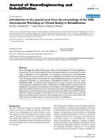
most of these corrections. As an example Figure 1 shows a recently
recorded fluorescence spectrum of Adenine at concentration 10- 5 M. At
present, a concentration ten times lower is therefore our limit.
Sample purity is an important limitation since traces of a highly
fluorescent impurity can give rise to perturbation in the fluorescence
spectrum. A number of commercially available bases, nucleosides and
www.pdfgrip.com
3
EXCITED STATES OF :-.IUCLEIC ACIDS
,-..
t·'"
. -..
....
..
>~
(/)
Z
......
-....
W
~
\.
Z
w
(,)
Z
W
.......
(,)
(/)
-
w
ex:
... 2.5
o
:3u..
..
..'-.
.'
>-1
'...
·;,1'
250
300
350
WAVELENGTH
400
(nm)
Figure 1. Room temperature fluorescence of Adenine at 10- 5 M in water. The left ,art of the figure shows the Raman scattering
on a different intensity scale. (.A exc . =255nm,lU exc . =6. Onm,
ll.A em. =3. Onm, counting time=30s per pOint).
nucleotides (Merck, Sigma, Calbiochem, Schwartz Bioresearch, Nutritional Biochemicals Corporation) have therefore been tested, some of
which have been shown to be unsuitable for fluorescence measurements.
Most of the reported fluorescence spectra are issued from products
purchased from Calbiochem (A grade). Suprasil quartz cells are carefully selected and the water is triple distilled from K Mn04 and Ba(OH)2.
Polynuclenotides were purchased from Miles Laboratories. They
can be more easily purified by extensive dialySis. However, due to their
structure, they may undergo photochemical reactions giving rise to
fluorescent adducts either during their preparation or during the record-
www.pdfgrip.com
4
P. VIGNY AND J. P. BALLIN I
ing of the spectra. The identification of the fluorescence spectrum of a
polynucleotide may therefore be troublesome.
III, EXCITED STATES OF MONONUCLEOTIDES
The corrected room temperature fluorescence spectra of the five
common nucleotides are given in Figure 2, As compared to the low-temperature spectra, they are broader and structureless but not very different. Except for GMP, the red-shifts when going from rigid samples
250
3111
350
4111
i'
rvn
250
w
AMP
Z
0
z
Q
W
I-
0
Q.
CI)
a:
w
a:
0
CI)
0
-'
m
<
:::I
II..
4,5
4,0
3,0
3.5
2.5
~o
~5
~o
CMP
Z
2.5
TMP
0
w
0
z
w
i=
0
Q.
CI)
a:
w
a:
0
CI)
0
-'
II..
m
<
:::I
4,5
4.0
3,5
3,5
2,5
4.5
250
z
i=
0-
4,0
3110
350
3,0
~5
4111
2.5
nm
w
I,D
0
z
0
w
0
CI)
w
a:
0
a:
CI)
0
:::I
-'
II..
m
<
4,5
4,0
3.5
3,0
2,5
fl fTT '
Figure 2. Absorption and fluorescence sEectra of the common nucleotides
at 300K, A comparison is made with fluorescence data obtained
at 77K (----) by Gueron et al (1) (Our experimental conditions
C=10- 4M, AexC. =248nm,AAexc =4. 2nm, ~=3, 2nm),
www.pdfgrip.com
EXCITED STATES OF NUCLEIC ACIDS
5
to fluid solutions are small (CMP, TMP) or negligible (AMP, UMP). The
most important feature lies in the quantum yields. Their values are
between 0.3 1O- 4 (UMP) and 1.2 10- 4 (CMP and TMP) (Table 1). It is of
interest to notice that addition of the ribose and phosphate group leaves
the quantum yields of C, T and U unchanged, whereas those of A and G
are decreased by a factor of five.
To interpret the difference between 77° and 300"1(, it is necessary to
postulate a very efficient Sr.... So internal conversion since other deactivation processes cannot quantitatively explain the low quantum yields
observed at room temperature (6). It is not possible to state whether this
quenching is intra or intermolecular or -more likely- have both origins.
Another interesting point about nucleotides is the knowledge of their
fluorescence lifetimes. Calculations derived from the room temperature
data and assuming that their entire low-energy absorption band is responsible for emission lead to singlet lifetimes of 1O- 12 s for bases (3)
and nucleotides (7), in agreement with experiments involving energy
transfer to Eu+ (8). No doubt that direct experimental determination of
these lifetimes in the future would be an important contribution in this
field.
IV. EXCITED STATE INTERACTIONS IN POLYNUCLEOTIDES
Bases are brought together in polynucleotides so that interactions
may occur. In addition to the well-known ground state interactions, can
excited state interactions also occur at room temperature? Such exciplexes and excimers have been proposed at 77K to explain the red-shift
observed in their emission spectra ( see reference (1) for a review).
Beside the monomer-like emission, the room temperature emission
spectrum of the dinucleotide ApA shows a new broad band at -420nm (9).
This emission can be thought to arise from an excimer formed between
two stacked bases. According to what is known about excimer emission,
its intenSity should be more or less intense, depending on the stacking of
the two bases. At room temperature ApA is supposed to be in a stacked
conformation. Moreover this stacking is very temperature dependent and
becomes less important when temperature is increased. Part a of Figure 3
shows that the second emission band is effectively temperature dependent
and notably increased when the temperature is lowered to 4°C. The same
interpretation has been proposed for C5 'pp5'C (10), whose second emission band ( A~~· =410nm) is strongly increased when ionic strength is
increased (Figure 3, part 3).
www.pdfgrip.com
6
P. VIGNY AND J. P. BALLINI
,\
1.10 •
i \
i
CD CACODYLATE 3.3l0-3M
'2' CACODYLATE 3.3 lO-3M
~ NaCI lO·l M
,;
>Iiii
;
I
z
W
I-
Z
0.5
i
j
z
w
(.)
~.
(/)
,-
w
II:
o::l
I.
300
350
400
I
\
\
\
,
\
\
0
i
w
(.)
...
"
I /;(®
\
I
...J
;
'
I
I
I
I
I
\
i
\
\
\
/
\
\.
\
\.
'.
450
WAVELENGTH
(nm)
Figure 3. Temperature and ionic strength dependence of the emission
spectra of dinucleotides. (experimental conditions .1exc . =248
nm,Ll.1 exc . =4. 2nm,fu i =6. 4nm, concentration of ApA 1. 5x10- 4 M
in monomer in phosphate buffer 10- 2M, concentration in
C 5'pp5'C 2x10- 4M in monomer. Uncorrected spectra).
A good example of excimer emission in polynucleotides at room
temperature is given by PolyC whose emission spectrum is strongly
dependent on the polymeric structure. At pH7 where PolyC is known to
be in a random coil, the emission spectrum is monomer-like
(.1 ~~. =343nm) with a weak contribution above 400nm. At pH4 on the
other hand, PolyC is known to be in a double stranded helix. The monomerlike emission is then very weak whereas an intense emission is observed
at 410nm with an excitation spectrum superimposable on the absorption
spectrum. Figure 4 shows other polynucleotides which can be thought to
form excimers. PolyA is known to have a locally organized structure and
shows a second emission band at 395nm which is strongly temperature
dependent. Such is also the case of Poly d (A-T) whose second emission
band (.1 W"JP." =415nm) is absent at 80°C when the double stranded polymer
is melted, a phenomenon which is reversible.
www.pdfgrip.com
7
EXCITED STATES OF NUCLEIC ACIDS
.
III
••
511
311
QI
WAVELENGTH
....
Figure 4. Temperature dependence of the emission spectra of dinucleotides. (optical density 6.6 at 260nm, in phosphate buffer O.15M.
Other experimental conditions are identical to those of Fig. 3).
A systematic study of the room temperature emission of all the
common polynucleotides clearly shows that all the observed second
emission bands cannot be understood in terms of excimers. From results
summarized in Table 1, three classes of polynucleotides are to be distinguished
i) class I contains those, already discussed, which are thought to form
excimers (polyA, Poly d (A-T) and acidic PolyC)
ii) class II contains those whose second emission band must be ascribed
to the fluorescence of photo-adducts that can be formed between residues
in well defined stacked positions. In these polynucleotides, the emission
is not related to the polymeriC structure but appears to be dependent on
irradiation time. That excimer emission may also be present cannot
be totally excluded ; an attractive idea would be that the excimer is a
common intermediate in both radiative and photochemical deactivation
processes. Most of the polynuc1eotides belonging to this class are pyrimidine derivatives (namely PolydT, PolyU, PolydG.PolydC ). However,
in addition to the excimer-like emission of PolyA, PolydA appears to
show a photoproduct emission ( 4. ~~. 345-360nm). This finding, already
www.pdfgrip.com
8
P. VIGNY AND 1. P. BALLIN I
AMP
ApA
PolyA
7
7
7
312
315 and 420
325 and 395
EO_o probable origin
of the 2nd emis
(cm- 1 )
sion band
0.5 10- 4 35550
1.410- 4 35300 excimer
3 10- 4 34800 excimer
GMP
GpG
PolyG
7
7
7
340
350
342
0.8 Hr4 33800
1.3 10- 4 33400
4.7 10- 4 32700
CMP
CpC
CppC
7
7
7
330
335
330 and 410
-400
343 and (weak)
1. 2 10- 4 34000
1.4 10- 4 34100
2.710- 4 34000 excimer
pH
7
PolyC
max.
(nm)
~330
4 (weak) and 415
TMP
PolydT
7
7
~f
1.310- 4 33500 excimer
8
10- 4
excimer
330
328 and 400
1. 2 10- 4 34100
0.3 10- 4 35100
10- 3
34200
UMP
7
320
PolyU
7
380
322 and (weak) 0.410- 4 35000
Polyd(A-T)
7
Poly dG. Poly dC 7
~330
and 415
(shoulder)
335 and 395
adduct(s)
adduct(s)
1. 8 10-4 34400 excimer
1. 3 10-4 33800
adduct(s)
Table 1. Fluorescent properties of nucleotides and polynucleotides at
300K. (The fluorescence quantum yields have been estimated
with reference to Adenine ~f=2. 6 10- 4 (3) with an excitation at
248nm. For nucleotides, the values are somewhat higher than
the previously reported ones (4), which were obviously underestimated. For polynucleotides the whole spectrum is taken
into account. Therefore the quantum yield of polynucleotides which
present a fluorescence due to adduct (s) is overestimated. The
0-0 energy has been determined by the absorption emission
intersection) .
mentioned in our previous work on PolyA (9) is probably related to the
specific photoreaction in PolydA observed by means of other techniques
(11) (12)
iii) class III contains polymers which only show the monomer-like emission spectrum. Only PolyG belongs to this class and one can wonder if
this observation can be related to the peculiar properties of Guanosine
www.pdfgrip.com
EXCITED STATES OF NUCLEIC ACIDS
9
already discussed (13).
V. ROOM TEMPERATURE LUMINESCENCE OF DNA
Going on with our investigation on polynucleotides, a characterization of the DNA emission was tempted. It was not hoped to get a complete
understanding of such a complicated system containing four bases in more
or less random fashion. A number of questions should be elucidated at the
monomeric and polymeric level before thinking to reach this ultimate
goal. Even at 77K is the DNA luminescence reported to be a difficult
study. Under such conditions, DNA quantum yield is about one tenth that
of an equimolar mixture of the four constituent nucleotides. Although the
emission is not well characterized, what comes out is that G-C base pairs
probably introduce quenching while the emission itself is mainly from
exciplexes involving A and T (1) (14).
Difficulties considerably increase at 300K since quantum yields are
two or three orders of magnitude lower. Highly purified samples are
needed and attention should be paid to fluorescent adducts that can be
formed by U. V. irradiation of DNA (15). A number of commercially
available DNAs have been extensively dialysed against phosphate buffer
and their fluorescence spectra have been recorded. All tested samples,
extracted respectively from Calf Thymus, Calf Spleen, Salmon Sperm,
Chicken Blood (Calbiochem.A grade) or from Calf Thymus, Micrococcus
Lysodeikticus (Sigma), show a maximum emission between 330 and 335
nm. Some of them also showed an emission at higher wavelength (around
400nm). This last observation, however, was not reproducible. Quantum
yields, relative to Adenine, were estimated between 0.6 and 0.8xl0- 4 ,
depending on the sample. These results are in agreement with those reported by Daniels (16). Unfortunately no excitation spectrum was given
by this author. We were surprised to find for the above mentioned DNAs
excitation spectra with maxima around 280nm, thus very different from
the absorption spectra. Before trying to give an explanation of this phenomenon, one must therefore ask the question whether commercial DNA
is suitable for refined fluorescence measurements.
We would prefer to focus our attention on the data obtained from a
highly purified DNA, extracted from Mouse Skin for other experiments
requiring very pure DNA (17). Its fluorescence characteristics are shown
in Figure 5. As in commercial DNA, the emission has a maximum at 335
nm, but a lower quantum yield has been found
(,/jf== 3 10- 5 .
Such a low value, lower than that of most nucleotides and polynucleotides
(Table 1) allows us to think that all excited bases in DNA do not emit.
Whether the observed emission is issued from only one or from several
www.pdfgrip.com
P. VIGNY AND 1. P. BALLINl
IO
250
nm
1.0
z
w
u
IC1.
U
0
Z
W
a::
en
til
«
0
en
w
a::
0
=>
0.5
...J
u.
4.5
10
1.5
Figure 5. Fluorescence characteristics of Mouse Skin DNA at 300K. The
fluorescence spectrum (right part of the figure) is obtained
with an excitation wavelength at 260nm (lu. exc . =6. Onm,
l:.Aem . =1. 5nm). Corrected excitation spectra are respectively
monitored at emission wavelength 350nm (-.-.- ), 330nm
( ----) and 310nm ( .... ). pH7 tris NaCl 10-2M buffer is used
and the optical density is 3.21 at 257. 5nm.
of the four bases is an important question. A first indication that the four
bases are probably not present is found in the fact that emission appears
to be less broad, specially in the red side region, than that of a mixture
of the four nucleosides at the same concentration. No answer can be drawn
from the position of the maximum emission. 335nm could correspond to
T reSidue, although C and G maxima, which are red-shifted in polymers
to respectively 343 and 342nm, cannot be excluded. Finally A residue
which emits at 312nm in aqueous solution is shifted to 325nm in PolyA
and should also be considered. This idea is corroborated by the 0-0 transition energy value
EO-O ~ 34 400cm-1
derived from Figure 5, a value which is near those of PolyA (34800cm- 1 ),
Polyd (A-T) (34400cm- 1) and PolydT (34200cm- 1 ). It has to be noticed
also that the blue-side shape of the emission spectrum of these polynucleotides is very close to that of DNA. More striking is the situation of the
excitation spectra, clearly different from DNA absorption. The fact that
they depend on the monitoring wavelength emission is another argument
in favour of the contribution of several residues to DNA emission. Cons i-
www.pdfgrip.com
EXCITED STATES OF NUCLEIC ACIDS
11
dering DNA as a sum of individual residues, one can compare the excitation spectra to the absorption spectra of the four bases. C and T residues
seem then ~o be involved. On the other hand taking DNA as an arrangement of A-T and G-C base pairs, one can compare the excitation spectra
to the absorption spectrum of the heteropolynucleotides Poly d (A-T) and
PolydG. PolydC. The excitation spectra clearly resemble that of Polyd
(A-T) absorption spectrum, the observed shifts being related to the
amount of A and T measured at different emiSSion wavelengths. If this
was true, A-T base pairs would be more important in DNA emission
than the G-C pairs, a situation which would not be distant from that
observed at low temperature (1). However, further work is still needed
to identify with certainty the residues involved in the room temperature
DNA emission.
VI. ENERGY TRANSFER IN NUCLEIC ACIDS
At this stage, the question of electronic energy transfer in nucleic
acids under phySiological conditions may be reinvestigated. From this
point of view, what comes out of our DNA study is somewhat disappointing since G which among the four residues has the lowest excited singlet
state (Table 1) and should act as an efficient energy trap in the case of
an important energy transfer, does not seem to play an important role in
DNA emission. However in view of DNA complexity, transfer studies
should be undertaken on simpler models such as di- and oligonucleotides.
No doubt that their interpretation will be difficult due to the overlap between the fluorescence spectra of the four bases.
Other nucleic acids such as tRNA are probably more suitable for
energy transfer studies because of spectroscopic and structural reasons.
tRNAs are much smaller molecules whose sequences are known nowadays.
For some of them, the crystallographic structure has been recently
established. On the other hand, they often possess odd nucleosides which
may have completely different spectroscopic properties and may therefore
be distinguished from the common bases. Such is the case of 4-Thiouridine which is present in position 8 of 70% of E. Coli tRNA. Its absorption
spectrum (
335nm) is shifted as compared to the normal nucleosides whereas it emits an unusual weak emission at 510nm in tRNA (18).
Moreover it can undergo a specific photoreaction which can be monitored
by the fluorescence of the reduced form of the product (19). In collaboration with A. Favre and G. Thomas, we have recently determined the
luminescence excitation spectrum in the range 230-380nm. The two spectra are identical but present a new peak around 260nm. At this wavelength
they are amplified by a factor of nine as compared with the absorption and
excitation spectra of the free nucleoside in aqueous solution. A detailed
,,\=..
www.pdfgrip.com
P. VIGNY AND J. P. BALLIN!
12
discussion of the possible origins of this peak led us to conclude that
electronic energy transfer does occur in native tRNA at room temperature, from the common bases to the 4-Thiouridine residue (20). Moreover, from the sets of atomic coordinates obtained on Yeast tRNAPhe
crystals a satisfactory account of this phenomenon can be obtained assuming a singlet-Singlet transfer.
Singlet-singlet energy transfer also occurs in tRNAs in which the 813 link has been photochemically introduced. The acceptor is not the 4Thiouridine in position 8 but the reduced 8-13 link. (to be published, in
collaboration with A. Favre and G. Thomas). Work is now in progress
on this subject following two directions i) a further investigation of the
transfer mechanism ii) the use of transfer properties as a tool in the
study of tRNA structure in aqueous solution, since significant differences
between the tRNA species are observed.
The last example shows that the understanding of the Excited States
of Nucleic Acids at 300K can be of help not only for photochemical and
photobiological problems but also for applications to the ground state
properties, in the field of Molecular Biology. From the photobiological
point of view, however, it is clear that such an understanding is far from
being solved and needs further exhaustive investigations. At the monomeric level a direct determination of the fluorescence lifetimes would be
an important contribution. At the polymeric level it is now important to
know whether the electronic energy transfer evidenced in tRNAs does
also occur between the common bases of DNA.
Acknowledgements - The authors wish to acknowledge Prof. M. Duquesne
for his help and encouragements in this work.
REFERENCES
1. GUERON, M., J. EISINGER and A.A. LAMOLA in Basic Principles
in Nucleic Acid Chemistry. P. O. P. Tslo Ed. Academic Press (1974).
2. EISINGER, J., A.A. LAMOLA, J. W. LONGWORTH and W. B.
GRATZER Nature, 226, 113 (1970).
3. DANIELS, M. and W. HAUSWIRTH Science, 171, 675 (1971),
HAUSWIRTH, W. and M. DANIELS. Photochem.Photobiol. 13, 157
(1971).
4. VIGNY, P., C.R. Acad. Sc. Paris D272, 2247 (1971), VIGNY, P.,
C. R. Acad. Sc. Paris D272, 3206 (1971), VIGNY, P., Proceedings
of the 5th Jerusalem Symposium: The Purines, theory and experi-