Tài liệu New Advances in Stem Cell Transplantation Edited by Taner Demirer ppt
Bạn đang xem bản rút gọn của tài liệu. Xem và tải ngay bản đầy đủ của tài liệu tại đây (15.76 MB, 594 trang )
NEW ADVANCES IN STEM
CELL TRANSPLANTATION
Edited by Taner Demirer
New Advances in Stem Cell Transplantation
Edited by Taner Demirer
Published by InTech
Janeza Trdine 9, 51000 Rijeka, Croatia
Copyright © 2012 InTech
All chapters are Open Access distributed under the Creative Commons Attribution 3.0
license, which allows users to download, copy and build upon published articles even for
commercial purposes, as long as the author and publisher are properly credited, which
ensures maximum dissemination and a wider impact of our publications. After this work
has been published by InTech, authors have the right to republish it, in whole or part, in
any publication of which they are the author, and to make other personal use of the
work. Any republication, referencing or personal use of the work must explicitly identify
the original source.
As for readers, this license allows users to download, copy and build upon published
chapters even for commercial purposes, as long as the author and publisher are properly
credited, which ensures maximum dissemination and a wider impact of our publications.
Notice
Statements and opinions expressed in the chapters are these of the individual contributors
and not necessarily those of the editors or publisher. No responsibility is accepted for the
accuracy of information contained in the published chapters. The publisher assumes no
responsibility for any damage or injury to persons or property arising out of the use of any
materials, instructions, methods or ideas contained in the book.
Publishing Process Manager Masa Vidovic
Technical Editor Teodora Smiljanic
Cover Designer InTech Design Team
First published February, 2012
Printed in Croatia
A free online edition of this book is available at www.intechopen.com
Additional hard copies can be obtained from
New Advances in Stem Cell Transplantation, Edited by Taner Demirer
p. cm.
ISBN 978-953-51-0013-3
Contents
Preface IX
Part 1 Basic Aspects of Stem Cell Transplantation 1
Chapter 1 Generation of Patient Specific Stem Cells:
A Human Model System 3
Stina Simonsson, Cecilia Borestrom and Julia Asp
Chapter 2 Importance of Non-HLA Gene Polymorphisms in
Hematopoietic Stem Cell Transplantation 25
Jeane Visentainer and Ana Sell
Chapter 3 Relevance of HLA Expression Variants in
Stem Cell Transplantation 39
Britta Eiz-Vesper and Rainer Blasczyk
Chapter 4 The T-Cells’ Role in Antileukemic Reactions -
Perspectives for Future Therapies’ 59
Helga Maria Schmetzer and Christoph Schmid
Chapter 5 Determination of Th1/Th2/Th17 Cytokines in
Patients Undergoing Allogeneic Hematopoietic
Stem Cell Transplantation 83
Adriana Gutiérrez-Hoya, Rubén López-Santiago,
Jorge Vela-Ojeda, Laura Montiel-Cervantes,
Octavio Rodríguez-Cortes and Martha Moreno-Lafont
Chapter 6 Licensed to Kill: Towards Natural Killer
Cell Immunotherapy 103
Diana N. Eissens, Arnold van der Meer and Irma Joosten
Chapter 7 Dendritic Cells in Hematopoietic Stem
Cell Transplantation 127
Yannick Willemen, Khadija Guerti, Herman Goossens,
Zwi Berneman, Viggo Van Tendeloo and Evelien Smits
Chapter 8 Mesenchymal Stem Cells
as Immunomodulators in Transplantation 143
Nadia Zghoul, Mahmoud Aljurf and Said Dermime
VI Contents
Chapter 9 Endovascular Methods for Stem Cell Transplantation 159
Johan Lundberg and Staffan Holmin
Chapter 10 Dynamic Relationships of Collagen Extracellular
Matrices on Cardiac Differentiation of Human
Mesenchymal Stem Cells 183
Pearly Yong, Ling Qian, YingYing Chung and Winston Shim
Part 2 Clinical Aspects of Stem Cell Transplantation 197
Chapter 11 Sources of Hematopoietic Stem Cells 199
Piotr Rzepecki, Sylwia Oborska and Krzysztof Gawroński
Chapter 12 Cryopreservation of Hematopoietic and Non-Hematopoietic
Stem Cells – A Review for the Clinician 231
David Berz and Gerald Colvin
Chapter 13 Hematopoietic Stem Cell Transplantation for
Adult Acute Lymphoblastic Leukaemia 267
Pier Paolo Piccaluga, Stefania Paolini, Francesca Bonifazi,
Giuseppe Bandini, Giuseppe Visani and Sebastian Giebel
Chapter 14 Treatment Options in Myelodysplastic Syndromes 289
Klara Gadó and Gyula Domján
Chapter 15 Mantle Cell Lymphoma:
Decision Making for Transplant 319
Yener Koc and Taner Demirer
Chapter 16 Autologous Peripheral Blood Purified Stem
Cells Transplantation for Treatment of
Systemic Lupus Erythematosus 345
Ledong Sun and Bing Wang
Chapter 17 Allogeneic Hematopoietic Cell Transplantation for
Paroxysmal Nocturnal Hemoglobinuria 355
Markiewicz Miroslaw, Koclega Anna,
Sobczyk-Kruszelnicka Malgorzata, Dzierzak-Mietla Monika,
Zielinska Patrycja, Frankiewicz Andrzej,
Bialas Krzysztof and Kyrcz-Krzemien Slawomira
Chapter 18 Intensified Chemotherapy with Stem Cell Support for
Solid Tumors in Adults: 30 Years of Investigations Can
Provide Some Clear Answers? 371
Paolo Pedrazzoli, Giovanni Rosti, Simona Secondino,
Marco Bregni and Taner Demirer
Chapter 19 Hematopoietic Stem Cell Transplantation
for Malignant Solid Tumors in Children 381
Toshihisa Tsuruta
Contents VII
Chapter 20 Stem Cells in Ophthalmology 405
Sara T. Wester and Jeffrey Goldberg
Chapter 21 Limbal Stem Cell Transplantation and
Corneal Neovascularization 443
Kishore Reddy Katikireddy and Jurkunas V. Ula
Chapter 22 Bone Marrow Stromal Cells for Repair
of the Injured Spinal Cord 471
D. S. Nandoe Tewarie Rishi, Oudega Martin and J. Ritfeld Gaby
Chapter 23 What Do We Know About the Detailed Mechanism on
How Stem Cells Generate Their Mode of Action 495
Peter Riess and Marek Molcanyi
Chapter 24 Autologous Stem Cell Infusion
for Treatment of Pulmonary Disease 505
Neal M. Patel and Charles D. Burger
Chapter 25 Neurologic Sequealae of Hematopoietic Stem
Cell Transplantation (HSCT) 517
Ami J. Shah, Tena Rosser and Fariba Goodarzian
Chapter 26 Adenoviral Infection – Common Complication Following
Hematopoietic Stem Cell Transplantation 533
Iwona Bil-Lula, Marek Ussowicz and Mieczysław Woźniak
Chapter 27 A Systematic Review of Nonpharmacological Exercise-Based
Rehabilitative Interventions in Adults Undergoing Allogeneic
Hematopoietic Stem Cell Transplantation 557
M. Jarden
Preface
This book documents the increased number of stem cell-related research, clinical
applications, and views for the future. The book covers a wide range of issues in cell-
based therapy and regenerative medicine, and includes clinical and preclinical
chapters from the respected authors involved with stem cell studies and research from
around the world. It complements and extends the basics of stem cell physiology,
hematopoietic stem cells, issues related to clinical problems, tissue typing,
cryopreservation, dendritic cells, mesenchymal cells, neuroscience, endovascular cells
and other tissues. In addition, tissue engineering that employs novel methods with
stem cells is explored. Clearly, the continued use of biomedical engineering will
depend heavily on stem cells, and this book is well positioned to provide
comprehensive coverage of these developments.
This book will be the the main source for clinical and preclinical publications for
scientists working toward cell transplantation therapies with the goal of replacing
diseased cells with donor cells of various organs, and transplanting those cells close to
the injured or diseased target. With the increased number of publications related to
stem cells and Cell Transplantation, we feel it is important to take this opportunity to
share these new developments and innovations describing stem cell research in the
cell transplantation field with our worldwide readers.
Stem cells have a unique ability. They are able to self renew with no limit, allowing
them to replenish themselves, as well as other cells. Another ability of stem cells is
that they are able to differentiate to any cell type. A stem cell does not differentiate
directly to a specialized cell however- there are often multiple intermediate stages. A
stem cell will first differentiate to a progenitor cell. A progenitor cell is similar to a
stem cell, although they are limited in the number of times they can replicate, and
they are also restricted in which cells they can further differentiate to. Serving as a
sort of repair system for the body, they can theoretically divide without limit in order
to replenish other cells for the rest of the person or animal's natural life. When a stem
cell divides, each new cell has the potential to either remain a stem cell, or become
another type of cell with a more specialized function, such as a muscle cell, a red blood
cell, or a brain cell.
Because of the unique abilities of stem cells, as opposed to a typical somatic cell, they
are currently the target of ongoing research. Research on stem cells is advancing in the
X Preface
knowledge about how an organism develops from a single cell and how healthy cells
replace damaged cells in adult organisms. This promising area of science is also
leading scientists to investigate the possibility of cell-based therapies to treat disease
such as diabetes or heart disease. It is often referred to as regenerative medicine or
reparative medicine.
During this last decade, the number of published articles or books investigating the
role of stem cells in cell transplantation or regenerative medicine increased remarkably
across all sections of the stem cell related journals. The largest number of stem cell
articles was published mainly in the field of neuroscience, followed by the bone,
muscle, cartilage, and hepatocytes. Interestingly, in recent years, the number of stem
cell articles describing the potential use of stem cell therapy and islet transplantation
in diabetes is also slowly increasing, even though this field of endeavor could have
one of the greatest clinical and societal impacts.
Stem cells could have the potential to diminish the problem of the availability of
transplantable organs that, today, limits the number of successful large-scale organ
replacements. Several different methods using stem cells are currently used, but there
are still several obstacles that need to be resolved before attempting to use stem cells in
the clinic. Regarding the transplantation of differentiated cells derived from stem cells,
one can argue that there are several regulatory, scientific, and technical issues, such as
cell manufacturing procedures, regulatory mechanisms for differentiation, and
developing screening methods to avoid developing inappropriate differentiated cells.
One of the next steps in stem cell therapy is the development of treatments that will
function not only at an early stage of transplantation, but will also remain intact
throughout the life of the host recipient.
It will be exciting and interesting for our readers to follow the recent developments in
the field of stem cells and cell transplantation, via this book, such as authors’ search
for the clues to what pathways are used by stem cells to repair tissue, or what can
trigger wound healing, bone growth, and brain repair. Although we are close to
finding pathways for stem cell therapies in many medical conditions, scientists need to
be careful how they use stem cells ethically, and should not rush into clinical trials
without carefully investigating the side effects. Focus must be on Good Manufacturing
Procedures (GMP) and careful monitoring of the long-term effects of transplanted
stem cells in the host.
In conclusion, Cell Transplantation is bridging cell transplantation research in a
multitude of disease models as methods and technology continue to be refined. The
use of stem cells in many therapeutic areas will bring hope to many patients awaiting
replacement of malfunctioning organs, or repairing of damaged tissues. We hope that
this book will be an important tool and reference guide for all scientists worldwide
who work in the field of stem cells and cell transplantation. Additionally, we hope that
it will shed a light upon many important debatable issues in this field.
Preface XI
I would like to thank all authors who contributed this book with excellent up to date
chapters relaying the recent developments in the field of stem cell transplantation to
our readers. I would like to give special thanks to Masa Vidovic, Publishing Process
Manager, and all InTech workers for their valuable contribution in order to make this
book available.
Taner Demirer, MD, FACP
Professor of Medicine, Hematology/Oncology
Dept. of Hematology
Ankara University Medical School
Ankara
Turkey
Part 1
Basic Aspects of Stem Cell Transplantation
1
Generation of Patient Specific Stem Cells:
A Human Model System
Stina Simonsson, Cecilia Borestrom and Julia Asp
Department of Clinical Chemistry and Transfusion Medicine,
Institute of Biomedicine, University of Gothenburg, Gothenburg
Sweden
1. Introduction
In 2006, Shinya Yamanaka and colleagues reported that only four transcription factors
were needed to reprogram mouse fibroblasts back in development into cells similar to
embryonic stem cells (ESCs). These reprogrammed cells were called induced pluripotent
stem cells (iPSCs). The year after, iPSCs were successfully produced from human
fibroblasts and in 2008 reprogramming cells were chosen as the breakthrough of the year
by Science magazine. In particular, this was due to the establishment of patient-specific
cell lines from patients with various diseases using the induced pluripotent stem cell
(iPSC) technique. IPSCs can be patient specific and therefore may prove useful in several
applications, such as; screens for potential drugs, regenerative medicine, models for
specific human diseases and in models for patient specific diseases. When using iPSCs in
academics, drug development, and industry, it is important to determine whether the
derived cells faithfully capture biological processes and relevant disease phenotypes. This
chapter provides a summary of cell types of human origin that have been transformed
into iPSCs and of different iPSC procedures that exist. Furthermore we discuss
advantages and disadvantages of procedures, potential medical applications and
implications that may arise in the iPSC field.
1.1 Preface
For the last three decades investigation of embryonic stem (ES) cells has resulted in better
understanding of the molecular mechanisms involved in the differentiation process of ES
cells to somatic cells. Under specific in vitro culture conditions, ES cells can proliferate
indefinitely and are able to differentiate into almost all tissue specific cell lineages, if the
appropriate extrinsic and intrinsic stimuli are provided. These properties make ES cells an
attractive source for cell replacement therapy in the treatment of neurodegenerative
diseases, blood disorders and diabetes. Before proceeding to a clinical setting, some
problems still need to be overcome, like tumour formation and immunological rejection of
the transplanted cells. To avoid the latter problem, the generation of induced pluripotent
stem (iPS) cells have exposed the possibility to create patient specific ES-like cells whose
differentiated progeny could be used in an autologous manner. An adult differentiated cell
has been considered very stable, this concept has however been proven wrong
experimentally, during the past decades. One ultimate experimental proof has been cloning
New Advances in Stem Cell Transplantation
4
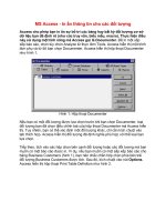
Fig. 1. Schematic picture of establishment of patient-specific induced pluripotent stem cells
(iPSCs), from which two prospective routes emerge1) in vivo transplantation 2) in vitro human
model system. Patient-specific induced pluripotent stem cells that are similar to embryonic
stem cells (ESCs) are produced by first 1) collecting adult somatic cells from the patient, for
example skin fibroblasts by a skin biopsy, 2) and reprogramming by retroviral transduction of
defined transcription factors (Oct4, c-Myc, Klf4 and Sox 2 or other combinations) in those
somatic fibroblast cells. Reprogrammed cells are selected by the detection of endogenous
expression of a reprogramming marker, for example Oct4. 3) Generated patient-specific iPSCs
can be genetically corrected of a known mutation that causes the disease. 4) Expansion of
genetically corrected patient-specific iPSCs theoretically in eternity. First prospective route
(Route 1): 5) upon external signals (or internal) iPSCs can theoretically be stimulated to
differentiate into any cell type in the body. 6) In this way patient-specific dopamine producing
nerve cells or skin cells can be generated and transplanted into individuals suffering from
Parkinson´s disease or Melanoma respectively. Second route (Route 2): Generated disease-
specific iPSCs can be used as a human in vitro system to study degenerative disorders or any
disease, cause of disease, screening for drugs or recapitulate development.
Generation of Patient Specific Stem Cells: A Human Model System
5
animals using somatic cell nuclear transfer (SCNT) to eggs. Such experiments can result in a
new individual from one differentiated somatic cell. The much more recent method to
reprogram cells was the fascinating finding that mouse embryonic fibroblasts (MEFs) can be
converted into induced pluripotent stem cells (iPSCs) by retroviral expression of four
transcription factors: Oct4, c-Myc, Sox2 and Klf4. iPSCs are a type of pluripotent stem cell
derived from a differentiated somatic cell by overexpression of a set of proteins. Nowadays,
several ways of generating iPSCs have been developed and includes 1) overexpression of
different combinations of transcription factors most efficiently in combination with
retroviruses (step 2 in Figure 1), 2) exposure to chemical compounds in combination with
the transcription factors Oct4, Klf4 and retroviruses, 3) retroviruses alone, 4) recombinant
proteins or 5) mRNA. The iPSCs are named pluripotent because of their ability to
differentiate into all different differentiation pathways. Generation of patient-specific iPSC
lines capable of giving rise to any desired cell type provides great opportunities to treat
many disorders either as therapeutic treatment or discovery of patient specific medicines in
human iPSC model systems (Figure 1). Here, some of this field’s fast progress and results
mostly concerning human cells are summarized.
2. Reprogramming-Induced Pluripotent Stem Cells (iPSCs)
Reprogramming is the process by which induced pluripotent stem cells (iPSCs) are
generated and is the conversion of adult differentiated somatic cells to an embryonic-like
state. Takahashi and Yamanaka demonstrated that retrovirus-mediated delivery of Oct4,
Sox2, c-Myc and Klf4 is capable of inducing pluripotency in mouse fibroblasts (Takahashi
and Yamanaka, 2006) and one year later was reported the successful reprogramming of
human somatic fibroblast cells into iPSCs using the same transcription factors (Takahashi et
al., 2007). Takahashi and Yamanaka came up with those four reprogramming proteins after
a search for regulators of pluripotency among 24 cherry picked pluripotency-associated
genes. These initial mouse iPSC lines differed from ESCs in that they had a diverse global
gene expression pattern compared to ESCs and failed to produce adult chimeric mice. Later
iPSCs were shown to have the ability to form live chimeric mice and were transmitted
through the germ line to offspring when using Oct4 or Nanog as selection marker for
reprogramming instead of Fbx15, which was used in the initial experiments (Meissner et al.,
2007; Okita et al., 2007; Wernig et al., 2007). Various combinations of the genes listed in table
1 have been used to obtain the induced pluripotent state in human somatic cells. The first
human iPSC lines were successfully generated by Oct4 and Sox2 combined with either, Klf4
and c-Myc, as used earlier in the mouse model, or Nanog and Lin28 (Lowry et al., 2008;
Nakagawa et al., 2008; Park et al., 2008b; Takahashi et al., 2007; Yu et al., 2007). Subsequent
reports have demonstrated that Sox2 can be replaced by Sox1, Klf4 by Klf2 and c-Myc by N-
myc or L-myc indicating that they are not fundamentally required for generation of iPSCs
(Yamanaka, 2009). Oct4 has not yet been successfully replaced by another member of the
Oct family to generate iPSCs which is logical due to the necessity of Oct4 in early
development. However, Blx-01294 an inhibitor of G9a histone methyl transferase, which is
involved in switching off Oct4 during differentiation, enables neural progenitor cells to be
reprogrammed without exogenous Oct4, although transduction of Klf4, c-Myc and Sox2
together with endogenous Oct4 was required (Shi et al., 2008). Recently, Oct4 has been
replaced with steroidogenic factor 1, which controls Oct4 expression in ESCs by binding the
New Advances in Stem Cell Transplantation
6
Oct4 proximal promoter, and iPSCs were produced without exogenous Oct4 (Heng et al.,
2010). Remarkably, exogenous expression of E-cadherin was reported to be able to replace
the requirement for Oct4 during reprogramming in the mouse system (Redmer et al.,
2011). iPSCs are similar to embryonic stem cells (ESCs) in morphology, proliferation and
ability to form teratomas. In mice, pluripotency of iPSCs has been proven by tetraploid
complementation (Zhao et al., 2009). Both ESCs and iPSCs can be used as the pluripotent
starting cells for the generation of differentiated cells or tissues in regenerative medicine.
However, the ethical dilemma associated with ESCs is avoided when using iPSCs since no
embryos are destroyed when iPSCs are obtained. Moreover, iPSCs can be patient-specific
and as such patient-specific drugs can be screened and in personalized regenerative
medicine therapies immune rejection could be circumvented. However the question
surrounding the potential immunogenicity remains unclear due to recent reports that
iPSCs do not form teratomas probably because iPSCs are rejected by the immune system
(Zhao et al., 2011).
Genes Description
Oct4
Transcription factor expressed in undifferentiated pluripotent
embryonic stem cells and germ cells during normal
development. Together with Nanog and Sox 2, is required for
the maintenance of
p
luri
p
otent
p
otential.
Sox2
Transcription factor expressed in undifferentiated pluripotent
embryonic stem cells and germ cells during development.
Together with Oct4 and Nanog, is necessary for the maintenance
of
p
luri
p
otent
p
otential.
Myc family
Proto-onco
g
enes, includin
g
c-M
y
c, first used for
g
eneration of
human and mouse iPSCs.
Klf family
Zinc-fin
g
er-containin
g
transcription factor Kruppel-like factor 4
(
KLF4
)
was first used for
g
eneration of human and mouse iPSCs
Nano
g
Homeodomai
n
-containin
g
transcription factor essential for
maintenance of pluripotency and self-renewal in embryonic
stem cells. Expression is controlled by a network of factors
includin
g
the ke
y
p
luri
p
otenc
y
re
g
ulator Oct4.
Lin 28
Conserved RNA bindin
g
protein and stem cell marker. Inhibitor
of microRNA processing in embryonic stem (ES) and carcinoma
(EC) cells.
Table 1. Combinations of the genes that have been used to obtain the induced pluripotent
state in human somatic cells
2.1 Differentiation of iPSCs into cells of the heart
After the cells have been reprogrammed, it will be possible to differentiate them towards a
wide range of specialized cells, using existing protocols for differentiation of hESCs.
Differentiation of beating heart cells, the cardiomyocytes, from hESCs has now been
achievable through various protocols for a decade (Kehat et al., 2001; Mummery et al., 2002).
In 2007, human iPSCs were first reported to differentiate into cardiomyocytes (Takahashi et
al., 2007), using a protocol including activin A and BMP4 which was described for
differentiation of hESCs the same year (Laflamme et al., 2007). A comparison between the
Generation of Patient Specific Stem Cells: A Human Model System
7
cardiac differentiation potential of hESCs and iPSCs concluded that the difference between
the two cell sources were no greater than the known differences between different hESC
lines and that iPSCs thus should be a viable alternative as an autologous cell source (Zhang
et al., 2009). Furthermore, a recent study demonstrated that reprogramming excluding c-
MYC yielded iPSCs which efficiently up-regulated a cardiac gene expression pattern and
showed spontaneous beating in contrast to iPSCs reprogrammed with four factors including
c-MYC (Martinez-Fernandez et al., 2010). On the transcriptional level, beating clusters from
both iPSCs and hESCs were found to be similarly enriched for cardiac genes, although a
small difference in their global gene expression profile was noted (Gupta et al., 2010). Taken
together, these results indicate that cardiomyocytes differentiated from both hESCs and
iPSCs are highly similar, although differences exist.
2.2 Additional methods to achieve reprogramming- 1.cloning = Somatic Cell Nuclear
Transfer (SCNT) 2.cell fusion 3.egg extract
In addition to the iPSC procedure other ways exist to reprogram somatic cells including: 1)
somatic cell nuclear transfer (SCNT), 2) cell fusion of somatic adult cells with pluripotent
ESCs to generate hybrid cells and 3) cell extract from ESCs or embryo carcinoma cells (ECs).
From the time when successful SCNT experiments, more commonly known as cloning, in
the frog Xenopus Laevis (Gurdon et al., 1958) to the creation of the sheep Dolly (Wilmut et al.,
1997), it has been proven that an adult cell nucleus transplanted into an unfertilized egg can
support development of a new individual, and researchers have focused on identifying the
molecular mechanisms that take place during this remarkable process. Even though SCNT
has been around for 50 years, the molecular mechanisms that take place inside the egg
remain largely unknown. The gigantic egg cell receiving a tiny nucleus is extremely difficult
to study. Single cell analysis are required and gene knock-out of egg proteins is very
challenging. In 2007 a report that the first primate ESCs were isolated from SCNT blastula
embryos of the species Rhesus Monkey was published (Byrne et al., 2007). The reason why it
took so long to perform successful SCNT in Rhesus Monkey was a technical issue; to
enucleate the egg, modified polarized light was used instead of traditional methods using
either mechanical removal of DNA or UV light mediated DNA destruction. The first reliable
publication of successful human SCNT reported generation of a single cloned blastocyst
(Stojkovic et al., 2005). Unfortunately, the dramatic advances in human SCNT reported by
Hwang and colleagues in South Korea were largely a product of fraud (Cho et al., 2006). In
human SCNT reports, left over eggs from IVF (in vitro fertilization) that failed to fertilize
have been used, indicating poor egg quality. However, human SCNT using 29 donated eggs
(oocytes) of good quality, and not leftovers from IVF, from three young women were
reported to develop into cloned blastocysts, at a frequency as high as 23% (French et al.,
2008). Theoretically, hESC lines can be derived in vitro from SCNT generated blastocysts.
However, so far no established hESC line using the SCNT procedure has been reported. The
shortage of donated high quality human eggs for research is a significant impediment for
this field.
Other methods that have been used to elucidate the molecular mechanism of
reprogramming are 2) fusion of somatic adult cells with pluripotent ESCs to generate hybrid
cells or 3) cell extract from ESCs or ECs (Bhutani et al., 2010; Cowan et al., 2005; Freberg et
al., 2007; Taranger et al., 2005; Yamanaka and Blau, 2010).
New Advances in Stem Cell Transplantation
8
3. Molecular mechanisms of reprogramming
The mechanisms of nuclear reprogramming are not yet completely understood. The crucial
event during reprogramming is the activation of ES- and the silencing of differentiation
markers, while the genetic code remains intact. Major reprogramming of gene expression
takes place inside the egg and genes that have been silenced during embryo development
are awakened. In contrast, genes that are expressed in, and are specific for, the donated cell
nucleus become inactivated most of the time, however some SCNT embryos remember their
heritage and fail to inactivate somatic-specific genes (Ng and Gurdon, 2008). It has been
reported that reprogramming involves changes in chromatin structure and chromatin
components (Jullien et al., 2010; Kikyo et al., 2000). Importantly, initiation of Oct4 expression
has been found to be crucial for successful nuclear transfers (Boiani et al., 2002; Byrne et al.,
2003) and important for iPSC creation; all other reprogramming iPSC transcription factors
have been replaced with other factors or chemical compounds, but only one report so far
could exclude Oct4. In murine ES cells, Oct4 must hold a precise level to maintain them as
just ES cells (Niwa et al., 2000) and therefore understanding the control of the Oct4 level will
be key if one wants to understand pluripotency and reprogramming at the molecular level.
A recent report demonstrated that Oct4 expression is regulated by scaffold attachment
factor A (SAF-A). SAF-A was found on the Oct4 promoter only when the gene is actively
transcribed in murine ESCs, depending on LIF, and gene silencing of SAF-A in ESCs
resulted in down regulation of Oct4 (Vizlin-Hodzic et al., 2011). Other Oct4 modulators have
been reported that in similarity with SAF-A are in complex with RNA polymerase II (Ding
et al., 2009; Ponnusamy et al., 2009). Post-translational modifications have been shown to be
able to modify the activity of Oct4, such as sumoylation (Wei et al., 2007) and ubiquitination
(Xu et al., 2004). During the reprogramming process epigenetic marks are changed such as
the removal of methyl groups on DNA (DNA demethylation) of the Oct4 promoter which
has been shown during SCNT (Simonsson and Gurdon, 2004) and has also been observed in
mouse (Yamazaki et al., 2006). The growth arrest and DNA damage inducible protein
Gadd45a and deaminase Aid was shown to promote DNA demethylation of the Oct4 and
Nanog promoters (Barreto et al., 2007; Bhutani et al., 2010). Consistent with those findings
is that Aid together with Gadd45 and Mbd4 has been shown to promote DNA
demethylation in zebrafish (Rai et al., 2008). Translational tumor protein (Tpt1) has been
proposed to control Oct4 and shown to interact with nucleophosmin (Npm1) during
mitosis of ESCs and such complexes are involved in cell proliferation (Johansson et al.,
2010b; Koziol et al., 2007). Furthermore, phosphorylated nucleolin (Ncl-P) interacts with
Oct4 during interphase in both murine and human ESCs (Johansson et al., 2010a). Core
transcription factors, Oct4, Sox2 and Nanog, were shown to individually form complexes
with nucleophosmin (Npm1) to control ESCs (Johansson and Simonsson, 2010). ESCs also
display high levels of telomerase activity which maintain the length of the telomeres. The
telomerase activity or Tert gene expression is rapidly down regulated during
differentiation and are much lower or absent in somatic cells. Therefore, reestablishment
of high telomerase activity (or reactivation of Tert gene) is important for reprogramming.
In SCNT animals, telomere length in somatic cells has been reported to be comparable to
that in normally fertilized animals (Betts et al., 2001; Lanza et al., 2000; Tian et al., 2000). A
telomere length-resetting mechanism has been identified in the Xenopus egg (Vizlin-
Hodzic et al., 2009).
Generation of Patient Specific Stem Cells: A Human Model System
9
When iPSCs first were introduced many thought that the molecular mechanism of
reprogramming was solved once and for all. It was soon shown that to generate iPSC
colonies one could use different combinations of transcription factors most efficiently
together with retroviruses or more recently, exposure to chemical compounds together with
the transcription factors, Oct4 and Klf4, and with retroviruses (Zhu et al., 2010) or
retroviruses alone (Kane et al., 2010). What retroviruses do for the reprogramming process is
unknown and the efficiency by which the egg reprograms the somatic cells is far more
efficient than the iPSC procedure. Moreover, mutagenic effects have been documented in
both laboratory and clinical gene therapy studies, principally as a result of a dysregulated
host gene expression in the proximity of gene integration sites. So the first question to ask is
whether all iPSC experiments so far forgot the obvious control of using only virus. The
answer is probably no because the efficiency is very low with viruses alone as compared to
using transcription factors combined with virus or identified reprogramming compounds.
Reprogramming an adult somatic frog cell nucleus to generate a normal “clonal“ new
individual is far less efficient (0.1-3%) than reprogramming to create a blastocyst, from
which ESCs are isolated (efficiency 20-40%) (Gurdon, 2008) and is comparable with blastula
formation after human SCNT (23%). This number could be compared with iPSC procedure
that has reported 0.5 % success rate at most with human cells (table 1). The low efficiency
and slow kinetics of iPSC derivation suggest that there are other procedures that are more
efficient, yet to decipher. There is a belief that there are different levels of pluripotency when
it comes to ESC and also that reprogramming follows an organized sequence of events,
beginning with downregulation of somatic markers and activation of pluripotency markers
alkaline phosphatase, SSEA-4, and Fbxo15 before pluripotency endogenous genes such as
Oct4, Nanog, Tra1-60 and Tra-1-80 become expressed and cells gain independence from
exogenous transcription factor expression (Brambrink et al., 2008; Stadtfeld et al., 2008a).
Only a small subset of somatic cells expressing the reprogramming factors down-regulates
somatic markers and activates pluripotency genes (Wernig et al., 2008a).
3.1 History of reprogramming
SCNT has been around for more than fifty years although it was already proposed in 1938
by Hans Spemann (Spemann, 1938), an embryologist who received the Nobel Prize in
Medicine for his development of new embryological micro surgery techniques. Spemann
anticipated that “transplanting an older nucleus into an egg would be a fantastic
experiment”. Later on, Robert Briggs and Thomas King were the first to put the nuclear
transfer technique into practice. However, they only managed to obtain viable offspring
through nuclear transfer of undifferentiated cells in the frog species Rana pipiens (Briggs and
King, 1952). During the 1950s to the 1970s a series of pioneering somatic nuclear transfer
experiments performed by John Gurdon showed that nuclei from differentiated amphibian
cells, for example tadpole intestinal or adult skin cells could generate cloned tadpoles
(Gurdon, 1962; Gurdon et al., 1958; Gurdon et al., 1975). In 1997, the successful cloning of a
mammal was first achieved. The sheep Dolly was produced by using the nuclei of cells
cultured from an adult mammary gland (Wilmut et al., 1997). Following the cloning of
Dolly, researchers have reported successful cloning of a number of species including cow,
pig, mouse, rabbit, cat (named Copycat) and monkey. In 2006, reprogrammed murine iPSCs
were reported by Takahashi and Yamanaka (Takahashi and Yamanaka, 2006) and in 2007
human iPSCs were reported (Takahashi et al., 2007; Yu et al., 2009).
New Advances in Stem Cell Transplantation
10
4. Producing iPSCs from other cell types than fibroblasts
The most studied somatic cell type that has been reprogrammed into iPSCs is fibroblasts.
The different human somatic cell types that have been transformed into iPSCs so far are
summarized in table 2. The efficiency of fibroblast reprogramming does not exceed 1-5% but
generally is extremely inefficient (0.001-0.1%) and occurs at a slow speed (> 2 weeks). In
order to use iPSCs in clinical applications, improved efficiency, suitable factor delivery
techniques and identification of true reprogrammed cells are crucial. In the fast growing
field of regenerative medicine, patient-specific iPSCs offer a unique source of autologous
cells for clinical applications. Although promising, using somatic cells of an adult individual
as starting material for reprogramming in this context has also raised concern. Acquired
somatic mutations that have been accumulated during an individual’s life time will be
transferred to the iPSCs, and there is a fear that these mutations may be associated with
adverse events such as cancer development. As an alternative, iPSCs have been generated
from human cord blood. These cells have been shown to differentiate into all three germ
layers including spontaneous beating cardiomyocytes (Haase et al., 2009). Reprogrammed
cells from cord blood have not only the advantage to come from a juvenescent cell source. In
addition, cord blood is already routinely harvested for clinical use.
Another issue that has been raised in this field is a wish to harvest cells for
reprogramming without surgical intervention. Therefore, reprogramming experiments
have also been performed using plucked human hair follicle keratinocytes. These iPSCs
were also able to differentiate into cells from all three germ layers including
cardiomyocytes (Novak et al., 2010).
Human Ori
g
in
Somatic Cell t
yp
e
Efficienc
y
Repro
g
rammin
g
Factors
Reference
Fibroblasts 0.02%
0.02%
0.002%
OKSM
OSLN
OKS
(Takahashi et al., 2007)
(Yu et al., 2007)
(
Naka
g
awa et al., 2008
)
He
p
atoc
y
tes 0.1% OKSM
(
Liu et al., 2010
)
Keratinoc
y
tes ND
ND
OKSM
OKS
(Aasen et al., 2008)
(
Aasen et al., 2008
)
Neural stem cells <0.004% O
(
Kim et al., 2008
)
Amniotic cells 0.05-1.5%
0.1%
OKSM
OSN
(Li et al., 2009)
(
Zhao et al., 2010
)
Adipose-derived stem cells 0.5%
<0.1%
OKSM
OKS
(Su
g
ii et al., 2010)
(
Aoki et al., 2010
)
Cord blood stem cells ND
<0.01%
OKSM
OS
(Eminli et al., 2009)
(
Gior
g
etti et al., 2009
)
Cord blood endothelial cells <0.01% OSLN
(
Haase et al., 2009
)
Mobilized
p
eri
p
heral blood 0.01% OKSM
(
Loh et al., 2009
)
Table 2. Different somatic cell types that human iPSCs have been generated from
4.1 iPSC as a disease model
The introduction of iPSC technology holds a great promise for disease modelling. By
differentiating iPSCs from patients into various cell lineages there is hope to be able to
follow the disease progression and to identify new prognostic markers as well as to use the
differentiated cells for drug screening in both toxicological testing and the development of
Generation of Patient Specific Stem Cells: A Human Model System
11
new treatment. This approach has already been tested for monogenic diseases using
genetically modified hESCs or hESCs from embryos carrying these diseases (reviewed in
(Stephenson et al., 2009)). However, diseases with a more complex genetic background
involving several or unknown genes have not been able to be studied in this way before
iPSCs became available. An additional advantage with iPSCs is that since many diseases
differ in both clinical symptoms and penetrance between patients, iPSCs derived from
patients will offer the opportunity to reveal a clinical history as well. It could also provide a
model for late-onset degenerative diseases such as Alzheimer’s disease or osteoarthritis.
Recent work on cardiac arrhythmias has fully shown the potential of disease modelling
using iPSCs. Long QT syndrome (LQTS) is characterized by rapid irregular heart beats due
to abnormal ion channel function and the condition can lead to sudden death. So far,
various mutations in at least 12 different genes have been associated with LQTS and the
disease is subdivided into different types depending on which gene is affected (reviewed in
(Bokil et al., 2010)). Fibroblasts from patients with LQTS1 (Moretti et al., 2010) and LQTS2
(Itzhaki et al., 2011; Matsa et al., 2011) were reprogrammed and differentiated into the
cardiac lineage. These cells displayed the electrophysiological pattern characteristic to the
disease. Moreover, the cells responded appropriately when treated with pharmacological
compounds, which further extends the usability of these cells.
iPSCs have also been generated from fibroblasts from patients suffering from the LEOPARD
syndrome, an autosomal-dominant developmental disorder where one of the major disease
phenotypes includes hyperthropic cardiomyopathy. The authors showed that
cardiomyocytes derived from those iPSCs were larger with another intracellular
organization compared to cardiomyocytes derived from hESCs or iPSCs generated from a
healthy sibling (Carvajal-Vergara et al., 2010). Today many laboratories and hospitals
worldwide are producing iPSC lines from patients with various diseases. Patient-specific
iPSC lines can be used as 1) a human modelling system for studying the molecular cause of,
and in the long run for 2) the treatment of, degenerative diseases with autologous
transplantation, which refers to the transplantation to a patient of his/her own cells. The
therapeutic potential of iPSCs in combination with genetic repair has already been
successfully shown in mouse models of sickle cell anemia (Hanna et al., 2007), Duchenne
muscular dystrophy (DMD) (Kazuki et al., 2010), hemophilia A (Xu et al., 2009) and, in a rat
model, Parkinson’s disease (Wernig et al., 2008c). For diseases where animal and human
physiology differ, disease-specific iPSC lines capable of differentiation into the tissue
affected by the disease could recapitulate tissue formation and thereby enable determination
of the cause of the disease and could provide cues to drug targets. Therefore iPSC lines from
patients suffering from a variety of genetic diseases with either Mendelian or complex
inheritance have been secured for future research, and include deaminase deficiency-related
severe combined immunodeficiency (ADA-SCID), Shwachman-Bodian-Diamond syndrome
(SBDS), Gaucher disease (GD) type III, Duchenne (DMD) and Becker muscular dystrophy
(BMD), Parkinson disease (PD), Huntington disease (HD), juvenile-onset (type1) diabetes
mellitus (JDM), Downs syndrome (DS)/trisomy21 and Lesch-Nyhan syndrome (Park et al.,
2008a). Furthermore, iPSCs derived from amyotrophic lateral sclerosis (ALS) patients were
terminally differentiated into motor neurons (Dimos et al., 2008).
4.2 Procedures to produce iPSCs
In the first iPSC reprogramming studies, retroviral or lentiviral vectors were used to
introduce the transcription factors into somatic cells. By using these viral delivery systems,
New Advances in Stem Cell Transplantation
12
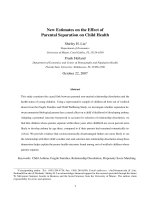
Fig. 2. Methods for producing induced pluripotent stem cells (iPSCs) by non-integrating
vectors. Several different methods exist to generate iPSCs by non-integrating vectors: for
Generation of Patient Specific Stem Cells: A Human Model System
13
example by plasmid, episomal, adenoviral minicircle vectors and mRNA. a) A combination
of expression plasmid vectors for defined reprogramming factors is transfected into somatic
cells. Plasmid vectors are not integrated into the genome of transfected cells and are
gradually lost during reprogramming. This method therefore requires multiple transfection
steps. b) Somatic cells can be transfected by episomal vectors expressing defined
reprogramming factors. These vectors can replicate themselves autonomously in cells
during reprogramming under drug selection and are not integrated into the genome. Upon
withdrawal of drug selection, the episomal vectors are lost. c) Adenovirus carrying defined
reprogramming factors can be infected into somatic cells to transiently express these factors.
This method requires multiple transductions since adenoviral vectors are lost upon
celldivision. d) The minicircle vector method is based on PhiC31-vector intra molecular
recombinant system that allows the bacterial elements of the vector to be degraded in
bacteria. Minicircle vector containing only defined reprogramming factors is not degraded
and is delivered into somatic cells by nucleofection. This strategy requires multiple
transfection steps since minicircle vectors are lost upon cell division. e) Reprogramming
using mRNA reprogramming factors have been achieved.
the transduced viral vectors and transgenes are randomly and permanently integrated
into the genome of infected somatic cells and remains in the iPSCs. The vector integration
into the host genome is a limitation of this technology if it is going to be used in human
therapeutic applications due to increased risk of tumor formation (Okita et al., 2007).
Approaches to derive transgene-free iPSCs are therefore critical. The first strategy was by
using non-integrating (Figure 2) vectors. Efforts have been made to derive iPSCs by
repeated plasmid transfections (Gonzalez et al., 2009; Okita et al., 2008) (Figure 2a),
adenoviral (Stadtfeld et al., 2008b) (Figure 2b) and episomal vectors (Yu et al., 2009)
(Figure 2c). Recently, minicircle vectors (Figure 2d) have been used to generate iPSCs (Jia
et al., 2010). Unfortunately, reprogramming with these techniques has extremely low
efficiency as compared to integrating viral vectors. Another promising alternative is the
use of excisable integrating vectors, allowing for the generation of transgene-free iPSCs. A
classical expression-excision system uses vectors with inserts flanked with recognition
sites, loxP sites, for Cre-recombinase (Figure 3a). Consequently, DNA is excised upon Cre-
recombinase expression in the cells. Cre-loxP-based approaches have been used to
reprogram human somatic cells from individuals with Parkinson’s disease by four
different vectors (Soldner et al., 2009) or by a single, polycistronic lentiviral vector
encoding reprogramming factors (Chang et al., 2009). Though, a potential limitation of
Cre-loxP-based approaches is that a long terminal repeat (LTR) will remain after Cre-
mediated excision which may interfere with the expression of endogenous genes. An
alternative integration-free strategy is based on the piggy-Bac transposon (Figure 3b), a
mobile genetic element from insects that integrates into the genome of mammalian cells
and, most importantly, can be entirely removed by a transposase. Two research teams
generated iPSCs using this system to deliver a single polycistron encoding four
reprogramming factors into somatic cells (Woltjen et al., 2009; Yusa et al., 2009).
Interestingly, the latest development indicates that gene transfection may not even be
needed for the generation of iPSCs and that direct delivery of four recombinant
reprogramming proteins that can penetrate the plasma membrane of somatic cells is
sufficient (Zhou et al., 2009), or mRNA (Angel &Yanik, 2010; Plews et al., 2010; Warren et
al. 2010; Yakoba et al., 2010; Zhou et al.,2009).